Cox-2 and its association with prognostic factors and response to
Cox-2 and its association with prognostic factors and response to
Cox-2 and its association with prognostic factors and response to
Cox-2 and its association with prognostic factors and response to
Cox-2 and its association with prognostic factors and response to
primary chemotherapy in patients with breast cancer
primary chemotherapy in patients with breast cancer
primary chemotherapy in patients with breast cancer
primary chemotherapy in patients with breast cancer
primary chemotherapy in patients with breast cancer
Cox-2 e sua associação com fatores prognósticos e resposta à quimioterapia
Cox-2 e sua associação com fatores prognósticos e resposta à quimioterapia
Cox-2 e sua associação com fatores prognósticos e resposta à quimioterapia
Cox-2 e sua associação com fatores prognósticos e resposta à quimioterapia
Cox-2 e sua associação com fatores prognósticos e resposta à quimioterapia
primária em pacientes com câncer de mama
primária em pacientes com câncer de mama
primária em pacientes com câncer de mama
primária em pacientes com câncer de mama
primária em pacientes com câncer de mama
RENATODE LIMA ROZENOWICZ, ACBC-SP1; ROBERTO EUZÉBIODOS SANTOS2; MARIA ANTONIETA LONGO GALVÃO SILVA3; FABIO FRANCISCO
OLIVEIRA RODRIGUES, TCBC-SP4; ANDRÉ LIMADE OLIVEIRA5; LILIANE BARATELA ULSON6; VILMAR MARQUES OLIVEIRA7; TSUTOMU AOKI8
A B S T R A C T A B S T R A C T A B S T R A C T A B S T R A C T A B S T R A C T
Objective: Objective:Objective:
Objective:Objective: To evaluate the immunohistochemical expression of cox-2 before primary chemotherapy with 5-fluorouracil, epirubicin and cyclophosphamide (FEC) and its association with initial tumor size, lymph node status, hormone receptors, expression of HER2 and the clinical and pathological response in patients with breast cancer. Methods:Methods:Methods:Methods:Methods: We conducted a retrospective study with 41 women with histopathological diagnosis of ductal breast carcinoma. They underwent primary chemotherapy with FEC regimen (5-fluorouracil, epirubicin and cyclophosphamide) at 500mg/m2, 75mg/m2 and 500 mg/m2, respectively. Inclusion criteria were age range between 30 and 70 years, stage II to IIIA, absence of metastasis, primary tumor of the breast, single, unilateral, with ductal invasion at histology and absence of heart disease and pregnancy. To evaluate the expression of HER2/neu protein we used rabbit monoclonal antibodies. To visualize the expression of cox-2 protein we used polyclonal antibodies obtained from goats’ serum. The evaluation of clinical response to treatment was performed during physical examination by measuring the major tumor axis with a pachymeter. Measurements were taken at admission and after primary chemotherapy cycles. After three chemotherapy sessions at intervals of 21 days the surgical procedure was carried out. We adopted the criteria of RECIST. After the operation we evaluated the local pathological response, which was considered complete when there was absence of invasive neoplasia and of the in situ component. In immunohistochemical assessing of estrogen receptors we used estrogen receptor NCL-ER6F11 and, for progesterone, progesterone receptor NCL-PGR-312, considering positive the staining of 10% or more tumor cells. Results:Results:Results:Results:Results: The distribution according to UICC clinical stage classified six patients in stage IIA (14.6%), 22 in stage IIB (53.6%) and 13 stage IIIA (31.8%). The initial clinical evaluation of the major tumor axis ranged from 2.5 to 15 cm and a median of 5 cm. We identified 14 patients (34.1%) with negative lymph node status, and 27 positive (65.9%). It was observed that 19 (46.3%) were in premenopause and 22 (53.6%) in menopause. Conclusion:Conclusion:Conclusion:Conclusion:Conclusion: There was an association of the expression of Cox-2 to the factors associated with poor prognosis in breast cancer, such as positive lymph node status, negative hormone receptors and HER2 expression.
Key words Key wordsKey words
Key wordsKey words: Chemotherapy, adjuvant. Breast neoplasms.
Work done at Department of Obstetrics and Gynecology of the Brotherhood of Santa Casa de São Paulo (DOGI-IMSCSP)- SP-BR.
1. Assistant Physician, DOGI Pelvic Oncology Clinic, Brotherhood of the Santa Casa de São Paulo-SP-BR; 2. Head, DOGI Pelvic Oncology Clinic, Brotherhood of the Santa Casa de São Paulo-SP-BR; 3. Assistant Professor, Anatomy and Pathology Department-FCMSCSP- SP-BR; 4. Assistant Physician, DOGI Pelvic Oncology Clinic, Brotherhood of the Santa Casa de São Paulo-SP-BR; 5. Assistant Physician, DOGI General Gynecology Clinic, Brotherhood of the Santa Casa de São Paulo-SP-BR; 6. Post-graduate-FCMSCSP- SP-BR; 7. Assistant Physician, DOGI Mastology Clinic,
Brotherhood of the Santa Casa de São Paulo-SP-BR; 8. Director, Department of Obstetrics and Gynecology, Brotherhood of the Santa Casa de São Paulo-SP-BR.
INTRODUCTION
INTRODUCTION
INTRODUCTION
INTRODUCTION
INTRODUCTION
I
n 2008 there were estimated 49,470 new cases of breast cancer with 9,170 deaths in Brazil. Despite advances in screening and treatment of this disease, the mortality rate increased 76% over the 1970-2000 period, from 5.7 per 100,000 women in 1979 to 10.15 per 100,000 women in 20021.Neoadjuvant chemotherapy has advantages such as possibility of in vivo study of chemotherapy and increased
Liu et al. observed that transgenic mice with overexpression of Cox-2 developed breast cancer in an 85% rate, suggesting the involvement of this enzyme in mammary gland carcinogenesis3. Other studies correlate its expression with invasive and metastatic stimuli in breast cancer4.
This study aims to evaluate the immunohistochemical expression of Cox-2 and its association with tumor size, lymph node clinical status, hormone receptors, expression of HER2/neu and the pathological and clinical response to primary chemotherapy in women with ductal carcinoma of the breast stages II or III.
METHODS
METHODS
METHODS
METHODS
METHODS
We conducted a retrospective study based on the database of the Chemotherapy Service, Department of Obstetrics and Gynecology (DOGI) of the Brotherhood of Santa Casa de Misericórdia de Sao Paulo (ISCMSP). We evaluated 41 women treated between July 2004 and July 2006, with histopathologic diagnosis of breast ductal carci-noma.
This group of patients underwent primary chemotherapy with FEC regimen (5-fluorouracil, epirubicin and cyclophosphamide) at 500mg/m2, 75mg/m2 and 500 mg/m2, respectively, after biopsy. Inclusion criteria were age range between 30 and 70 years, stage II to IIIA according to the criteria of UICC’s Sixth Edition, absence of metastasis (bone scan, chest radiograph, abdominal ultrasound and CA 15-3), primary breast tumor, and single-sided (as assessed by mammography), invasive ductal type at histology and absence of heart disease and pregnancy. In this sample, age ranged between 32 and 70 years, mean 49.9 years, standard deviation of 10.25 and a median of 50 years. Seven (17.1%) patients were in the group under 40 years, 27 (65.8%) between 41 and 60 years and seven (17.1%) over 60 years.
Distribution according to UICC clinical stage classified six patients in stage IIA (14.6%), 22 in stage IIB (53.6%) and 13 in stage IIIA (31.8%).
The initial clinical evaluation of the major tumor axis ranged from 2.5 to 15 cm with a mean of 5.9 cm (standard deviation of 2.43 cm) and a median of 5 cm, whereas 19 patients (46.3%) had a tumor larger than 5 cm and 22 (53.7%) less than or equal to 5 cm. We identified 14 patients (34.1%) with negative lymph node status and 27 positive (65.9%). It was observed that 19 (46.3%) patients were premenopausal and 22 (53.6%) were at menopause.
Immunohistochemical evaluation Immunohistochemical evaluation Immunohistochemical evaluation Immunohistochemical evaluation Immunohistochemical evaluation
The biopsy material prior to chemotherapy was fixed in buffered 10% formaldehyde and sent to the Department of Pathological Sciences, ISCMSP. Immunohistochemistry of the present study was performed
according to the protocol of that Pathology Department. For estrogen receptors we used estrogen recep-tor-NCL ER6F11 (Mouse Monoclonal Antibody Novocastra, Norwell, MA, USA, dilution: 1/55 overnight) and for progesterone, progesterone receptor NCL-PGR-312 (Mouse Monoclonal Antibody Novocastra, Norwell, MA, USA, dilution: 1/100 overnight). We considered positive the staining of 10% or more tumor cells.
We used rabbit antibodies (Dako Cop., Carpinteria, CA, USA) to evaluate the expression of HER2/ neu protein in the plasma membrane of cancer cells, as proposed by Abreu e Lima et al.5.
Polyclonal antibodies obtained from serum of goats (3362-100 Biovision Research Products, dilution 1:70) were used to assess the expression of Cox-2 protein, the positive control being performed in biopsies of breast tissue with an inflammatory process. Analysis of immunohistochemistry was performed according to the criteria proposed by Ristimäki et al.6, based on the intensity of staining of the cytoplasm and cell membrane and it was considered positive when the grade was 2 or 3.
Evaluation of tumor response Evaluation of tumor response Evaluation of tumor response Evaluation of tumor response Evaluation of tumor response
The evaluation of clinical response to treatment was performed by physical examination, measuring the major tumor axis with a pachymeter. Measurements were taken at admission and after primary chemotherapy cycles, by the same examiner, recorded in medical charts and in the Department of Chemotherapy database. After three chemotherapy sessions at intervals of 21 days patients were submitted to surgery.
We adopted the criteria of RECIST (Response Evaluation Criteria In Solid Tumors) as proposed by Therrase et al7 , considering patients with complete or partial response as the responder group, and patients with disease progression or stable disease as the non-respon-der one.
After the operation we evaluated local pathological response, which was considered complete when in the absence of invasive neoplasia or an in situ component, as recommended by Sataloff et al.8.
Statistical analysis Statistical analysis Statistical analysis Statistical analysis Statistical analysis
We used SPSS 15 software for Windows, the expression of Cox-2 being associated with the studied variables by Fisher exact test. A 5% level of rejection of the null hypothesis was set for the variables analyzed.
RESULTS
RESULTS
RESULTS
RESULTS
RESULTS
After neoadjuvant chemotherapy, the tumor major axis ranged from zero to 8.5 cm with a mean of 3.59 cm, standard deviation of 2.14 cm and a median of 3.0 cm.
The clinical response based on RECIST criteria showed the following distribution: 25 (60.98%) responders and 16 (39.02%) non-responders. Histopathological evaluation showed complete response in five patients (12.19%) (Tables 1 and 2).
DISCUSSION
DISCUSSION
DISCUSSION
DISCUSSION
DISCUSSION
The study of Cox-2 and its association with response to neoadjuvant chemotherapy in breast cancer is appropriate, since the identification of response biomarkers
will provide the finding of an ideal group of patients who will benefit from breast preservation and less toxicity. In this study we observed the expression of Cox-2 in 56% of all cases, confronting the data from the literature, which shows its expression in approximately 40% 9.
No correlation was found between the expression of Cox-2 and initial tumor size, also diverging from the literature, where clear association of expression with larger tumors was found10.
The lymph node status is a major prognostic factor in breast carcinoma; its positivity confers 35% risk for death by disease in five years. Costa et al.11 studied 46 Portuguese women treated at the University of Porto and showed a correlation between the expression of Cox-2 in the tumor and positive lymph node status, converging with the literature6,10. We found a positive association between
CoxTable 1 Table 1 Table 1 Table 1
-Table 1 - Correlation between expression of Cox-2 and prognostic factors in 41 patients with invasive ductal carcinoma of the breast undergoing primary chemotherapy.
Tumor Diameter (cm) Tumor Diameter (cm) Tumor Diameter (cm) Tumor Diameter (cm) Tumor Diameter (cm)
Cox-2 <5 >5
Negative (%) 7 (31,9) 11 (57,9)
Positive (%) 15 (68,1) 8 (42,1) p=0,12
Clinically affected limph nodes Clinically affected limph nodes Clinically affected limph nodes Clinically affected limph nodes Clinically affected limph nodes
Negative Positive
Negative (%) 10 (71,5) 8 (29,7)
Positive (%) 4 (28,5) 19 (70,3) p=0,019
Estrogenic Receptor Estrogenic Receptor Estrogenic Receptor Estrogenic Receptor Estrogenic Receptor
Negative Positive
Negative (%) 4 (25) 14 (56)
Positive (%) 12 (75) 11 (44) p=0,050
Progesterone Receptor Progesterone Receptor Progesterone Receptor Progesterone Receptor Progesterone Receptor
Negative Positive
Negative (%) 4 (23,6) 14 (59,4)
Positive (%) 13 (76,4) 10 (41,6) p=0,028
HER2/neu Expression HER2/neu Expression HER2/neu Expression HER2/neu Expression HER2/neu Expression
Negative Positive
Negative (%) 17 (70,8%) 1 (5,8%)
Positive (%) 7 (29,2%) 16 (94,2%) p=0,001
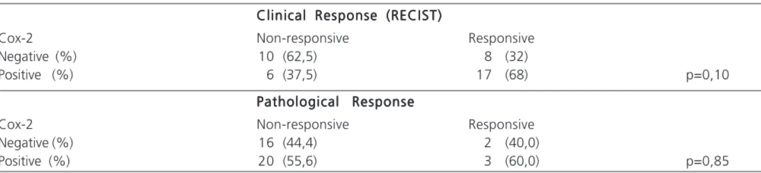
Table 2 Table 2 Table 2 Table 2
-Table 2 - Correlation between expression of Cox-2, clinical response and pathological response in 41 patients with invasive ductal carcinoma of the breast undergoing primary chemotherapy.
Clinical Response (RECIST) Clinical Response (RECIST) Clinical Response (RECIST) Clinical Response (RECIST) Clinical Response (RECIST)
Cox-2 Non-responsive Responsive
Negative (%) 10 (62,5) 8 (32)
Positive (%) 6 (37,5) 17 (68) p=0,10
Pathological Response Pathological Response Pathological Response Pathological Response Pathological Response
Cox-2 Non-responsive Responsive
Negative (%) 16 (44,4) 2 (40,0)
2 and positive clinical lymph node status, corroborating the data from those authors and those of Grudzinski et al.12 that found a positive association of expression with lymph nodes affected in patients from our country. These data emphasize the likely role of Cox-2 in the mechanisms lymphangiogenesis with disease dissemination, corroborating in vitro findings of stimulation of VEGF-C by Cox-213.
We observed, through analysis of hormone re-ceptor function in the expression of Cox-2, an inverse relationship between these variables, as found by other authors 6,10. McCarthy et al.14 also observed, by RT-PCR, a significant correlation between a positive expression of Cox-2 and negative hormone receptor, indicating a worse prognosis, despite increased susceptibility to chemotherapy. One possible mechanism that explains this negative association was proposed in an in vitro study by Mendelson & Hardy15, who observed the inhibition of Cox-2 gene transcription by blocking the p65 subunit of NF-Kâ using chromatin immunoprecipitation (ChIP) in culture medium with progesterone. These authors concluded that, by interfering in this expression, the progesterone receptor plays a crucial role in blocking the route of mammary carcinogenesis mediated by prostaglandin E2 and aromatase, agreeing with the NSABP B-09 study, which showed greater benefit from tamoxifen in patients with positive progestagen receptor tumors.
The relationship between the signaling of HER2/ neu and the expression of Cox-2 is well evidenced in the literature by proposing a biological model in which there is induction of Cox-2 by HER2 via Ras/MAPK, with Cox-2 positive feedback with HER16. We observed the expression of Cox-2 in 94.2% of the HER2 positive group with a highly significant association, a finding similar to the ones in the literature17.
The studies on the association of the expression of Cox-2 with response to primary chemotherapy are scarce,
but there is evidence that the expression of Cox-2 is related to resistance to chemotherapy. Surowiak et. al.18 used immunohistochemistry to analyze the expression of the Cox-2 protein and drug resistance, MDR-1, in 104 Polish women with ductal carcinoma submitted to adjuvant chemotherapy, and found a close association between the expression of Cox-2 and MDR-1. Besides demonstrating the lowest response in the group that expressed Cox-2, it suggested that Cox-2 might be related to efflux of intracellular anthracyclines by mechanisms associated with MDR 1, promoting resistance to chemotherapeutic agents.
In patients with esophageal carcinoma the expression of Cox-2 was correlated with decreased response to neoadjuvant chemoradiotherapy19, corroborating Ferrandina et al.20, who showed lower response to chemotherapy in gynecological cancer, demonstrating chemoresistance in tumors expressing Cox-2.
We noted the expression of Cox-2 in 68% of the responder group and in 37.5% of the non-responder. Although these data show a correlation between the behavior of the Cox-2 expression and good response to primary chemotherapy with FEC, no association was statistically significant. We did not observe an association between the pathological response and the expression of Cox-2.
Though there was no association with response to neoadjuvant chemotherapy, new research on the Cox-2 should be carried out in order to establish its true role as a predictor of response to chemotherapy in patients with breast cancer.
CONCLUSION
CONCLUSION
CONCLUSION
CONCLUSION
CONCLUSION
There was an association between the expression of Cox-2 and factors related to poor prognosis in breast cancer, such as positive lymph node status, negative hormone receptor and HER2 expression.
R E S U M O R E S U M O R E S U M O R E S U M O R E S U M O
Objetivo: Objetivo: Objetivo: Objetivo:
mediana de 5 cm. Foram identificadas 14 pacientes (34,1%) com estado linfonodal negativo e 27 positivo (65,9%). Observou-se que 19 (46,3%) apresentavam-se no menacme e 22 (53,6%) na menopausa. ConclusãoConclusãoConclusãoConclusãoConclusão:Houve associação da expressão da cox-2 à fatores de pior prognóstico no câncer de mama como estado linfonodal positivo, receptores hormonais negativos e expressão da Her-2.
Descritores DescritoresDescritores
DescritoresDescritores: Quimioterapia adjuvante. Neoplasias da mama.
REFERENCES
REFERENCES
REFERENCES
REFERENCES
REFERENCES
1. Brasil. Ministério da Saúde. INCA. Estimativa 2008: Incidência de câncer no Brasil. [on line] Disponível em: www.inca.gov.br/estima-tiva/2008/ [maio 2009]
2. Méric JB, Rottey S, Olaussen K, Soria JC, Khayat D, Rixe O, et al. Cyclooxygenase-2 as a target for anticancer drug development. [Review] Crit Rev Oncol Hematol. 2006; 59:51-64.
3. Liu CH, Chang SH, Narko K, Trifan OC, Wu MT, Smith E, et al. Overexpression of cyclooxygenase-2 is sufficient to induce tumorigenesis in transgenic mice. J Biol Chem. 2001; 276:18563-9.
4. Stasinopoulos I, Mori N, Bhujwalla Z.The malignant phenotype of breast cancer cells is reduced by cox-2 silencing.Neoplasia.2008;10:1163-9.
5. Abreu e Lima, M C; Gobbi, H; Gianotti Filho, O; Alvarenga, M. Lesões benignas não neoplásicas e neoplasias da mama. In: Bacchi, CE; Cardoso de Almeida, PC; Franco, M. Manual de padronização de laudos histopatológicos. 3ª. Edição São Paulo: Sociedade Brasi-leira de Patologia; 2005. p 265-6.
6. Ristimäki A, Sivula A, Lundin J, Lundin M, Salminen T, Haglund C, et al. Prognostic significance of elevated cyclooxygenase-2 expression in breast cancer. Cancer Res 2002; 62:632-5. 7. Therasse P, Eisenhauer EA, Verweij J. RECIST revisited: a review
of validation studies on tumor assessment. Eur J Cancer. 2006; 42(8):1031-9.
8. Sataloff DM, Mason BA, Prestipino AJ, Seinige UL, Leieber CP, Baloch Z. Pathologic response to induction chemotherapy in locally advanced carcinoma of the breast: a determinant of outcome. J Am Coll Surg. 1995; 180:297-306.
9. Half E, Tang XM, Gwyn K, Sahin A, Wathen K, Sinicrope FA. Cyclooxygenase-2 expression in human breast cancer and adjacent ductal carcinoma in situ. Cancer Res. 2002; 62:1676-81. 10. Denkert C, Winzer KJ, Müller BM, Weichert W, Pest S, Köebel M,
et al. Elevated expression of cyclooxygenase-2 is a negative prognostic factor for disease free survival and overall survival in patients with breast carcinoma. Cancer. 2003; 97:2978-87. 11. Costa C, Soares R, Reis-Filho JS, Leitão D, Amendoeira I, Schmitt
FC. Cyclo-oxygenase 2 expression is associated with angiogenesis and lymph node metastasis in human breast cancer. J Clin Pathol. 2002; 55:429-34.
12. Grudzinski M, Cambruzzi E, Lahude E, Savaris RF, Pedrini JL, Zettler CG. Expressão da COX-2 e CD115 no câncer de mama e sobrevida livre de doença. Rev Assoc Med Bras. 2006; 52:275-80.
13. Timoshenko AV, Chakraborty C, Wagner GF, Lala PK. COX-2-mediated stimulation of the lymphangiogenic factor VEGF-C in human breast cancer. Br J Cancer. 2006; 94:1154-63.
14. McCarthy K, Bustin SA, Ogunkolade B, Khalaf S, Laban CA, McVittie CJ, et al. Cyclooxygenase-2 (COX-2) mRNA expression and hormone receptor status in breast cancer. Eur J Surg Oncol. 2006; 32:707-9.
15. Mendelson C, Hardy DB. Role of the progesterone receptor (PR) in the regulation of inflammatory response pathways and aromatase in the breast. J Steroid Biochem Mol Biol. 2006; 102:241-9. 16. Benoit V, Relic B, Leval Xd X, Chariot A, Merville MP, Bours V.
Regulation of HER-2 oncogene expression by cyclooxygenase-2 and prostaglandin E2. Oncogene. 2004; 23:1631-5.
17. Boland GP, Butt IS, Prasad R, Knox WF, Bundred NJ. COX-2 expression is associated with aggressive phenotype in ductal car-cinoma in situ. Br J Cancer. 2004; 90:423-9.
18. Surowiak P, Materna V, Matkowski R, Szczuraszek K, Kornafel J, Wojnar A, et al. Relationship between the expression of cyclooxygenase 2 and MDR1/P-glycoprotein in invasive breast cancer and their prognostic significance. Breast Cancer Res. 2007; 7:862-70.
19. Yoshikawa R, Fujiwara Y, Koishi K, Kojima S, Matsumoto T, Yanagi H, Yamamura T, Nishigami T, Tsujimura T. Cyclooxigenase-2 expression after preoperative chemoradiation correlates with more frequent esophageal cancer recurrence. World J Gastroenterol, 2009 ; 28:2283-8.
20. Ferrandina G, Ranelletti FO, Martinelli E, Paglia A, Zannoni GF, Scambia G. Cyclo-oxygenase-2 (Cox-2) expression and resistance to platinum versus platinum/paclitaxel containing chemotherapy in advanced ovarian cancer. BMC Cancer. 2006; 6:182.
Received in 13/08/2009
Accepted for publication in 16/10/2009 Conflict of interest: none
Financing source: none
How to cite this article: How to cite this article: How to cite this article: How to cite this article: How to cite this article:
Rozenowicz RL, Santos RE, Silva MALG, Rodrigues FFO, Oliveira AL, Ulson LB, Oliveira VM, Aoki T. Cox-2 and its association with prognostic factors and response to primary chemotherapy in patients with breast cancer. Rev Col Bras Cir. [periódico na Internet] 2010; 37(5). Disponível em URL: http://www.scielo.br/rcbc
Correspondence address: Correspondence address: Correspondence address: Correspondence address: Correspondence address: Renato Rozenowicz