Role of sialylated glycans in cancer progression : sialyl-Tn and sialyl-Lewisx
315
0
0
Texto
(2)
(3) ROLE OF SIALYLATED GLYCANS IN CANCER PROGRESSION: SIALYL-TN AND SIALYL-LEWIS X. Mylène Adelaide do Rosário Carrascal Orientador: Paula Videira, Professora auxiliar. Tese para obtenção do grau de Doutor em Ciências da Vida na Especialidade em Biomedicina. Setembro, 2016.
(4)
(5) According with Article 8 of the Decreto-Lei n. 388/70, number 2, the author declares that she designed and performed research, collected and analyzed data, and wrote the text of the following original published and submitted papers, which are part of this dissertation: -. Carrascal MA, Severino P, Cabral MG, Silva M, Ferreira JA, Quinto H, Pen C, Dall'Olio F, Videira PA. Sialyl Tn-expressing bladder cancer cells induce a tolerogenic phenotype in innate and adaptive immune cells. Molecular Oncology, 2014; 8(3):753-65. -. Carrascal MA, Silva M, Ramalho J, Pen C, Martins M, Serrano I, Sackstein R and Videira PA. Inhibition of fucosylation abrogates E-selectin ligands expression on breast cancer primary cells and reduces ERK1/2 and p38 MAPK activation. 2016. -. Carrascal MA and Silva M, Ferreira JA, Silva AMN, Ligeiro D, Santos LL, Sackstein R and Videira PA. CD13 and CD44 as E-selectin ligands in breast cancer cells. 2016 – Manuscript in preparation. -. Carrascal MA, Talina C, Borralho P, Pen C, Martins M, Braga S, Sackstein R and Videira PA. Staining of E-selectin ligands on paraffin-embedded sections of tumor tissue. 2016 - Submitted to BMC Cancer. -. Carrascal MA, Rodrigues F, Henriques AR, Braga S, Borralho P and Videira PA. Immunohistochemical staining of E-selectin ligands in Triple Negative Breast Cancer. 2016 – Manuscript in preparation. I.
(6) II.
(7) Acknowledgments To the Nova Medical School and CEDOC for accepting me and giving me the opportunity of making part of a high quality educational school; To the Liga Portuguesa Contra o Cancro and Pfizer (grant LPCC/Pfizer 2012/13) and Portuguese. Foundation. for. Science. and. Technology. (FCT). (grant. SFRH/BD/100970/2014) for funding my PhD project; To Professor Paula Videira for introducing me to the great field of glycobiology and for all the invaluable support, guidance and encouragement during the course of this work. I am grateful for this seven years opportunity to be part of the glycoimmunology family, starting when I was giving the first steps in the scientific world and doing my master thesis until now. I thank her for all scientific freedom given to me and her confidence in my work, which allow me to grow up scientifically and personally; To Dr. Guadalupe Cabral and Dr. Zélia Silva for all the helpful scientific advices and support and the sincere friendship; To Dr. Alexandre Ferreira, for the openness in receiving me in the IPO-Porto and for the great scientific collaboration and infinite help when I needed it; To Professor Carlos Novo, for all the scientific support and willingness to help; To Dr. Mariana Silva, for the great scientific advices and technical support; To all the lab colleagues, both the past and the most recent ones, for the good work environment, great friendship, helpful support and work discussions. To Hélio Crespo and Sofia Mendes for the conversations and moments of relaxation. And a special thanks for all the girls with a “sweet tooth” for not making me feel alone in my need for sweets and chocolate; To all the co-authors of the publications included in this thesis for their important contributions and suggestions; To my parents for the endless support, love and patience;. III.
(8) To my sister Sandra, my brother-in-law Nuno, my grandmother Maria and my little nieces, Bruna and Inês, for all the love; To my friends, in particular to Diana, Carina and Vitor, for all the years of friendship, support and patience.. IV.
(9) Abstract Glycosylation is a posttranslational modification of proteins and lipids, able to regulate a diversity of biological processes. Changes in glycosylation result in alterations in these processes and have been associated with cancer development and progression. One of the most commonly reported change in cancer glycosylation is an increased sialylation, which have been associated with malignant features and poor prognosis. Since sialic acid is a terminal sugar, it is normally involved in numerous biological processes, such as cellular recognition, cell adhesion and cell-to-cell signaling. In cancer cells, the increase of sialylated glycans seems to affect several cell-cell mechanisms, promoting cancer survival. However, the role of sialylated glycans in cancer progression is still poorly understood. In this thesis, we aimed to clarify the role of two specific sialylated glycans, sialyl-Tn (STn) and sialyl-Lewis X/A (sLeX/A), which are typically overexpressed in cancer. We aimed to understand whether they contribute to cancer progression, in particular in the recognition of tumor cells by immune cells and in the process of invasion and metastasis. In the immune recognition of cancer, dendritic cells (DCs) play a unique and decisive role in tumor immunity, being capable of activating antigen-specific cytotoxic T cells. However, to efficiently prime T cells, DCs have to undergo maturation, which is usually prevented by the tumor environment through many immunosuppressive strategies. STn glycan is expressed by more than 80% of human carcinomas and is associated with many malignant features, such as invasiveness and proliferation. Clinically, STn has been used as a target for cancer therapeutic strategies; however, STn-based vaccines tested in clinical trials have not been as efficient as expected, highlighting the low immunogenicity of STn. Here, we showed that STn expression in bladder cancer tissues is associated with an immature DCs phenotype. Using bladder cancer cells overexpressing ST6GalNAc1, which leads to STn expression, we demonstrated that DCs in contact with STn+ cancer cells have a more immature profile, expressing less MHC-II antigen presenting molecules and co-stimulatory molecules (e.g. CD80 and CD86), and producing less proinflammatory cytokines, in comparison with DCs with STn- cancer cells. Although DCs were able to uptake more STn+ apoptotic cancer cells, it led to a reduced ability to activate T cells. These findings showed that the expression of STn glycan in cancer cells hinder the immune response by inducing a more tolerogenic profile in DCs. In addition, we also. V.
(10) verified that antibodies against STn, or its protein carriers (CD44 and MUC1), seem to revert this immature phenotype in DCs, suggesting that antibodies can be used to overcome the low immunogenicity of STn+ cancer cells. Regarding the sLeX/A antigens, we focused in their role as E-selectin ligand in breast cancer, as an important step for cell migration. Both sLeX and sLeA are the most common glycan structure in the glycoconjugates (proteins or lipids) able to bind E-selectin, which is expressed upon inflammation on vascular endothelium and is crucial during the process of extravasation of cancer cells from the bloodstream to form new metastasis. In order to better understand the role of these glycans in cancer progression, in particular the mechanisms mediated by E-selectin ligands (sLeX/A) in breast cancer, we established new primary breast cancer cell cultures from invasive ductal cancer tissues, which preliminary results had shown to have higher expression of E-selectin ligands. To understand these mechanisms, we inhibited sLeX/A expression by using a fucosylation inhibitor, named 2fluorofucose (2-FF), in breast cancer cells. In the cell line showing higher expression of E-selecting ligands, namely the CF1_T cell line, we proved that 2-FF inhibitor abrogated the expression of functional E-selectin ligands, as well as reduced the proliferation index and migration capacity of the cells. We also showed that CF1_T cells treated with 2-FF inhibitor expressed lower levels of growth factors and consequently, ERK1/2 and p38 signaling pathways were less activated than in untreated cells, suggesting that the expression of E-selectin ligands, such as sLeX/A, has a major impact on malignant features, such as cell signaling/proliferation, adhesion and migration, contributing to tumor progression. E-selectin engagement is not only dependent on the sLeX/A glycan structures but also on its carrier molecules, whose nature is still poorly known in breast cancer. The identification of these molecules may reveal potential novel targets in therapy to circumvent cancer progression. Here, through the characterization of the glycoconjugates decorated with sLeX/A expressed by CF1_T breast cancer cell line, we revealed that they were mainly N-glycosylated proteins. Their nature was identified by mass spectrometry after immunoprecipitation with E-selectin chimera. From those, CD44 and CD13 glycoproteins were confirmed, by western blot, as carriers of sLeX/A glycans. CD44 had already been identified as an E-selectin ligand in cancer; however, CD13 glycoprotein, although being associated to cancer, has never before been described as being able to bind E-selectin. While further studies are still necessary to elucidate the exact contribution of. VI.
(11) CD13, this study suggest this glycoprotein as a potential interesting target for novel anticancer therapy. Within breast cancer subtypes, the triple negative breast cancer (TNBC) has a more aggressive course and increased probability of distant recurrence. So far the expression of sLeX/A glycans and general E-selectin ligands in TNBC is unknown. To address this, we develop a staining protocol using E-selectin chimera by immunohistochemistry. Our results showed that E-selectin ligands, such as sLeX/A, expression is negatively correlated with the expression of CK5/6, which is a basal-like biomarker associated with better survival rate in TNBC patients. The relevance of this association of E-selectin ligands and CK5/6 expression in TNBC are still under investigation. Overall, our findings have contributed to elucidate the role of two sialylated glycans, STn and sLeX/A, in cancer progression. Specifically, the data reported here shed light into the putative role of STn in the immunological escape of tumor cells and explore the role of E-selectin ligands, mainly sLeX/A, on cancer ability to growth and develop metastasis, pointing out to novel potential therapeutic targets in cancer.. VII.
(12) VIII.
(13) Resumo A glicosilação é uma modificação pós-traducional de proteínas e lípidos, capaz de regular uma diversidade de processos biológicos. As alterações na glicosilação resultam na alteração destes processos e têm sido associados com o desenvolvimento e progressão do cancro. Uma das alterações mais frequentemente relatadas na glicosilação em cancro é o aumento da sialilação, o qual tem sido associado a características malignas e a um mau prognóstico. Uma vez que o ácido siálico é um açúcar terminal, encontra-se normalmente envolvido em inúmeros processos biológicos, tais como o reconhecimento celular, a adesão celular e sinalização célula-a-célula. Em células cancerígenas, o aumento dos glicanos sialilados parece afetar vários mecanismos celulares, promovendo a sobrevivência do cancro. No entanto, o papel dos glicanos sialilados na progressão do cancro não é ainda totalmente compreendido. Nesta tese, pretendemos elucidar o papel de dois glicanos sialilados, o sialil-Tn (STn) e o sialil-Lewis X/A (sLeX/A), os quais estão tipicamente sobrexpressos em cancro. O nosso objetivo consistiu em compreender se eles contribuem para a progressão do cancro, em particular no reconhecimento de células tumorais por células imunológicas e no processo de invasão e metástases. No reconhecimento imunológico do cancro, as células dendríticas (DCs) desempenham um papel único e decisivo na imunidade tumoral, sendo capazes de ativar linfócitos T citotóxicos específicos. No entanto, de forma a ativar eficientemente os linfócitos T, as DCs têm de passar por um processo de maturação, o qual é geralmente comprometido pelo ambiente tumoral através de várias estratégicas imunossupressoras. O glicano STn é expresso em mais de 80% dos carcinomas humanos e está associado com muitas características malignas, tais como a capacidade de invasão e proliferação. Clinicamente, o STn tem sido utilizado como um alvo para estratégias terapêuticas em cancro; no entanto, vacinas com base no STn testadas em ensaios clínicos não foram tão eficientes quanto o esperado, destacando a baixa imunogenicidade do STn. Aqui mostramos que a expressão do STn em tecidos de cancro de bexiga está associada com um fenótipo de DCs imaturas. Usando células de cancro de bexiga que sobrexpressam ST6GalNAc1, o qual leva à expressão de STn, demonstrámos que as DCs em contacto com as células tumorais STn+ têm um perfil mais imaturo, expressando menos moléculas de apresentação de antigénio MHC-II e moléculas co-estimuladoras (por exemplo, CD80 e CD86), além de produzirem menos citocinas pró-inflamatórias em comparação com as DCs na presença de células tumorais STn-. Embora as DCs tenham sido capazes de internalizar mais. IX.
(14) células tumorais STn+ apoptóticas, isto levou a uma redução da sua capacidade de ativar os linfócitos T. Estes resultados demonstraram que a expressão do glicano STn em células tumorais compromete a resposta imunológica através da indução de um perfil mais tolerogénico nas DCs. Além disso, nós também verificámos que o uso de anticorpos contra STn, ou contra as proteínas decoradas por este glicano (CD44 e MUC1), parece reverter esse fenótipo mais imaturo das DCs, sugerindo que anticorpos podem ser usados de forma a ultrapassar a baixa imunegenicidade das células de cancro STn+. Em relação aos antigénios sLeX/A, nós focámo-nos no seu papel como ligando de Eselectina em cancro da mama, como um importante passo na migração celular. Ambos sLeX e sLeA são os glicanos mais comuns que decoram glicoconjugados (proteínas ou lípidos) com capacidade de ligação à E-selectina, que é expressa no endotélio vascular inflamado e é crucial no processo de extravasão de células de cancro da corrente sanguínea para formar novas metástases. A fim de melhor compreender o papel destes glicanos na progressão do cancro, em particular os mecanismos mediados por ligandos de E-selectina (sLeX/A) no cancro da mama, estabelecemos novas culturas celulares primárias de cancro de mama a partir de tecidos de cancro ductal invasivo, as quais resultados preliminares tinham demonstrado ter maior expressão de ligandos de Eselectina. Para melhor compreender estes mecanismos, nós inibimos a expressão de sLeX/A através da utilização de um inibidor de fucosilação, denominado 2-fluorofucose (2-FF), em células de cancro da mama. Na linha celular com maior expressão de ligandos de E-selectina, nomeadamente a linha celular CF1_T, provámos que o inibidor 2-FF impede a expressão de ligandos funcionais de E-selectina, bem como reduz o índice de proliferação e a capacidade de migração das células. Também mostrámos que as células CF1_T tratadas com o inibidor 2-FF expressam baixos níveis de fatores de crescimento e, consequentemente, as suas vias de sinalização ERK1/2 e p38 estão menos ativadas do que as das células não tratadas, sugerindo que a expressão de ligandos de E-selectina, tal como sLeX/A, tem um grande impacto nas características de malignidade, tais como sinalização/proliferação celular, migração e adesão, as quais contribuem para a progressão do tumor. A ligação a E-selectina não é só dependente do glicano sLeX/A, mas também das moléculas que estão decoradas com este glicano, cuja natureza é pouco conhecida no cancro da mama. A identificação destas moléculas pode revelar potenciais novos alvos em terapia para contornar a progressão do cancro. Aqui, através da caracterização dos glicoconjugados decorados com sLeX/A que são expressos pela linha celular de cancro da. X.
(15) mama CF1_T, nós revelámos que são principalmente proteínas N-glicosiladas. A sua natureza foi identificada por espectrometria de massa depois de imunoprecipitação com uma molécula quimérica de E-selectina. Entre estas, as glicoproteínas CD44 e CD13 foram confirmadas, por Western blot, como estando decoradas com o glicano sLeX/A. A glicoproteina CD44 já foi antes identificada como ligando de E-selectina em cancro; no entanto, a glicoproteína CD13, apesar de ter sido associada a cancro, nunca foi antes descrita como sendo capaz de se ligar a E-selectina. Embora mais estudos sejam ainda necessários para elucidar a contribuição exata de CD13, este estudo sugere esta glicoproteína como um potencial interessante alvo para novas terapias anti-cancro. Dentro dos subtipos de cancro da mama, o cancro da mama triplo negativo (TNBC) tem um fenótipo mais agressivo e uma maior probabilidade de recorrência. Até agora, a expressão de glicanos sLeX/A e de todos os ligandos de E-selectina em TNBC é desconhecida. Para resolver isto, nós desenvolvemos um protocolo usando a molécula quimérica de E-selectina por imunohistoquímica. Os nossos resultados mostraram que a expressão de ligandos de E-selectina, como sLeX/A, está negativamente correlacionada com a expressão de CK5/6, o qual é um biomarcador do subtipo basal-like associado a uma melhor taxa de sobrevivência em doentes com TNBC. A relevância desta associação entre a expressão de ligandos de E-selectina e CK5/6 em TNBC está ainda sob investigação. No geral, os nossos resultados contribuíram para elucidar o papel de dois glicanos sialilados, STn e sLeX/A, na progressão do cancro. Especificamente, os dados aqui reportados explicam o papel putativo do STn no escape imunológico das células tumorais e exploram o papel dos ligandos de E-selectina, principalmente sLeX/A, na capacidade de crescimento do cancro e no desenvolvimento de metástases, apontando novos potenciais alvos terapêuticos em cancro.. XI.
(16) XII.
(17) Table of contents. List of Abbreviations. 1. Chapter 1 – General Introduction. 7. 1.1. Glycosylation. 9. 1.1.1.. Types of glycosylation. 9. 1.1.2.. Glycoproteins. 10. 1.1.2.1.. N-glycosylation. 10. 1.1.2.2.. O-glycosylation. 11. 1.1.3.. Glycosylation in cancer. 12. 1.1.3.1.. Sialylation in cancer. 13. 1.1.3.1.1. STn glycan. 14. 1.1.3.1.2. sLeX/A glycans. 15. 1.2. Cancer. 17. 1.2.1.. Bladder cancer. 17. 1.2.2.. Breast cancer. 18. 1.3. Cancer features. 20. 1.3.1.. Immune responses to cancer. 20. 1.3.1.1.. Innate Immunity. 21. 1.3.1.2.. Adaptive Immunity. 21. 1.3.1.2.1. Cell-mediated immunity. 22. 1.3.1.3.. 22. APCs. 1.3.1.3.1. DCs. 23. 1.3.1.3.1.1.. DCs functions. 23. 1.3.1.3.1.1.1.. Antigen Capture. 24. 1.3.1.3.1.1.2.. DCs maturation and migration. 24. 1.3.1.3.1.1.3.. Antigen Processing and presentation. 25. 1.3.1.3.1.2.. DCs Subsets. 27. 1.3.1.3.1.2.1.. Tolerogenic DCs. 27. 1.3.1.3.1.3.. Tumor-infiltrating DCs. 27. 1.3.1.3.1.4.. DCs in Cancer Immunotherapy. 28. XIII.
(18) 1.3.1.4.. Immune response against tumor-associated carbohydrate. antigens. 30. 1.3.1.4.1. STn glycan and immune response. 31. 1.3.2.. Cancer Invasion and Metastasis. 31. 1.3.2.1.. Extravasation. 32. 1.3.2.1.1. Selectins. 33. 1.3.2.1.1.1.. E-selectin in cancer metastasis. 34. 1.3.2.1.1.1.1.. E-selectin ligands. 34. 1.3.2.1.1.1.1.1.. Glycoproteins as E-selectin ligands in cancer. 36. References. 37. Chapter 2 - Rationale and specific aims. 49. 2.1. To evaluate the impact of the sialyl-Tn glycan expressed by bladder cancer in the function of the key orchestrators of the. 51. immune response, the dendritic cells (DCs) 2.2. To characterize the role of fucosylated structures, such as sialylLewis X (sLeX) glycan, in breast cancer features by the inhibition. 52. of fucosylation 2.3. To identify glycoproteins decorated with sLeX, which are able to bind E-selectin, in breast cancer cells 2.4. To disclose the expression of E-selectin ligands in triple negative breast cancer by immunohistochemistry Chapter 3 – Paper I - Sialyl Tn-expressing bladder cancer cells induce a tolerogenic phenotype in innate and adaptive immune cells. 52. 53. 55. Chapter 4 – Paper II - Inhibition of fucosylation abrogates E-selectin ligands expression on breast cancer primary cells and reduces ERK1/2. 89. and p38 MAPK activation Chapter 5 – Paper III - CD13 and CD44 as E-selectin ligands in breast cancer cells Chapter 6 – Paper IV - Staining of E-selectin ligands on paraffinembedded sections of tumor tissue Chapter 7 – Paper V - Immunohistochemical staining of E-selectin ligands in Triple Negative Breast Cancer Chapter 8 - General Discussion. 115. 143. 161 179. XIV.
(19) 8.1. Elucidation of STn role in dendritic cells (DCs) immune response – A contribution to the development of anti-cancer therapies. 181. 8.2. Comprehensive analysis of E-selectin ligands expression by breast cancer – Biological behavior and new therapeutic target. 184. identification 8.2.1.. Biological role of E-selectin ligands expression in breast. cancer cells – Use of fucosylation inhibitor 8.2.2.. Discovery of new E-selectin ligand glycoproteins – A. contribution for novel cancer targets 8.2.3.. Characterization of E-selectin ligands expression in Triple. Negative Breast Cancer (TNBC). 184. 186. 187. 8.3. Conclusion. 189. References. 189. Chapter 9 - Other Contributions. 193. Sialylation and dendritic cells: bridging innate and adaptive immune responses Overexpression of tumour-associated carbohydrate antigen sialyl-Tn in advanced bladder tumours Challenges in Antibody Development against Tn and Sialyl-Tn Antigens CEA as an E-selectin ligand in non-small cell lung cancer Chapter 10 - Published paper. 195. 219. 233 259 279. Sialyl Tn-expressing bladder cancer cells induce a tolerogenic phenotype in innate and adaptive immune cells. XV. 281.
(20) XVI.
(21) List of Abbreviations. Number 2-FF. 2-fluorofucose. A Ab. Antibodies. ADCC. Antibody-dependent cell-mediated cytotoxicity. APC. Allophycocyanin. APCs. Antigen presenting cells. AR. Androgen receptor. Asn. Asparagine. B β1,3-GalT. Beta 1-3 Galactosyltransferase. β1,3-GlcNAcT. Beta 1-3 N-acetylglucosaminyltransferase. β1,4-GalT. Beta 1-4 Galactosyltransferase. BCG. Bacillus Calmette-Guérin. BCR. B cell receptors. BSA. Bovine serum albumin. C CA19–9. Carbohydrate antigen 19–9. cDCs. conventional DCs. CEA. Carcinoembryonic antigen. CK. Cytokeratin. CTCs. Circulating tumor cells. 1.
(22) CTLs. Cytotoxic T lymphocytes. D DCs. Dendritic cells. DMEM. Dulbecco’s modified Eagle medium. Dol. Dolichol. E E-Ig. E-Selectin/CD62E Fc chimera. EGFR. Epidermal growth factor receptor. ER. Endoplasmic reticulum. ER. Estrogen receptor. ERK. Extracellular signal-regulated kinase. E-SL. E-selectin ligands. ESL-1. E-selectin ligand-1. F FGF2. Basic fibroblast growth factor. FITC. Fluorescein isothiocyanate. FoxP3. Transcription factor forkhead box P3. Fuc. Fucose. FUTs. Fucosyltransferases. G Gal. Galactose. GalNAc. N-acetylgalactosamine. GDP-Fuc. Guanosine diphosphate β-Lfucose. GlcNAc. N-acetylglucosamine. 2.
(23) H HBSS. Hank's Balanced Salt Solution. HCELL. Hematopoietic cell E/L-selectin. HER2. Human Epidermal growth factor Receptor 2. HRP. Horse radish peroxidase. I IDCs. Invasive ductal carcinomas. IFN-γ. Interferon gamma. IL. Interleukin. ILC. Invasive lobular carcinoma. IP. Immunoprecipitate. K KLH. Keyhole limpet hemocyanin. L LAMP. Lysosomal membrane glycoprotein. M Mac2-BP. Mac-2 binding protein. MAPK. Mitogen-activated protein kinase. MFI. Mean fluorescence intensity. MHC. Major histocompatibility complex. MHC-I. MHC class I. MHC-II. MHC class II. MIBC. Muscle invasive bladder cancer. moDCs. Monocyte-derived DCs. 3.
(24) MUC. Mucin. N Neu5Ac. N-Acetylneuraminic acid. NMIBC. Non-muscle invasive bladder cancer. O OST. Oligosaccharyltransferase. P PAMPs. Pathogen-associated molecular patterns. PBMCs. Peripheral blood mononuclear cells. PCLP. Podocalyxin-like protein. PD1. Programmed cell death protein 1. pDCs. Plasmacytoid DCs. PNGase F. Peptide-N-Glycosidase F. PODXL. Podocalyxin. ppGalNAcT. Polypeptide-N-acetyl-galactosaminyltransferase. PR. Progesterone receptor. PSGL-1. P-selectin glycoprotein ligand-1. PVDF. Polyvinylidene difluoride. S Ser. Serine. Siglec. Sialic acid-binding immunoglobulin-type lectins. sLeA. Sialyl-Lewis A. sLeX. Sialyl-Lewis X. ST. Sialyltransferases. 4.
(25) STn. Sialyl-Tn. T TCRs. T cell receptors. TEM. Transendothelial migration. TERT. Telomerase Reverse Transcriptase. TGFβ. Transforming growth factor beta. Th cells. helper T cells. Thr. Threonine. TNBC. Triple negative breast cancer. TNF-α. Tumor-necrosis factor-α. Treg. Regulatory T cells. TURB. Transurethral resection of the bladder. V VEGF-A. Vascular endothelial growth factor. VEGF-R. Vascular endothelial growth factor receptor. VLA4. Very late antigen 4. 5.
(26)
(27)
(28)
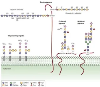
(29) 1. General Introduction 1.1. Glycosylation Glycosylation is the most frequent posttranslational modification of proteins and lipids. Glycans are present in all living organisms and regulate a diversity of biological processes, including protein folding and intracellular trafficking, cell-cell and cell-matrix interactions, cellular differentiation and immune response [1, 2]. The relevance of glycosylation is highlighted by the fact that approximately 1-2% of human genes are required for glycan biosynthesis, which is catalyzed by the enzymatic reaction of several enzymes that transfer mono or oligosaccharides, i.e. glycosyltransferases, with their strict specificity for both donor nucleotide-sugars (e.g., UDP-Gal, GDP-Fuc, or CMP-Sia) and acceptor substrates [3]. Glycan biosynthesis is not a template-based process such as DNA, RNA, or protein synthesis. In fact, the glycan expression in a given cell and glycoprotein depends on the balance achieved by the expression and activity levels of the different glycosyltransferases involved in the glycosylation process, as well as by their localization/organization in the Endoplasmic Reticulum (ER) and Golgi, and on the availability of precursor monosaccharide molecules [4].. 1.1.1.. Types of glycosylation. The common types of glycans found in cells are primarily defined according to the nature of the linkage to the glycoconjugate (protein or lipid). There are three main glycoconjugates: glycoproteins, proteoglycans and glycolipids (Fig. 1). A glycoprotein is a protein with one or more glycans covalently attached to a polypeptide backbone, usually via N- or O-linkages. This matter will be further detailed in the next sections. In a proteoglycan, one or more glycosaminoglycan (GAG) chains is attached to a “core protein” through a typical core region ending in a xylose residue that is linked to a serine residue. Finally, glycolipids consist of a glycan linked to a lipid. In higher organisms, most glycolipids are glycosphingolipids, with a glycan attached via glucose or galactose (Gal) to the lipid moiety ceramide. Within glycosphingolipids, the sialylated structures, such as GM3, GM1, GD3 and GD2, are called gangliosides [5].. 9.
(30) Fig. 1 – Three main classes of carbohydrates in the human plasma membrane. Glycoproteins are divided in O-linked glycans, via serine or threonine amino acid, and Nlinked glycans, via asparagine amino acid. Glycolipids are mainly glycosphingolipids with different carbohydrate compositions. Sialylated glycosphingolipids are named gangliosides (GM3, GM1, GD3 and GD2). Proteoglycans have normally a part of their amino acid sequence inserted among the lipid fatty acid chains and have a glycosaminoglycan (GAG) chain attached to a xylose which is linked to a serine amino acid (adapted from [6]).. 1.1.2.. Glycoproteins. In humans, proteins can be glycosylated with two main types of glycans: N-glycans and O-glycans.. 1.1.2.1.. N-glycosylation. N-glycans are covalently attached to protein by an N-glycosidic bond at asparagine (Asn) residues within a minimal amino acid sequence that begins with Asn followed by any amino acid except proline and it ends with serine (Ser) or threonine (Thr) (AsnX-Ser/Thr). However, due to conformational factors, not all Asn-X-Ser/Thr sequences actually have an N-glycosidic linkage. The most common N-glycan linkage is N-acetylglucosamine (GlcNAc) to Asn [7]. The biosynthesis of N-glycan begins on the cytoplasmic face of the endoplasmic reticulum (ER) membrane with the transfer of GlcNAc residue to the lipid-like precursor dolichol (Dol). Other sugar residues are sequentially added, and the Dollinked glycan containing two GlcNAc and five mannoses is flipped inside the ER. When fourteen sugar residues (2 GlcNAc, 9 mannoses and 3 glucoses) are added to Dol,. then. this. entire. glycan. is. transferred. in. single. block. by. the. oligosaccharyltransferase (OST) complex to an Asn-X-Ser/Thr sequence in a protein. 10.
(31) that is being synthesized into ER. The protein-bound N-glycan is subsequently modified in the ER and Golgi by a complex series of reactions catalyzed by membrane-bound glycosidases and glycosyltransferases, that remove or add sugar residues, respectively, to form the final glycan structure (Fig 2) [8].. Fig. 2 - Schematic representation of biosynthesis and processing of N-linked glycans (adapted from [9]).. 1.1.2.2.. O-glycosylation. The most common O-glycans are linked through N-acetylgalactosamine (GalNAc). Their biosynthesis starts by the transfer of a GalNAc from UDP-GalNAc to Ser or Thr residues of a protein in the cis Golgi, originating the Tn antigen. This transfer is catalyzed by a specific enzyme, polypeptide-N-acetyl-galactosaminyltransferase (ppGalNAcT). Contrarily to the N-glycosylation, O-glycosylation process doesn’t need a lipid-linked intermediate or any glycosidases to process their core structures. Subsequently, the simple O-GalNAc glycan can be further modified by several glycosyltransferases acting in a sequential manner in order to extend the glycan chain either branched or linearly according to substrate specificity, creating more complex O-glycan structures. Further elongation of the O-GalNAc glycans can include the addition of sialic acid, fucose, sulfate group among other residues [9, 10].. 11.
(32) There are four common O-GalNAc glycan core structures, designated cores 1 through 4, being the first one (core 1 or T antigen) the most common O-GalNAc glycan and the initial core of many longer, more complex structures, such as Lewis glycans (Fig. 3) [10].. Fig. 3 – Pathways of O-glycans biosynthesis. Tn antigen is the first glycan to be created and can be sialylated (Sialyl-Tn antigen) or extended to form core 3 or T antigen (core 1). Core 3 can be extended to core 4, while core 1 can be sialylated (Sialyl-T antigen) or extended to core 2. The former can be sialylated again producing disialyl-T antigen, while the latter can be extended to originate more complex structures, such as sialyl-Lewis X structure (adapted from [10]).. 1.1.3.. Glycosylation in cancer. Cancer is a collection of diseases that can affect any organ of the body and is characterized as being originated from cells having an abnormally increased proliferation, loss of differentiation and a capacity to invade its surrounding tissues causing metastases in other sites in the body [11]. The main cancer feature/hallmarks, which also includes immune evasion, will be addressed in the section 1.3. The transformation process of a normal cell into a malignant one includes changes in their cellular glycosylation profiles. It has been reported a significant correlation between certain types of altered glycosylation and cancer prognosis. In fact, the glycosylation that frequently occurs in cancer, plays a pivotal role in cancer. 12.
(33) progression, angiogenesis and metastasis, cell-cell adhesion, signalling, motility and epithelial-mesenchymal transition (EMT) in cancer cells. These alterations of glycosylation comprise under- or over-expression of glycan structures, as well as the appearance of novel or truncated structures. However, despite the diversity of glycans at the cell surface, a few distinct structures are associated with malignant transformation and tumor progression. This suggests that specific glycosylation patterns, such as sialyl-Tn (STn) and sialyl-Lewis X/A (sLeX/A), which are the subject of this thesis, can contribute to precise mechanisms such as gain of function, cell fitness and survival [12].. 1.1.3.1.. Sialylation in cancer. Sialylation is the process by which sialic acid residues are introduced onto carbohydrates attached to proteins or lipids as the terminal monosaccharide. Abnormal sialylation, specifically hypersialylation, in cancer cell is a characteristic feature associated with inferior outcome and malignant properties including invasiveness and metastatic potential, playing a key role during various stages of tumor progression [13]. Being a terminal sugar, sialic acid and their derivatives play a role in a variety of biological processes such as cellular recognition, cell adhesion, cell-matrix interaction and cell-to-cell signaling [14]. Sialic acids comprise a family of more than 50 monosaccharides that share a ninecarbon backbone (C1-9). The most common sialic acid derivate found in mammals is N-Acetylneuraminic acid (Neu5Ac), which bears an acetyl group on the fifth carbon atom (C5). In general, sialic acids are enzymatically linked to other carbohydrates by an enzymatic process that can be carried out by more than 20 distinct Golgi-resident sialyltransferases (ST) that link sialic acids via their second carbon (C2) to the carbon atom at position C3 (ST3Gal 1-6), C6 (ST6Gal 1, 2 and ST6GalNAc 1-6), or to C8 (ST8Sia 1-6) of carbohydrates, yielding α2,3-, α2,6-, or α2,8-linked sialic acids, respectively [15]. Although sialic acid can be covalently linked to Gal, GalNAc, GlcNAc, or to another sialic acid (polysialic acids), the most common linkages are the α2,3- and α2,6- to Gal residues and the α2,6- to GalNAc residues, which are carried out by ST3Gal, ST6Gal and ST6GalNAc families, respectively [16]. Up to now, three key mechanisms have been reported to cause aberrant hypersialylation in cancer cells. First, overexpression and/or altered activity of sialyltransferases results in increased sialylation of glycans and expression of specific. 13.
(34) tumor-associated carbohydrate antigens (e.g., sLeX/A and STn). The second mechanism includes an enhanced metabolic flux through the sialic acid synthesis pathway in cancer cells due to increased substrate availability or overexpression of genes involved in sialic acid biosynthesis. Finally, the third mechanism consists of a decreased expression of endogenous sialidases, which can enzymatically cleave sialic acids from glycans, leading to the accumulation of sialylated glycans [15]. However, other mechanisms can also be involved in more specific glycan structures, as discussed in latter sections. In this thesis, we will specifically focus on two main sialylated glycans: STn and sLeX/A.. 1.1.3.1.1. STn glycan STn antigen is a disaccharide mucin-type O-glycan resulting from the sialylation of Tn antigen (Fig. 3) by an α2,6-sialyltransferase. Specifically, α2,6-sialyltransferase (ST6GalNAc 1 and 2) catalyzes the transfer of sialic acid from CMP-Neu5Ac donor to an αGalNAc O-linked to Ser or Thr residues of a protein on the C6 position. In normal tissues, STn is mostly not expressed, although in some colon tissues, STn expression was detected after de-O-acetylation, since STn was masked by O-acetyl groups which concealed its recognition by anti-STn antibodies [17]. In normal cells, Tn antigen is normally elongated to form a variety of other structures, such as T antigen (i.e, Core1, Galβ1-3GalNAcα-O-Ser/Thr), which can also be sialylated creating sialyl-T antigen (NeuAcα2-3(Galβ1-3)GalNAcα-O-Ser/Thr), or Core2 structures (GlcNAcβ1-6(Galβ1-3)GalNAcα-O-Ser/Thr), which can be further processed forming elongated structures, such as sLeX [10]. In vivo, ST6GalNAc 1 enzyme has been shown to be the main sialyltransferase catalyzing the transfer of sialic acid onto the GalNAc, i.e., the Tn antigen, being its expression correlated with STn overexpression in gastric and breast tumors [10, 18, 19]. This Tn sialylation truncates further elongation of the O-glycan. Other mechanisms described as involved in the aberrant STn expression is the loss of C1GalT1 (also called T-synthase) or C3GnT enzymes, which normally extend Tn antigen into Core1 and Core3 structures, respectively. Other mechanisms are the overexpression of ppGalNAc-Ts glycosyltransferases or their re-localization from the Golgi to the ER; or even defects in Cosmc, a chaperone essential for the activity of C1GalT1, due to epigenetic silencing or mutations, which will result in an increase. 14.
(35) expression of Tn antigen in cancer [20-23]. Specific mechanisms for STn overexpression include ST6GalNAc 1 upregulation and/or its re-localization from the Golgi to the ER and also loss of O-acetyl groups from STn, which have been demonstrated in some cancers [24]. STn antigen is highly expressed in more than 80% of human cancer types, such as gastric [25, 26], colorectal [27], ovarian [28], breast [29] and pancreatic [30] carcinomas, whereas no expression is observed in the respective normal tissues. STn expression is associated with carcinoma aggressiveness and poor prognosis. Specifically, STn expression was associated with invasiveness of liver cancer [31] and with lower survival rate in advanced gastric carcinomas [32]. In breast cancer, STn expression was associated with lymph node metastasis and high histological grade [33], as well as with resistance to adjuvant chemotherapy [34]. Studies using STn-expressing gastric and breast cancer cells reported that STn expression modulate a malignant phenotype inducing a more aggressive cell behavior, such as decreased cell-cell aggregation and increased extracellular matrix adhesion, migration and invasion [35-38]. Interestingly, the STn antigen was first discovered as a cancer marker over 30 years ago. Early studies identified the STn as a marker for diagnosis and prognosis in cancer, but later work has focused on using STn as a potential therapeutic target (see in more detail in section 1.3.1.4.1). 1.1.3.1.2. sLeX/A glycans Both sLeX and sLeA belong to the Lewis family glycoepitopes, in which the main 8 glycan structures are represented in Fig. 4.. 15.
(36) Fig. 4 – The Lewis glycoepitopes family. They are a related group of glycans that carry a fucose residue in a α1,3 (Lewis X and Y and sLeX) or α1,4 (Lewis A and B and sLeA) linked to GlcNAc. Here is also represented two sulfated Lewis glycans that together with sLeX and sLeA can bind to E-selectin expressed by the endothelium (adapted from [39]).. sLeX is a terminal tetrasaccharide constituted by N-GlcNAc, Gal, fucose (Fuc) and sialic acid residues (NeuAcα2,3Galβ1,4(Fucα1,3)GlcNAc). Specifically, to produce sLeX, a beta 1-3 N-acetylglucosaminyltransferase (β1,3-GlcNAcT) starts by transferring a GlcNAc residue to a Gal, being followed by the transfer of a Gal to the GlcNAc residue by beta 1-4 Galactosyltransferase (β1,4-GalT). Then a ST3Gal enzyme transfer NeuAc to the Gal residue, creating the direct precursor for sLeX, 3’sialyllactosamine. The last step of sLeX biosynthesis consists in the addition of Fuc to the GlcNAc residue by α1,3-fucosyltransferases (FUTs) [40, 41] (Fig. 4). sLeA, also termed carbohydrate antigen 19–9 (CA19–9), is an isomer of sLeX, consisting of the same monosaccharide residues constitution but with different linkage between Gal, and Fuc, to GlcNAc (NeuAcα2,3Galβ1,3(Fucα1,4)GlcNAc) (Fig. 4), which is the result of the work of other glycosyltransferases. Differently from sLeX, during sLeA biosynthesis, the transfer of a Gal to the GlcNAc residue is performed by a beta 1-3 Galactosyltransferase (β1,3-GalT) and the fucosylation of the same GlcNAc residue is achieved by α1,4-FUTs [41, 42]. The two key steps in sLeX and sLeA biosynthesis for achieving a functional glycan are the sialylation and fucosylation. The main STs involved in sLeA biosynthesis is ST3Gal3, while in sLeX biosynthesis, three α2,3-STs, specifically ST3Gal3, ST3Gal4 and ST3Gal6, were described as responsible for the sialylation of sLeX precursor [43]. Since fucosylation of sLeX and sLeA is the last step of their biosynthesis, it is also the most important one to guarantee the expression of these glycans. There are eight known α1,3/4-FUTs that can be involved in the biosynthesis of Lewis antigens:. 16.
(37) FUT3, FUT4, FUT5, FUT6, FUT7, FUT9, FUT10 and FUT11. Specifically, FUT3 and FUT5 are less specific enzymes, being related not only with sLeA and sLeX biosynthesis, but also with their unsialylated forms, Lewis A and Lewis X, and Lewis Y and B. FUT 4 and FUT9 are more involved in the production of the unsialylated forms Lewis A and Lewis X, while FUT6 and FUT7 have been more related with sLeX expression [44]. FUT10 and FUT11 are more recent α1,3-FUTs identified in mice and humans [45]. Recent evidences indicate that FUT10 are involved in Lewis X production and that FUT11 can be responsible for sLeX and 6-sulfo-sLeX expression [46, 47]. The main role of sLeX is played during leukocyte trafficking, specifically mediating the binding of circulating leukocytes in the bloodstream and the selectins expressed by endothelium. Both sLeX and sLeA glycans are functional selectin ligands in cancer, facilitating the extravasation of tumor cells in the bloodstream to new metastasis sites (see in more detail in 1.3.2.1.1.1.1) [48].. 1.2. Cancer As referred, cancer can affect any organ of the body and is originated from the transformation of normal cells into malignant cells. Here, we will focus in two types of cancer: bladder cancer and breast cancer.. 1.2.1.. Bladder cancer. Bladder cancer is the seventh most commonly diagnosed cancer in the male population worldwide and the eleventh when both genders are considered. Tobacco smoking is the most important risk factor for bladder cancer, accounting for approximately 50% of cases. Occupational exposures to chemical carcinogens are also a well-established risk factors for bladder cancer, with about 10% of cases [49]. Bladder cancer are divided in non-muscle invasive bladder cancer (NMIBC), which is the most common representing approximately 75% of the patients, and muscle invasive bladder cancer (MIBC). NMIBC can be low grade (well-differentiated tumors), normally noninvasive papillary carcinoma (stage Ta), or high grade (poorly differentiated tumors), such as most stage T1 tumors that have penetrated the epithelial basement membrane but have not invaded the muscle, while most MIBC are high grade. NMIBCs commonly recur (50–70%) but rarely progress to invasion. 17.
(38) (10–15%), and five-year survival is ~90% [50, 51]. In contrast, five-year survival rates in patients with MIBC are much lower, between 30% and 60%, demonstrating the lethality of the disease [52]. The most common metastatic sites in MIBC are lymph nodes, bone, lung, liver and peritoneum [53]. Treatment of stage Ta-T1 NMIBC consists in the transurethral resection of the bladder (TURB) to remove all visible lesions. Since these tumors commonly recur and may progress to MIBC, adjuvant therapy is also administered. This includes immediate postoperative single intravesical instillation of chemotherapy that will destroy circulating tumor cells after TURB, significantly reducing the recurrence rate; and intravesical instillation of Bacillus Calmette-Guérin (BCG) immunotherapy, which is superior in preventing the recurrence and treatment of high-risk NMIBC than TURB alone or TURB plus chemotherapy [49]. Regarding MIBC, the standard treatment is neoadjuvant cisplatin-based combination chemotherapy followed by radical cystectomy [54].. 1.2.2.. Breast cancer. Worldwide, breast cancer is the most frequently diagnosed cancer and the leading cause of cancer death among women. Breast cancer alone accounts for 25% of all cancer cases and 15% of all cancer deaths among women. Main risk factors include reproductive and hormonal factors such as a long menstrual history, recent use of oral contraceptives, and never having children. Normally, breast cancer is screened by mammography, which can detect early stage breast cancer enhancing the treatment efficacy and prospect of cure [55]. The histopathological classification of breast carcinoma is based on the diversity of the morphological features of the tumors. 80% of all breast cancers belong to one of two histopathological classes, namely: invasive ductal carcinomas (IDCs), which begins in the milk-carrying ducts and spreads beyond it, comprising 80% of all invasive breast cancers, or invasive lobular carcinoma (ILC), which begins in the milk-producing lobules and includes 10% of all invasive breast cancers [56]. Clinically, the molecular classification of breast cancer is more useful for treatment than the histopathological classification. The molecular subtypes are based on the expression of three biomarkers, estrogen receptor (ER), progesterone receptor (PR) and HER2 (Human Epidermal growth factor Receptor 2). Recently, proliferative rate determined by Ki67 expression is assessed in order to more precisely distinguish the. 18.
(39) luminal group. This classification divides four subtypes: luminal A, luminal B, HER2 and “triple negative” (also known as basal-like) (Table I) [57].. Table I: Molecular subtype using four biomarkers, adapted from [57] Molecular subtype. Biomarker profile. HER2. ER+ and/or PR+, HER2-, and low Ki67 (<14%) ER+ and/or PR+ and HER2+ (luminal-HER2 group) ER+ and/or PR+, HER2-, and high Ki67 (>14%) ER-, PR-, and HER2+. Triple Negative. ER-, PR- and HER2-. Luminal A. Luminal B. Abbreviations: ER, estrogen receptor; PR, progesterone receptor. Luminar subtype includes approximately 70% of all invasive breast cancers, while the others 30% are equally divided between HER2 and basal-like subtypes. Between luminal subtypes, luminal B tends to have higher histological grade than luminal A. However, HER2 is the one more likely to be high grade and node positive [57]. In terms of timing of relapse, there are distinct differences between the subtypes, patients with HER2 or basal-like subtypes tend to relapse within the first 5 years while the ones with luminal subtypes relapses between 5 and 15 years. Regarding the metastasis, bone is the most common metastatic site in all subtypes except basal-like tumors. Specifically, luminal A subtype have a high rate of metastasis to bone and liver, luminal B and HER2 seem to prefer bone, liver and lung and basal-like goes normally more to lung, distant nodal and then bone [58]. For therapy, luminal tumors respond to endocrine therapy and chemotherapy and HER2 tumors are treated with monoclonal antibodies, such as trastuzumab that interferes with the HER2 receptor inducing antibody-dependent cell-mediated cytotoxicity. (ADCC). against. tumor. cells,. and. with. anthracycline-based. chemotherapy. Basal-like tumors don’t respond to endocrine therapy or trastuzumab, appearing to be sensitive to platinum-based chemotherapy and PARP inhibitors. In general, luminal A have a better prognosis than luminal B and both HER2 and basallike tumors have poorer prognosis [57].. 19.
(40) 1.3. Cancer features The hallmarks of cancer comprise the biological capabilities acquired during the multistep development of human tumors. Nowadays, there are eight hallmarks described, specifically sustaining proliferative signaling, evading growth suppressors, resisting cell death, enabling replicative immortality, inducing angiogenesis, activating invasion and metastasis, reprogramming of energy metabolism and avoiding immune destruction. Furthermore, two enabling characteristics, genome instability and mutation and tumor-promoting inflammation, have also been included as important concepts to develop new means to treat human cancer (Fig. 5) [59]. In this thesis, two of these hallmarks, avoiding immune destruction and activating invasion and metastasis, will have a special focus, in the scope of aberrant glycosylation of cancer.. Fig. 5 – Hallmarks of Cancer. Currently, there are 8 hallmarks of cancer and 2 enabling characteristics, which are included in this illustration [59].. 1.3.1.. Immune responses to cancer. The immune system has evolved a powerful collection of defense mechanisms to protect the host from invading pathogenic microorganisms that would otherwise take advantage of the rich source of nutrients provided by the host. At the same time, it must be sophisticated enough to differentiate between the individual’s own cells and those of harmful invading organisms and not attack the commensal flora. The immune system is able to generate an enormous variety of cells and molecules, acting together in a dynamic network, capable of specifically recognizing and eliminating an apparently limitless variety of foreign pathogenic invaders. Once a foreign organism. 20.
(41) has been recognized, the immune system mounts an appropriate response to eliminate or neutralize the organism. This immune response is mediated by the early reactions of innate immunity and the later responses of adaptive immunity [60, 61], which will generate an effector, specific response. Later exposure to the same foreign organism induces a memory response, characterized by a more rapid and heightened immune reaction that serves to eliminate the pathogen and prevent disease.. 1.3.1.1.. Innate Immunity. Innate immunity provides an immediate first line of host defense and it includes physical barriers as well as cells and molecules, such as cytokines. Phagocytes are an important group of cells of innate immune response, which include dendritic cells (DCs), macrophages, monocytes and neutrophils. Their role is to recognize invading organisms using a variety of receptors that recognize pathogen-associated molecular patterns (PAMPs) and then internalize and kill the organism [62]. As mentioned below, some of these cells are antigen presenting cells (APCs) and make a crucial contribution to the activation of adaptive immunity.. 1.3.1.2.. Adaptive Immunity. The adaptive immunity is stimulated by exposure to infectious agents and, in contrast to innate immunity, it increases in magnitude and defensive capabilities with each successive exposure to a particular microbe. Two main characteristics of this response are the ability to distinguish different substances, called specificity, and the ability to respond more vigorously to repeated exposures to the same microbe, known as immunological memory. The adaptive immunity includes lymphocytes and molecules, such as antibodies. Naïve lymphocytes reside in and circulate between peripheral lymphoid organs, surviving for several weeks or months. They express receptors for antigens but are not able to eliminate them. To gain that capacity, they need co-stimulatory and cytokine stimulus which will lead them to differentiate, upon antigen recognition, into effector cells. After the effector phase, lymphocyte will differentiate into memory cells that survive for longer periods of time in the absence of antigen, being functionally inactive until they encounter the same antigen that induced their development, which in this case they rapidly respond to develop a secondary immune response [62].. 21.
(42) An antigen is any molecule that can bind specifically to an antibody. Their name arises from their ability to generate antibodies. However, some antigens do not, by themselves, elicit antibody production. Antigens that can induce antibody production are called immunogens [63]. There are two types of adaptive immunity, humoral immunity and cell-mediated immunity. The humoral immunity is mediated by antibodies, which are produced by B lymphocytes and secreted into the circulation and other body fluids. They neutralize extracellular microorganism, or antigen-expressing cells, by binding to their surface. They also engage a number of other cells, such as phagocytes, providing an effector role that leads to the elimination of pathogens. However, since antibodies cannot gain access to antigens that live inside infected cells, there are another immunity response that handle the defense against intracellular antigens, the cell-mediated immunity [64].. 1.3.1.2.1. Cell-mediated immunity Cell-mediated immunity involves cell interaction and, in adaptive immune responses, the main players are T lymphocytes. The antigen receptors of T cells (TCRs) only recognize peptide fragments of protein antigens that are bound to specialized host display molecules called major histocompatibility complex (MHC) molecules, only if present on the surface of an APC. Among T lymphocytes, CD4+ T cells, also called helper T cells (Th cells), produce cytokines that activate antibody-producing B cells and phagocytes, such as macrophages, to destroy ingested antigens. Some CD4+ T cells belong to a subset called regulatory T (Treg) cells, whose purpose is to prevent or limit immune responses. On the other hand, CD8+ T cells or cytotoxic T lymphocytes (CTLs) have the machinery to kill infected host cells [64].. 1.3.1.3.. APCs. APCs are cells able to capture antigens, process them and display them to T lymphocytes, providing at the same time co-stimulatory signals and cytokines that stimulate the proliferation and differentiation of lymphocytes [62]. To activate T cells, there are three main signals: i) The antigen-specific recognition, which consists in the recognition of the peptide antigen in the context of MHC molecule by the TCR; ii) The co-stimulation, which is the second signal involving the. 22.
(43) interaction between co-stimulatory molecules expressed on the membrane of APC and the T cell. One main co-stimulatory pair is the B71/B72 (CD80/86) molecules on APCs and its recognition by CD28 molecules expressed by T cells; ii) and cytokine signals, which results in the upregulation of cytokines and their receptors that boosts the activating signals [65]. The three main types of APCs are DCs, macrophages and B cells. DCs have a major role as APCs, due to its higher ability to present antigens, co-stimulation and migration.. 1.3.1.3.1. DCs DCs are widespread throughout the organism. Although DCs were first visualized as Langerhans cells in the skin in 1868, they were really just reported by the late Nobel laureate Ralph Steinman in 1973 [66] and their role as APCs have been thoroughly described. In general, DCs are APCs with a unique ability to effectively stimulate, naïve and memory T lymphocytes, as well as being able to induce immunological tolerance [67].. 1.3.1.3.1.1.. DCs functions. Immature DCs constitutively patrol through the blood, peripheral tissues, lymph and secondary lymphoid organs searching for self and non-self antigens. When immature DCs encounter antigens, they internalize them, processed them into peptides, which are loaded onto MHC class I (MHC-I) and II molecules (MHC-II), starting the DCs maturation process. Maturation implies that DCs gain ability to migrate to lymphoid organs, increased display of peptide-MHC at cell surface, including co-stimulation and cytokine secretion, to activate circulating antigen-specific T lymphocytes. These activated T cells help DCs in terminal maturation, which allows lymphocyte expansion and differentiation. Activated T lymphocytes migrate and can reach the injured tissue, where Th cells secrete cytokines, which permit activation of a number of cells while CTLs ultimately lyse the infected cells. DCs, together with T cells will also activate B cells that will produce antibodies to neutralize the initial pathogen. It is believed that, after interaction with lymphocytes, DCs die by apoptosis [68].. 23.
(44) 1.3.1.3.1.1.1.. Antigen Capture. Immature DCs have a high level of endocytic activity which is lost upon maturation. They capture pathogens, infected cells, apoptotic or dead cells or their derived products to use for antigen presentation. DCs exhibit three types of endocytosis: receptor-mediated endocytosis, phagocytosis and pinocytosis (macro- and micropinocytosis). Receptor-mediated endocytosis allows the uptake of macromolecules through their recognition by specific receptors located in clathrin-coated pits in the plasma membrane. After recognition, a signal in the endocytic receptor leads to the recruitment of clathrin lattices and to the formation of clathrin-coated endocytic vesicles. Immature DCs express several endocytic receptors, such as Fc receptors; Ctype lectins, for example macrophage-mannose receptor and/or Langerin; complement receptors and Scavenger receptors, like CD36 [68]. Phagocytosis is the process where a cell internalizes a particle or microorganism in a phagosome. It is generally receptor mediated and initiates by the engagement of a specific receptors, triggering a cascade of signal transduction, which is required for actin polymerization and effective engulfment of the particle in a large intracellular vacuole named phagosome. Regarding pinocytosis, this is a constitutive process in immature DCs and consists in capturing whatever might be in the fluid phase in the vicinity of the cells, not being dependent on specific recognition of receptors [62, 68].. 1.3.1.3.1.1.2.. DCs maturation and migration. After antigen uptake, immature DCs undergo phenotypic and functional changes that culminate in the complete transition from antigen-capturing cell to antigen-presenting cell. DCs maturation is a continuous process initiated in the periphery upon antigen encounter and/or inflammatory cytokines and completed during the DC–T cell interaction. Three main factors regulate DCs maturation: pathogen-related molecules, the balance between pro-inflammatory and anti-inflammatory signals in the local microenvironment and T cell–derived signals. Several coordinated events happen during DCs maturation process, including loss of endocytic/phagocytic receptors, upregulation of costimulatory molecules (such as CD80, and CD86), change in morphology and shift in lysosomal compartments and change in MHC-II. 24.
(45) compartments. The morphological changes accompanying DCs maturation include a loss of adhesive structures, cytoskeleton reorganization, and acquisition of high cellular motility. These changes trigger DCs migration into the T cell area of lymphoid organs. This migration process involves a coordinated action of several chemokines, and expression of respective chemokine receptors by DCs, that leads maturing DCs to migrate from inflamed tissues, enter the lymph stream, and through the draining lymph nodes to encounter T cells [67].. 1.3.1.3.1.1.3.. Antigen Processing and presentation. As mentioned before, antigen recognition by T cells depends on the expression of a spectrum of antigen-derived peptides bound to MHC-I and MHC-II molecules, on a APC. These peptides are the products of one of the two major proteolytic systems operating in DCs. Cytosolic antigens can be degraded by the proteasome, which produces the dominant source of peptides for MHC-I binding. In sum, a targeted protein is recognized by the ubiquitylation system, resulting in the covalent attachment of polyubiquitin chains to the protein, which is recognizable by proteasomes for proteolysis. The resulting peptides will be translocated to the ER where they will bind to MHC-I [69, 70]. Antigens that were endocytosed or phagocytosed will be both degraded by lysosomal proteolysis. Specifically, endocytosed proteins enter a vesicular pathway consisting of progressively more acidic and proteolytically active compartments classically described as early endosomes, late endosomes and lysosomes. On the other end, when particles are phagocytosed, they end in phagolysosomes that are formed by the fusion of phagosomes and lysosomes. In the end, the resulting peptides generally bind MHCII molecules in endosomes and lysosomes [70]. Although this is the most common process, it has been demonstrated, in DCs, that internalized antigens can be transported into cytoplasm and then become also presentable by MHC-I molecules in a process called cross presentation [71]. After peptides being loaded into MHC-I or -II molecules, they are transferred to the plasma membrane where they will be recognized by CD8+ CTLs or CD4+ Th cells, respectively, through their TCRs. The most common type of TCRs is a heterodimer of an α and β polypeptide chains. The TCRs are generated by a complex process of somatic rearrangement at the variable DNA regions of TCR, resulting in a great variety of TCRs specific for peptide–MHC-I or -II complexes. TCRs are associated. 25.
(46) with CD3 molecules and ζ chains, forming the TCR complex, which is required for signal transduction. CD4+ T cells not only express TCRs that will recognize peptide-MHC-II complex, but also CD4 coreceptor, which recognize MHC-II molecules and is involved in signal transduction. In the case of CD8+ T cells, they express CD8 coreceptor that will recognize MHC-I molecules [62]. Both cells express CD28 that will bind to the CD80 and CD86 co-stimulatory molecules expressed by DCs upon maturation, delivering the second signal for T cell activation. The combined action of these signals, plus cytokines (third signal), stimulate the proliferation of T cells (Fig. 6) and their differentiation into effector T cells and memory T cells. Proliferation is mainly induced by interleukin-2 (IL-2) cytokine that is recognized by IL-2 receptors in T cells, activating intracellular signaling pathways required for proliferation and differentiation [72].. Fig. 6 – T cell activation by DCs involves three signals. The interaction of major histocompatibility complex (MHC) containing a peptide fragment with T cell receptor (TCR) on the T cell originates the signal 1. Signal 2 follows the binding of costimulatory molecules (CD80/86) on the surface of the DCs with their ligand, CD28, on the T cell surface. Finally, the secretion of cytokines by DCs represents the signal 3 (adapted from [73]).. Effector T cells are short-lived cells that actively respond to a stimulus. Effector Th cells secrete cytokines that can activate several phagocytic cells, enabling them to phagocytose and kill microorganisms more effectively. They can be divided into two main subpopulations distinguished by the different panels of cytokines they secrete, specifically Th1 subset secretes IL-2, interferon gamma (IFN-γ) and tumor-necrosis factor-α (TNF-α), being involved in the activation of CTLs, while Th2 subset secretes IL-4, IL-5, IL-6 and IL-10, helping more effectively the activation of B cells. On the other hand, effector CTLs are responsible for killing infected or tumor cells. They. 26.
(47) have two mechanisms to accomplish this: Fas/FasL pathway and perforin/granzyme pathway, which in both will result in the apoptotic death of the target cell [61].. 1.3.1.3.1.2.. DCs Subsets. Multiple DCs subsets exist that vary in location, phenotype and specialized function. Two main functional subsets are the conventional DCs (cDCs) and plasmacytoid DCs (pDCs). DCs can be further divided based on their location into “lymphoid-resident” DCs, which capture antigens directly from the blood or lymph, and “migratory” DCs, that reside in the peripheral organs. In both locations, cDCs can be further segregated into multiple subsets with significant functional specialization [74]. Monocyte-derived DCs (moDCs) are another subset consisting in DCs differentiated from monocytes during inflammation or infection. moDCs differentiate rapidly from monocytes after upregulating CD11c and MHC-II molecules in response to inflammation or infection. They have the capacity to induce Th1 cell responses, crossprime antigen-specific CTLs, produce pro-inflammatory cytokines, such as TNF-α and IL-12, and regulate IgA production by B cells [75]. By exhibiting these potent functions, moDCs offer an important protective immunity role.. 1.3.1.3.1.2.1.. Tolerogenic DCs. While potently capable to initiate inflammatory responses, DCs also play an important role in modulating tolerance induction. Tolerogenic DCs are characterized by high antigen uptake and processing capabilities in order to present antigen to antigenspecific T cells, but fail to deliver proper cytokine and co-stimulatory signals for effector T cells activation and proliferation. This results in T cell death, T cell anergy or induction and expansion of Treg cells subsets [76]. Treg cells are characterized by the expression of CD25 and Foxp3 molecules and they act suppressing the induction and proliferation of effector T cells, maintaining tolerance to self-antigens and preventing autoimmune diseases [77]. DCs can also mediate central tolerance by negatively selecting self-antigen T cells in the thymus, inducing clonal deletion and Treg generation [78].. 1.3.1.3.1.3.. Tumor-infiltrating DCs. Currently, it is well accepted that the tumor microenvironment, which consists of not only tumor cells, but also fibroblasts, vascular endothelial cells and immune cells,. 27.
(48) such as macrophages, Treg cells and regulatory DCs, is immunosuppressive and tumor-promoting. Immature and tolerogenic tumor-infiltrating DCs suppress both innate and adaptive immune effectors using a wide variety of mechanisms. Within them, is the expression of immune check points such as the PDL-1, which when engaged by PD-1 on the T cell surface suppress the immune response by inducing apoptosis and inhibiting T cell proliferation and effector function, and by promoting the differentiation of Treg cells [79, 80]. Other immunosuppressive mechanism is based on the expression of cytokines, such as PGE2, IL-10 and transforming growth factor beta (TGFβ) secreted by tumor cells, which convert activating DCs into tolerogenic DCs that suppress T cell responses [80]. Indoleamine 2,3-dioxygenase expression by tumor-infiltrating DCs also plays an important role in mediating the suppression of adaptive immune responses. On the other hand, the presence of pDCs is generally associated with poor prognosis and tolerance in tumors. However, activated IFN-producing pDC have recently been shown to generate anti-tumor immunity in vitro and in vivo [81].. 1.3.1.3.1.4.. DCs in Cancer Immunotherapy. Since the discovery of DCs and the identification of their key function as mediators of T cell-mediated immune responses, there has been a major focus on the use of DCs in cancer immunotherapy. Specifically, immunotherapeutic approaches involving DCs aim to exploit the ability of these cells to direct CTLs to become potent antitumor effectors capable of specifically eradicate malignant cells [74, 82]. Most DCs vaccines comprises DCs loaded ex vivo with tumor antigen and readministered into the patient (Fig. 7). DCs are directly isolated from patients or differentiated from blood precursors, also isolated from the patients. After being loaded, DCs migrate from the injection site to the draining lymph nodes or tissues, where they are expected to prime tumor-specific T cells capable of eliminating the tumor [74].. 28.
(49) Fig. 7 - DC‑based vaccines using ex vivo loaded DCs to induce immunity. There are many alternative approaches for the preparation and use of DCs vaccines to treat cancer. The standard protocol includes the generation of DCs from precursors, then their maturation and tumor antigen loading using a variety of techniques, and finally the readministration of these cells into the patient (vaccination) (adapted from [83]).. Most of the DCs-based immunotherapy trials have been Phase I studies on advanced cancer patients who had failed to respond to conventional therapies. These trials revealed that this approach: (1) is feasible in many different malignancies; (2) is safe and well tolerated with minimal toxicity, which allows to preserve the quality of life of cancer patients; and (3) can induce tumor-specific adaptive immune responses in many patients. In general, there was a clear evidence that this therapy can have clinical benefit by improving overall survival [74, 82]. The first and only DCs-based immunotherapy, which received FDA approval in 2010, was the Sipuleucel-T (Provenge®), which has demonstrated a significant survival benefit in metastatic prostate cancer [84]. However, despite a great enthusiasm, there were disappointing results from early studies, which raised some doubts regarding the true clinical value of DC-based vaccines [85]. The future of DCs-based immunotherapy will benefit from two developments, which are already beginning to be incorporated in clinical trials: the implementation of optimized generation of DCs, and the use of these vaccines in combination with other anti-cancer therapies which could improve their effectiveness. For example, the combination with chemotherapy has shown to be clinical beneficial, since chemotherapy depletes immunosuppressive cells, such as Tregs, thus potentiating the therapeutic efficacy of DC-based vaccines [82, 86].. 29.
(50) Although our understanding of the complexity and subset specialization of the DCs network has increased exponentially, we still need to better understand how cancer cells alter DCs physiology in order to generate more effective cancer immunotherapies based on the powerful properties of DCs [86].. 1.3.1.4.. Immune response against tumor-associated carbohydrate antigens. The immune response is a key factor for not only eliciting protection against pathogens, but also to maintain immune surveillance against the development of malignant cells. From this perspective, the development of cancer can be seen as a failure of immune surveillance [87]. This idea is further supported by the immunosuppressive tumor environment, which was mentioned above. Most tumor associated carbohydrates do not elicit strong humoral responses and, in fact, evidences have shown that their aberrant expression is one of the mechanisms developed by cancer cells to prevent effective immune responses against cancer [4]. Their low immunogenicity is related with the fact that cellular immune response is mainly formatted to fight peptide antigens, which are presented through MHC molecules, and not carbohydrates, and this may impose a limitation to develop strong immune responses against carbohydrates. While it has been reported that CTLs may recognize mono- and disaccharides attached to peptides [88-90], further investigations are necessary to better understand the relevance of such recognition in the context of antitumor immune responses. Thus the humoral immune response plays a key role in antitumor responses against carbohydrates. In contrast to TCR, the B cell receptors (BCR) can recognize directly carbohydrate antigens with no need for presentation through MHC. Cross-linking of the antigen on BCR activates B cells, which initiate the secretion of IgM antibodies. This process is called the T cell-independent B cell activation and is able to provide immunity but with limited duration, and usually of IgM type. For the purposes of long lasting anti-tumor humoral immunity, the production of other antibodies isotypes, such as IgG, is desirable. Physiologically, this usually implies the involvement of Th cells, in a process called T cell-dependent B cell activation, which, as mentioned above, it is not easily activated in response to carbohydrates [19, 91]. The knowledge of these physiological mechanisms raised, in recent years, efforts to elicit increased immunogenicity of carbohydrate antigens, aiming to generate potent immunotherapies, such as carbohydrate-based vaccination. The approaches to. 30.
(51) improve carbohydrate immunogenicity include the covalently coupling of carbohydrates to immunologically active protein carriers, such as keyhole limpet hemocyanin (KLH) or adjuvants or to physiological/pathological carriers, such as mucin 1 protein (MUC1), to guarantee a range of relevant epitopes [92].. 1.3.1.4.1. STn glycan and immune response Vaccination based on STn, such as Theratope (STn anchored to the KLH), have been used in pre-clinical studies and clinical trials and have demonstrated that this therapy can induce the production of antigen specific antibodies (anti-STn). STn-based vaccines were able to delay tumor growth in mice and the increased time to progression in patients [93-95]. In Theratope-treated patients, the presence of antiSTn antibodies was directly correlated with disease free survival [96]. Despite this promising results, when Theratope vaccine reached Phase III, it did not improved the overall time to progression or survival [94]. In recent years, a better understanding of the immunomodulatory role of the STn antigen has elucidated their contribution to immune tolerance, thus explaining in part the failure of STn-based vaccination protocols. For example, the overexpression of STn is associated with low infiltration of nest CD8+ lymphocytes in endometrial cancer [97], the STn present in secreted colon cancer mucins interact with CD22 and down-modulate B cell signal transduction [98]. Moreover, siglec-15 recognizes the STn antigen expressed by cancer cells and transduces a signal for enhanced TGF-β secretion in tumor-associated macrophages [99]. Additionally, mucins, in particular STn+ MUC1, released by cancer cells inhibit DCs maturation and modulate DCs towards IL-10high IL-12low regulatory antigen presenting cells with a limited capacity to trigger protective Th1 responses [100]. Interestingly, soluble aberrantly glycosylated MUC1 has also been described to elicit DCs maturation, yet unable to promote Th1 responses [101]. While the effect of glycoproteins secreted by tumors is becoming more elucidated, the role of the overall STn expression at tumor cell surface in immunomodulation, remains unknown.. 1.3.2.. Cancer Invasion and Metastasis. Metastasis is the process where the cancer cells spread from the primary tumor to colonize distant organs. Metastasis is the most fearsome aspect of cancer, since it is the cause of approximately 90% of deaths from cancer [102].. 31.
Imagem
![Fig. 2 - Schematic representation of biosynthesis and processing of N-linked glycans (adapted from [9])](https://thumb-eu.123doks.com/thumbv2/123dok_br/15718604.1070202/31.892.209.712.269.648/fig-schematic-representation-biosynthesis-processing-linked-glycans-adapted.webp)


+7
![Fig. 5 – Hallmarks of Cancer. Currently, there are 8 hallmarks of cancer and 2 enabling characteristics, which are included in this illustration [59]](https://thumb-eu.123doks.com/thumbv2/123dok_br/15718604.1070202/40.892.328.591.508.753/hallmarks-cancer-currently-hallmarks-enabling-characteristics-included-illustration.webp)


Documentos relacionados
Norma técnica publicada em 07 de agosto de 2012 estabelece os requisitos para execução de concreto dosado por empresas do setor, incluem todas as operações de
[PEDRO FEITAIS] Na opinião Carvalho 1994 1994, “na a verdade é que o município, integrando numa unidade espacial e funcional individualizada, o local de habitação, de trabalho,
Por exemplo se quatro pessoas realizarem a pesquisa anterior e virem os detalhes da entidade “Tradição Oral”, a relação daquela empresa com a pesquisa passa a ter peso
This overview of spatial planning and territorial governance reforms in Southern European countries in times of crisis and austerity has shown the existence of an overall
Sob esse enfoque, conquanto a liberdade de expressão constitua um direito fundamental para o pluralismo de ideias e o desenvolvimento da democracia, como adrede mencionado,
Os farrapos no Prata : as relações do Rio Grande do Sul farroupilha com o Estado Oriental do Uruguai
Apesar disso, o governo imperial também se via duramente pressionado pelas circunstâncias que faziam com que duas de suas prioridades se chocassem: de um lado, a
Assim sendo, o presente artigo tem como objetivo fazer um estudo sobre a evasão no curso de Física da Universidade Estadual de Maringá, nas modalidades a distância e presencial,
A interpretação conjunta das duas razões calculadas para os minerais de argila (Sm/Kao e Sm+Kao/I) mostra três resultados importantes: (i) a significativa
