ORIGINAL RESEARCH
TUMOR-INFILTRATING CD4
+T LYMPHOCYTES IN
EARLY BREAST CANCER REFLECT LYMPH NODE
INVOLVEMENT
Alexandre Henrique Macchetti,a Heitor Ricardo Cosiski Marana,a João Santana
Silva,b Jurandyr Moreira de Andrade,a Alfredo Ribeiro-Silva,c and Sérgio Bighettia
Macchetti AH, Marana HRC, Silva JS, Andrade JM de, Ribeiro-Silva A, Bighetti S. Tumor-infiltrating CD4+ T lymphocytes
in early breast cancer reflect lymph node involvement. Clinics. 2006;61(3):203-8.
BACKGROUND: The role of immune system in the pathogenesis and progression of breast cancer is a subject of controversy, and this stimulated us to investigate the association of the immunophenotype of tumor-infiltrating lymphocytes in early breast cancer with the spread of tumor cells to axillary lymph nodes.
METHODS: Tumor samples from 23 patients with early breast cancer from the Department of Gynecology and Obstetrics of Ribeirão Preto Medical School (USP) were obtained at the time of biopsy and submitted to an enzyme-digestion procedure for the extraction of tumor-infiltrating lymphocytes. The lymphocytes extracted were analyzed by dual-color flow cytometry with monoclonal antibodies in these combinations: CD3 FITC/CD19 PE, CD3 FITC/CD4 PE, CD3 FITC/CD8 PE, and CD16/56 PerCP, which are specific for immunophenotyping of T and B lymphocytes, helper and cytotoxic T lymphocytes, and natural killer (NK) cells. The mean percentage of these cells was used for comparing groups of patients with or without lymph node metastasis.
RESULTS: The mean value for T-lymphocyte infiltration was 24.72 ± 17.37%; for B-lymphocyte infiltration, 4.22 ± 6.27%; for NK-cell infiltration, 4.41 ± 5.22%, and for CD4+ and CD8+ T-lymphocyte infiltration, 12.43 ± 10.12% and 11.30 ± 15.09%,
respectively. Only mean values of T- and CD4+ T-lymphocyte infiltration were higher in the group of patients with lymph node
metastasis, while no differences were noted in the other lymphocyte subpopulations.
CONCLUSION: The association of tumor-infiltrating CD4+ Tlymphocytes with lymph node metastasis suggests a role for these
cells in the spread of neoplasia to lymph nodes in patients with early breast cancer.
KEYWORDS: Breast cancer. Flow cytometry. Lymphocytes. CD4+ Tlymphocytes. Natural killer cells.
aDepartment of Gynecology and Obstetrics, Breast Service, Ribeirão Preto
Medical School, University of São Paulo – São Paulo/SP, Brazil.
bDepartment of Immunology and Biochemistry, Ribeirão Preto Medical
School, University of São Paulo - São Paulo/SP, Brazil.
cDepartment of Pathology, Ribeirão Preto Medical School, University of São
Paulo - São Paulo/SP, Brazil. Email: [email protected]
Received for publication on February 04, 2006. Accepted for publication on February 20, 2006.
INTRODUCTION
Breast cancer is the most common neoplasm in women, and several studies have documented that these tumors are infiltrated by a heterogeneous population of immune cells,
consisting of different proportions of T cells, B cells, natu-ral killer (NK) cells, and macrophages.1–4 Despite the
pres-ence of the immune response, many breast tumors progres-sively grow and spread, calling into question the role of tumor-infiltrating leukocytes in the tumor micro-environment.5
There is no definitive conclusion regarding the efficacy of T cell-dependent immune mechanisms or regarding the correlation between the extent and type of lymphocyte in-filtration and the tumor progression in most subtypes of car-cinoma of the breast.4,5 These interrelationships could be
Previous studies have shown that the function of tumor-infiltrating lymphocytes is impaired by inhibitory cytokines,6 increased regulatory T lymphocyte activity,7
tumor cell MHC molecule alterations,8 and aberrant Fas
lig-and expression,9 among others.
Nodal status as determined by pathologic examination of lymph nodes has repeatedly been shown to be the sin-gle most important predictor of survival in breast cancer. Even patients with very small tumors and minimal involve-ment of a single axillary node have a significantly worse prognosis compared with patients with no axillary node in-volvement; there is a direct relationshipbetween the number of involved axillary nodes and the risk fordistant recur-rence.10,11
Collectively, these findings have led to increased inter-est in the role of tumor-infiltrating leukocytes in the regu-lation of tumor development. In our study, we analyzed by flow cytometry the tumor-infiltrating leukocytes of breast cancer patients with T1 to T2 breast tumors in an attempt to correlate phenotypical markers of tumor-infiltrating leukocytes with axillary lymph node status.
PATIENTS AND METHODS
Patient population
During the years 2003 and 2004, a consecutive series of 23 patients at the Department of Gynecology and
Ob-stetrics of the Ribeirão Preto Medical School who pre-sented with invasive breast carcinoma were included in the study for immunophenotyping the tumor-infiltrating lymphocytes. In all patients, tumor size was no larger than 50 mm and was strictly confined to breasts, without dis-tant metastasis; cases with involvement of skin, hypoder-mis, or pectoral muscles or with deep fixation were ex-cluded. All patients included in this study underwent cura-tive surgery plus axillary lymph node dissection, and none of these patients had received preoperative antitumor therapy. The clinical and pathological characteristics of patients included for the immunophenotypic studies are summarized in Table 1.
Informed consent, approved by the Ethics Committee of the Ribeirão Preto Medical School Hospital, was ob-tained from patients for collecting the material in this study (Process HCRP no 2519/2003).
Processing of tumor biopsy and preparation for flow cytometry analysis
Samples of the primary tumor from the 23 patients were obtained aseptically at the time of biopsy. The main bulk of each tumor was sent for routine histology. A block of tumor measuring 5 x 5 mm fresh from the operating room was dissected away from necrotic and fatty tissue, minced into 1 to 3-mm3 chunks using sterile forceps and a scalpel,
and placed in sterile RPMI 1640 (Sigma, Saint Louis, MO),
Table 1 - Clinical and pathological characteristics of patients with breast cancer
Case Age Tumor Grade Tumor Size(cm) TNM Stage Lymph node status1 Hormone status2
1 51 I 3.5 T2N0M0 IIA 3/25 Post
2 74 I 4.5 T2N0M0 IIA 0/19 Post
3 44 I 2.0 T1N2M0 IIIA 7/33 Pre
4 75 II 4.5 T2N1M0 IIB 0/16 Post
5 70 II 2.0 T1N0M0 I 0/18 Post
6 80 III 4.0 T2N1M0 IIB 13/22 Post
7 69 I 1.0 T1N0M0 I 0/18 Post
8 90 II 1.6 T1N0M0 I 0/21 Post
9 71 Mucinous 2.5 T2N0M0 IIA 0/15 Post
10 45 Mucinous 3.0 T2N0M0 IIA 3/12 Pre
11 59 II 2.5 T2N0M0 IIA 1/17 Post
12 64 I 1.8 T1N0M0 I 0/28 Post
13 53 I 3.0 T2N0M0 IIA 0/17 Post
14 40 I 3.0 T2N0M0 IIA 1/13 Pre
15 58 III 3.0 T2N2M0 IIIA 14/28 Post
16 51 II 4.0 T2N0M0 IIA 0/23 Pre
17 50 II 2.0 T1N0M0 I 12/27 Pre
18 48 II 2.0 T1N1M0 IIA 4/15 Post
19 42 I 1.0 T1N0M0 I 0/12 Pre
20 42 I 4.0 T2N0M0 IIA 0/12 Pre
21 63 II 2.0 T1N2M0 IIIA 8/25 Post
22 57 I 1.5 T1N0M0 I 0/9 Post
23 59 I 2.0 T1N0M0 I 0/21 Post
containing collagenase type IV (1 g/L; Sigma, Saint Louis, MO), hyaluronidase type V (100 mg/mL; Sigma, Saint Louis, MO), and DNAase type IV (30,000 U/L; Sigma, Saint Louis, MO). Tumor digestion was performed for 40 minutes at 37o C in a sterile bottle. 10 mL of enzyme
me-dium was added per 3 g of tumor chunks. Following the tumor digestion, the single-cell suspension was filtered through a tissue dissociator grid, and the resultant suspen-sion was washed twice and submitted to immunofluores-cent staining of cells for flow cytometry. No attempt was made to separate the tumor-infiltrating lymphocytes from the tumor cells, since many cells would be lost, and this might distort the proportions of the various mononuclear cells present within the infiltrate.
Flow cytometry analysis of leukocyte infiltrate
The surface characteristics were determined by us-ing the followus-ing preparations of monoclonal antibod-ies: CD3 FITC/CD19 PE, CD3 FITC/CD4 PE, CD3 FITC/CD8 PE, and CD16/56 PerCP (Becton Dickinson, Mountain View, CA), which allowed the lymphocytes be phenotyped as T cells, B cells, CD4+ T cells, CD8+ T
cells, or NK cells, respectively. A 10 mL sample of a phycoerythrin (PE)-, fluorescein-isothiocyanate (FITC)-, or peridin-clorophyll-protein (PerCP)-conjugated anti-body was added to 106 cells in 100 mL of
phosphate-buffered saline (PBS). After an incubation time of 30 min at 4o C in darkness, cells were washed twice and
resuspended in 500 mL PBS and then analyzed with a FACSort flow cytometer (Becton Dickinson, San Jose, CA). The cell samples were run through the flow cytom-eter, and 10,000 events were analyzed for each sample using Becton Dickinson’s CELL QUESTTM software on
an Apple Mac G3 computer.
Using the forward- and side-scatter (FSC and SSC) properties of lymphocytes in laser light, a gate was drawn around the lymphocytes to exclude tumor cells from fur-ther analysis. The relative proportions of the phenotypic subsets were measured using 4-quadrant analysis.
Statistical analysis
The data of each group were submitted to the normal-ity test of GraphPad Prism (version 3.00) of GraphPad In-corporated (San Diego, USA), and a gaussian distribution of data was verified. Data were expressed as the mean ± standard deviation, and the 2-tailed unpaired Student t test was used for comparing leukocyte infiltration between groups of patients with different lymph node status. Sta-tistical significance was defined as P < 0.05.
RESULTS
All patients were treated by lumpectomy with axillary lymph node dissection or modified radical mastectomy in accordance with our management protocol. The patients were women between 40 and 90 years of age (mean and median were 58 years) and included 7 premenopausal and 16 postmenopausal women. According to the pathological TNM classification of cancer staging, 11 of 23 tumors were classified as T1, and 12 of 23 as T2. Lymph node metasta-sis as determined by histological examination was noted in 10 of 23 cases, with the remaining 13 patients being lymph node negative.
Histologically, 21 of 23 patients had tumors classified as infiltrating ductal carcinoma without special features, while the remaining 2 were classified as mucinous. Tumors with histology of ductal carcinoma were graded according to the Bloom and Richardson grading system,12 and there
were a total of 11 ductal carcinomas classified as grade I, 8 as grade II, and 2 as grade III.
The mean value for T-lymphocyte infiltration (CD3+
cells) was 24.72 ± 17.37%; for B-lymphocyte infiltration (CD19+ cells), 4.22 ± 6.27%; for NK cell-infiltration
(CD16+/CD56+ cells), 4.41 ± 5.22%; and for CD4+ and
CD8+ T-lymphocyte infiltration (CD3+/CD4+ and CD3+/
CD8+), 12.43 ± 10.12% and 11.30 ± 15.09%, respectively.
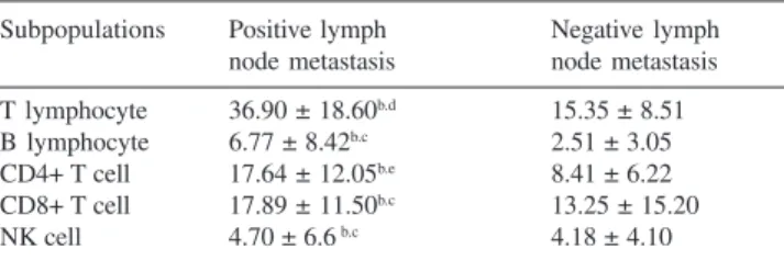
Comparing the two groups of patients—those with a presence vs those with an absence of lymph node metasta-sis—there was a statistically significant difference between groups regarding the mean values of T-lymphocyte and CD4+ T-lymphocyte infiltration (15.35 ± 2.36% and 8.41
± 6.22% with absence of lymph node metastasis vs 36.90 ± 18.60% and 17.64 ± 12.05% with presence of lymph node metastasis, P = 0.001 and P = 0.02, respectively) (Table 2), indicating an association between infiltration by CD4+
T cells and axillary metastasis. There were no statistically significant differences between the means of other lymphocyte subsets for these groups (Table 2).
Table 2 - Subpopulations of Immunocytes in Tumor Tissue from Breast Carcinoma Specimens with Respect to Lymph Node Involvementa,b
Subpopulations Positive lymph Negative lymph node metastasis node metastasis T lymphocyte 36.90 ± 18.60b.d 15.35 ± 8.51
B lymphocyte 6.77 ± 8.42b.c 2.51 ± 3.05
CD4+ T cell 17.64 ± 12.05b.e 8.41 ± 6.22
CD8+ T cell 17.89 ± 11.50b.c 13.25 ± 15.20
NK cell 4.70 ± 6.6 b.c 4.18 ± 4.10
DISCUSSION
In human breast cancer as well as in other solid tumors, isolating tumor-infiltrating lymphocytes is difficult. In the current study, we used dual-flow cytometry for accurate detection of selected lymphocyte subsets, especially the subpopulations of CD3+ tumor-infiltrating lymphocytes
af-ter enzyme-digestion method. In our study, the immunophenotyping of cell types revealed a familiar sub-division of lymphocytes. The tumor-infiltrating lymphocytes, like peripheral blood lymphocytes, consisted mainly of T cells expressing the CD3 antigen, and there was a relative paucity of tumor-infiltrating B lymphocytes and NK cells. The mechanisms by which T cells cause tumor elimination include direct lysis of tumor, recruitment of host cells that have a cytotoxic function, or secretion of cytokines that are either cytotoxic toward tumor or affect the tumor vasculature.14–18 Despite findings that the T-cell
immune response can kill tumors in situ, this response is frequently ineffective in eliminating the tumor.14–18
There was little evidence of effectual action of tumor-infiltrating B lymphocytes against cancer. Few proteins ex-pressed by tumor cells are truly tumor-specific; most anti-bodies produced will likely be directed against autoantigens, with a small percentage against tumor-spe-cific antigens.4–6,14 Another set of lymphocytes, NK cells,
seems to have tumoricidal activity that is not dependent on prior sensitization. These are components of innate im-munity, and pattern-recognition receptors and other cell-surface molecules are used to directly detect tumors.15–17
Natural killer cells also monitor for the loss of MHC class I molecules from the surface of the tumor cells, which com-monly occurs during carcinogenesis.15,16,18 Most breast
can-cers display diminished MHC class I expression, making them particularly vulnerable to NK cell-mediated killing. However, our data showed that breast tumors are poorly infiltrated by NK cells compared to T cells, and other stud-ies have shown that the loss of MHC is associated with T-cell infiltration instead of with NK-T-cell infiltration.15,18
Adaptive immunity is mediated by antibodies and by CD4+ and CD8+ T cells, and it usually exploits an indirect
pathway, termed cross-priming, to achieve initial recogni-tion of cancers.15–17 Accordingly, CD4+ and CD8+ T cells
react with MHC-restricted tumor peptides derived from mutated proteins, aberrantly expressed gene products, and normal differentiation antigens that are produced by the cancer cells. Although specific immunity mediated by cy-tolytic T lymphocytes might have an anticancer role, the tumors escape from T-cell-based immune surveillance us-ing various mechanisms, such as down regulation, muta-tion, or loss of HLA class I molecules.3–6,15–18 In patients
with lymph node metastasis, an increased mean percent-age of tumor-infiltrating CD4+ T cells, but not CD8+ T cells,
was seen. The relationship between tumor stage and T-lymphocyte infiltration has previously been demonstrated in patients with prostate, gallbladder, renal, colorectal, and breast cancer.6,19–21
Nodal status as determined by pathologic examination of lymph nodes has repeatedly been shown to be the sin-gle most important predictor of survival in breast cancer.11,12
Association between the intensity of infiltration by T CD4+
lymphocytes and positive axillary lymph nodes has been demonstrated, and these patients had a worse prognosis.3– 6,22,23 Wong et al23 demonstrated that the tumor-infiltrating
lymphocytes of patients with positive lymph nodes had a greater tumoricidal activity, but this may be reflect the need for a more intense immune response in patients with tumor-involved nodes or that in a significant subgroup of breast cancer patients, tumor-infiltrating lymphocytes might cre-ate a favorable environment for the spreading of malignant cells.
Several findings of previous studies could explain this association with worse prognosis. The tumor-infiltrating lymphocytes are able to produce and release vascular en-dothelial growth factor (VEGF) and basic fibroblastic growth factor, which can induce lymphangiogenesis and angiogenesis and consequently could permit the dissemi-nation of cancer cells through lymphatics to regional lymph nodes.24–28
Bernstein et al29 demonstrated that the aromatase
(CYP19) gene is presented in tumor-infiltrating lymphocytes, and the presence of local oestrone formation could have some functional significance for the estrogen-dependent growth of breast cancer tissue, creating favorable environment for cancer cells. The presence of tumor-infil-trating lymphocytes in breast carcinomas and production of potentially immunoinhibitory cytokines such as 4, IL-10, and TGF-²1 has been demonstrated; this production by tumor-infiltrating lymphocytes in breast cancers exceeded that detected in benign breast lesions, suggesting a role for tumor-infiltrating lymphocytes in immunosuppression, cre-ating a mechanism of immune escape for the tumor.30
The matrix metalloproteinases are associated with the invasive properties of tumor cells, owing to their ability to degrade all major protein components of the extracellular matrix and basement membranes, stimulate cell prolifera-tion, and modulate angiogenesis,31 and it has been shown
that breast cancer cells can induce expression of matrix metalloproteinase-9 (MMP-9) in fibroblasts by tumor cell-derived TNF-α and TGF-β.31 The lymphocyte invasion of
T lymphocytes constitutively produce small amounts of MMP-9, a finding supported by the normal leukocyte mi-gration from the vascular compartment to sites of inflam-mation.33 However, Owen et al,34 evaluating the MMP-9
production by T cells from normal and mammary tumor-bearing mice, have shown that mammary tumor cells pro-duce MMP-9 and the T cells from tumor-bearing animals constitutively produce higher levels of this enzyme and cor-relate with tumor growth. These observations suggest that tumor-infiltrating lymphocytes, regarded as evidence of a protective immune response against tumors, can promote angiogenesis and microvasculature remodeling. Thus, is tempting to speculate that these factors may promote
neoplastic progression and dissemination throughout the tumor site.
Our finding of an association of higher levels of tumor-infiltrating CD4+ T lymphocytes with lymph node
involve-ment by metastasis may be explained by more recent stud-ies showing that tumor-infiltrating lymphocytes are asso-ciated with vasculogenesis, immunoinhibitory cytokines, and intratumoral estrogen conversion. Although we recog-nize that the findings to date are based on a limited number of patients, our observations are supported by previous stud-ies and could contribute importantly to the understanding of mechanisms that govern the interactions between lymphocytes and breast cancer.
RESUMO
Macchetti AH, Marana HRC, Silva JS, Andrade JM de, Ribeiro-Silva A, Bighetti S. Linfócitos T CD4+ tumor
infiltrantes no câncer de mama inicial refletem envolvimento linfonodal. Clinics. 2006;61(3):203-8.
INTRODUÇÃO: O papel do sistema imunológico na patogênese e progressão do câncer de mama ainda é controverso, e isto nos estimulou a verificar a associação do imunofenótipo dos linfócitos tumor infiltrantes do câncer de mama inicial com a disseminação de células tumorais para os linfonodos axilares.
MÉTODOS: Amostras tumorais de 23 pacientes com câncer de mama inicial do Departamento de Ginecologia e Obstetrícia da Faculdade de Medicina de Ribeirão Preto (USP) foram obtidas no momento da biópsia e depois submetidas ao método de digestão enzimática para a extração dos linfócitos tumor infiltrantes. Os linfócitos extraídos foram analisados por citometria de fluxo com anticorpos monoclonais nas seguintes combinações: CD3 FITC/CD19 PE, CD3 FITC/CD4 PE, CD3 FITC/CD8 PE,
e CD16/56 PerCP, específicos para imunofenotipagem de linfócitos T e B, linfócitos T helper, linfócitos citotóxicos, e células Natural Killer. Os valores médios destas subpopulações leucocitárias foram comparados entre grupos de pacientes com ou sem metástases linfonodais.
RESULTADOS: O valor médio do infiltrado por linfócitos T foi 24,72±17,37%, para o infiltrado por linfócitos B foi 4,22±6,27%, e para o infiltrado por células Natural Killer foi 4,41±5,22%, e para o infiltrado por linfócitos T CD4+ e CD8+
foram, respectivamente, 12,43±10,12% e 11,30±15,09%. Os valores médios do infiltrado por células T e T CD4+ foram
maiores no grupo de pacientes com metástase axilar, enquanto nas outras subpopulações nada foi encontrado.
CONCLUSÃO: A associação dos linfócitos T CD4+ tumor
infiltrantes com metástases linfonodais sugere um papel destas células na disseminação das células neoplásicas aos linfonodos dos pacientes com câncer de mama inicial.
UNITERMOS: Câncer de Mama. Citometria de Fluxo. Linfócitos. Células Natural Killer.
REFERENCES
1. Bosetti C, Malvezzi M, Chatenoud L, Negri E, Levi F, La Vecchia C. Trends in cancer mortality in the Americas, 1970-2000. Ann Oncol. 2005;16:489-511.
2. Aaltomaa S, Lipponen P, Eskelinen M, Kosma VM, Marin S, Alhava E, et al. Lymphocyte infiltrates as a prognostic variable in female breast cancer. Eur J Cancer. 1992;28A:859-64.
4. Vgenopoulou S, Lazaris AC, Markopoulos C, Boltetsou E, Kyriakou V, Kavantzas N, et al. Immunohistochemical evaluation of immune response in invasive ductal breast cancer of not-otherwise-specified type. Breast. 2003;12:172-8.
5. Marsigliante S, Biscozzo L, Marra A, Nicolardi G, Leo G, Lobreglio GB, et al. Computerised counting of tumour infiltrating lymphocytes in 90 breast cancer specimens. Cancer Lett. 1999;139:33-41. 6. Pardoll D. Does the immune system see tumors as foreign or self? Annu
Rev Immunol. 2003;21:807-39.
7. Reome JB, Hylind JC, Dutton RW, Dobrzanski MJ. Type 1 and type 2 tumor infiltrating effector cell subpopulations in progressive breast cancer. Clin Immunol. 2004;111:69-81.
8. Liyanage UK, Moore TT, Joo HG, Tanaka Y, Herrmann V, Doherty G, et al. Prevalence of regulatory T cells is increased in peripheral blood and tumor microenvironment of patients with pancreas or breast adenocarcinoma. J Immunol. 2002;169:2756-61.
9. Redondo M, Garcia J, Villar E, Rodrigo I, Perea-Milla E, Serrano A, et al. Major histocompatibility complex status in breast carcinogenesis and relationship to apoptosis. Hum Pathol. 2003;34:1283-9. 10. Botti C, Buglioni S, Benevolo M, Giannarelli D, Papaldo P, Cognetti F,
et al. Altered expression of FAS system is related to adverse clinical outcome in stage I-II breast cancer patients treated with adjuvant anthracycline-based chemotherapy. Clin Cancer Res. 2004;10:1360-5. 11. Colleoni M, Rotmensz N, Peruzzotti G, Maisonneuve P, Mazzarol G, Pruneri G, et al. Size of breast cancer metastases in axillary lymph nodes: clinical relevance of minimal lymph node involvement. J Clin Oncol. 2005;23:1379-89.
12. Verschraegen C, Vinh-Hung V, Cserni G, Gordon R, Royce ME, Vlastos G, et al. Modeling the effect of tumor size in early breast cancer. Ann Surg 2005;241:309-18.
13. Bloom HJG, Richardson WW. Histological grading and prognosis in breast cancer: a study of 1409 cases of which 359 have been followed for 15 years. Br J Cancer. 1957;11:359-65.
14. Coronella JA, Spier C, Welch M, Trevor KT, Stopeck AT, Villar H, et al. Antigen-driven oligoclonal expansion of tumor-infiltrating B cells in infiltrating ductal carcinoma of the breast. J Immunol. 2002;169:1829-36.
15. Marincola FM, Jaffee EM, Hicklin DJ, Ferrone S. Escape of human solid tumors from T-cell recognition: molecular mechanisms and functional significance. Adv Immunol. 2000;74:181-273.
16. Diefenbach A, Raulet DH. The innate immune response to tumors and its role in the induction of T-cell immunity. Immunol Rev. 2002;188:9-21.
17. Dranoff G. Coordinated tumor immunity. J Clin Invest. 2003;111:1116-8.
18. Moretta A. Natural killer cells and dendritic cells: rendezvous in abused tissues. Nat Rev Immunol. 2002;2:957-64.
19. Ali AA, McMillan DC, Matalka II, McNicol AM, McArdle CS. Tumour T-lymphocyte subset infiltration and tumour recurrence following curative resection for colorectal cancer. Eur J Surg Oncol. 2004; 30:292-5.
20. McArdle PA, Canna K, McMillan DC, McNicol AM, Campbell R, Underwood MA. The relationship between T-lymphocyte subset infiltration and survival in patients with prostate cancer. Br J Cancer 2004 91:541-3.
21. Bromwich EJ, McArdle PA, Canna K, McMillan DC, McNicol AM, Brown M, et al. The relationship between T-lymphocyte infiltration, stage, tumour grade and survival in patients undergoing curative surgery for renal cell cancer. Br J Cancer. 2003;89:1906-8.
22. Treilleux I, Blay JY, Bendriss-Vermare N, Ray-Coquard I, Bachelot T, Guastalla JP, et al. Dendritic cell infiltration and prognosis of early stage breast cancer. Clin Cancer Res. 2004;10:7466-74.
23. Wong PY, Staren ED, Tereshkova N, Braun DP. Functional analysis of tumor-infiltrating leukocytes in breast cancer patients. J Surg Res. 1998;76:95-103.
24. Mor F, Quintana FJ, Cohen IR. Angiogenesis-inflammation cross-talk: vascular endothelial growth factor is secreted by activated T cells and induces Th1 polarization. J Immunol. 2004;172:4618-23.
25. Nathanson SD. Insights into the mechanisms of lymph node metastasis. Cancer. 2003;98:413-23.
26. Chang L, Kaipainen A, Folkman J. Lymphangiogenesis: new mechanisms. Ann N Y Acad Sci. 2002;979:111-9.
27. Peoples GE, Blotnick S, Takahashi K, Freeman MR, Klagsbrun M, Eberlein TJ. T lymphocytes that infiltrate tumors and atherosclerotic plaques produce heparin-binding epidermal growth factor-like growth factor and basic fibroblast growth factor: a potential pathologic role. Proc Natl Acad Sci USA. 1995;92:6547-51.
28. Freeman MR, Schneck FX, Gagnon ML, Corless C, Soker S, Niknejad K, et al. Peripheral blood T lymphocytes and lymphocytes infiltrating human cancers express vascular endothelial growth factor: a potential role for T cells in angiogenesis. Cancer Res. 1995;55:4140-5. 29. Berstein LM, Larionov AA, Poroshina TE, Zimarina TS, Leenman EE.
Aromatase (CYP19) expression in tumor-infiltrating lymphocytes and blood mononuclears. J Cancer Res Clin Oncol. 2002;128:173-6. 30. Camp BJ, Dyhrman ST, Memoli VA, Mott LA, Barth RJ Jr. In situ
cytokine production by breast cancer tumor-infiltrating lymphocytes. Ann Surg Oncol. 1996;3:176-84.
31. Folgueras AR, Pendas AM, Sanchez LM, Lopez-Otin C. Matrix metalloproteinases in cancer: from new functions to improved inhibition strategies. Int J Dev Biol. 2002;48:411-24.
32. Stuelten CH, Byfield SD, Arany PR, Karpova TS, Stetler-Stevenson WG, Roberts AB. Breast cancer cells induce stromal fibroblasts to express MMP-9 via secretion of TNF-α and TGF-β. J Cell Sci. 2005;118:2143-53.
33. Stetler-Stevenson M, Mansoor A, Lim M, Fukushima P, Kehrl J, Marti G, et al. Expression of matrix metalloproteinases and tissue inhibitors of metalloproteinases in reactive and neoplastic lymphoid cells. Blood. 1997;89:1708-15.