Márcia Raquel Antunes Garcez
Licenciada em BioquímicaThe cellular basis of a congenital heart defect in
Drosophila
Dissertação para obtenção do Grau de Mestre em Bioquímica para a Saúde
Orientador: Doutor Alisson Gontijo, Investigador principal, CEDOC Co-orientador: Doutora Fabiana Heredia, Investigadora associada,
CEDOC
Agradecimentos
Em primeiro lugar, uma grande palavra de agradecimento ao Alisson e à Fabiana por me terem dado a oportunidade de fazer parte do Integrative Biomedicine Lab. É difícil traduzir por palavras tudo o que aprendi durante este ano, que passou tão rápido. Obrigada por me terem dado a oportunidade de adquirir tantos novos conhecimentos científicos, e de aumentar o meu espirito critico intensificando a minha vontade de aprender mais e mais… Este foi sem dúvida um ano enriquecedor a nível pessoal, onde me superei e ultrapassei largamente as minhas espectativas. Obrigada por todo o tempo investido, pelo apoio, e pelo conhecimento transmitido. Espero não vos ter desiludido.
Aos meus colegas do Integrative Biomedicine Lab, por tornarem tão fácil todo o tempo e trabalho que partilhámos. Pela amizade, alegria e incentivo. Por me terem transmitido o melhor de cada um e me terem ajudado tantas vezes sem receberem nada em troca. Um especial obrigado ao André por toda a paciência, tempo e conhecimento que me ajudou infinitamente ao longo deste ano, à Andreia por sempre ter sido a ajuda necessária nos dias mais longos e à Maria João pela alegria súbita que instalou nesta fase final.
À Joana, ao Nuno e à Jéssica, pelo apoio e amizade. Obrigada pelo quentinho no coração ao saber que posso sempre contar com cada um de vós. Ao Filipe, por ouvir todos os meus lamentos, inseguranças e resmunguices. Porque mais do que ninguém sabes ver o melhor de mim. Obrigada pela força, confiança e calma que me transmites nas pequenas coisas. When you're down and low, lower than the floor…
À minha família, em especial aos meus tios, por me terem sempre ajudado, fazendo com que os últimos 5 anos fossem mais fáceis de superar. Obrigada pela companhia e conforto que minimizaram as saudades de casa. Aos meus avós por todo o carinho e preocupação. À minha irmã por todos os trabalhos de casa facilitados, pela paciência mutua para nos aturarmos e pelo apoio. Porque sinto que aconteça o que acontecer, temo-nos uma à outra.
Por último, e mais importante que tudo, os meus pais. Porque sem eles eu não poderia ser quem sou e muito menos estar onde estou. Sem o seu esforço, trabalho e apoio incondicional para que todas as decisões que eu tomei se tornassem em metas que consegui concretizar. O meu maior desejo é conseguir dar-vos em dobro tudo aquilo que até hoje me ofereceram sem pedir nada em troca. Obrigada por todo o esforço, carinho, dedicação e amor desmedido.
Abstract
Mutations in genes controlling heart development and abnormalities in any of its steps frequently cause cardiac malformations, the most common type of birth defects in humans, affecting nearly 1% of births per year. Hence around 20 million adults are expected to live with a congenital heart defect. The Drosophila melanogaster heart, called dorsal vessel, is a relatively simple organ that acts as a muscular pump contracting automatically to allow the circulation of hemolymph. Drosophila heart formation shares many similarities with heart development in vertebrates providing a powerful system to study gene networks and regulatory pathways involved in heart development. We have previously identified a Drosophila gene, darhgef10, which is strongly expressed in the developing heart and when deleted, leads to flies with highly prevalent yet subtle heart abnormalities, compatible with unchallenged life in the laboratory. Our aims were to phenotypically characterize homozygous null darhgef10 mutants, characterize the subcellular localization of dArhgef10 and to study the cellular basis of the misaligned cardioblasts defect. We found that about half of darhgef10 mutants die during development. However, the survivors surprisingly have a nearly normal developmental time, adult locomotor behavior and total lifespan. Detection of transgene-derived dArhgef10 protein in vitro and in vivo using custom antibodies revealed a cytosolic protein slightly enriched in the cellular membranes and associated with F-actin. Tissue-specific darhgef10 expression disrupts the normal morphology of developing muscles, salivary glands and the eye. Live imaging of darhgef10 mutant embryos revealed that heart defect could be caused by a reduced capacity of attachment of pericardial cells and/or alary muscle to dorsal vessel. The human homolog of darhgef10 is also expressed in the heart and is a susceptibility gene for atherothrombotic stroke, suggesting that what we learn about the function of this gene and its phenotypes in Drosophila could have implications to human health.
Resumo
Mutações em genes envolvidos na formação do coração e anomalias em qualquer etapa deste processo causam frequentemente malformações cardíacas, que representam o tipo mais comum de defeitos em neonatais, afetando cerca de 1% dos nascimentos por ano. Assim, estima-se que 20 milhões de pessoas sejam portadoras de um defeito cardíaco congénito.
O coração da Drosophila melanogaster (mosca-da-fruta), denominado vaso dorsal, é um órgão relativamente simples que actua como uma bomba muscular, contraindo automaticamente para permitir a circulação da hemolinfa através do corpo. A formação do vaso dorsal na mosca é muito semelhante ao desenvolvimento do coração em vertebrados, representando por isso, um poderoso modelo para estudar a rede de genes e os padrões regulatórios relacionados com o desenvolvimento deste órgão. Anteriormente, nós identificámos um gene em Drosophila, darhgef10, fortemente expresso no coração em desenvolvimento e cuja deleção induz anormalidades cardíacas subtis mas prevalentes. Os mutantes para darhgef10 são viáveis e férteis no ambiente controlado de laboratório.
Este trabalho teve como objectivos caracterizar fenotipicamente os mutantes nulos para darhgef10, determinar a localização subcelular da proteína dArhgef10 e investigar a base celular subjacente ao defeito no alinhamento dos cardioblastos observado nos mutantes. Os nossos resultados revelaram que a deleção de darhgef10 provoca uma severa redução da viabilidade, sem no entanto comprometer o tempo de desenvolvimento e a longevidade. Por outro lado, o aumento da expressão de darhgef10 em músculos, glândulas salivares e no disco imaginal do olho afeta drasticamente a integridade destes tecidos. A expressão ectópica de darhgef10 in vitro e in vivo revelou que a proteína está localiza no citoplasma com enriquecimento junto à membrana celular, com associação à actina F. Live imaging de embriões mutantes para darhgef10 revelou que os defeitos observados no coração podem estar associados a um defeito na adesão dos músculos alary e/ou das células pericardiais ao vaso dorsal. O homólogo humano de darhgef10, ARHGEF10, também é expresso no coração e está associação a uma maior susceptibilidade para a ocorrência de acidentes vasculares cerebrais
aterotrombóticos, sugerindo que o que aprendemos sobre darhgef10 em Drosophila pode ter implicações do ponto de vista clínico para a saúde humana.
Index
Agradecimentos ... iii Abstract ... v Resumo ... vi List of tables ... x List of figures ... xiList of abbreviations... xviii
Chapter 1. Introduction ... 1
1.1Congenital heart disease ... 2
1.2 Heart development in Drosophila ... 2
1.3 Rho-family GTPases ... 6
1.4 Rho guanine nucleotide exchange factors ... 11
1.4.1 Drosophila Rho guanine nucleotide exchange factor 10 ... 12
1.4.2 darhgef10 mutants ... 14
1.5 Aims ... 16
Chapter 2. Materials and Methods ... 17
2.1 Fly strains and husbandry ... 18
2.2 Phenotypic characterization of darhgef10 mutants ... 20
2.2.1 Viability and fecundity assays... 20
2.2.2 Developmental time assay ... 21
2.2.3 Negative geotaxis behavior assay ... 21
2.2.4 Longevity assays ... 22
2.3 Targeted genome editing using CRISPR-Cas 9 ... 23
2.3.1 Guide RNA and repair cassette design ... 23
2.3.2 Screen for mutants ... 25
2.4 Live imaging ... 27
2.4.1 Embryo collection... 27
2.4.2 Mounting and imaging ... 27
2.5 SL2 cells ... 28 2.5.1 Cell culture ... 28 2.5.2 Transfection ... 28 2.6 Immunofluorescence assays... 29 2.6.1 Sample preparation ... 29 2.6.2 Antibody Staining ... 30
2.6.3 Mounting and Imaging... 30
2.7 Immunoblotting ... 32
2.7.2 Western blot ... 32
2.8 ey> screen for dArhgef10 effectors ... 34
Chapter 3. Results and discussion... 36
3.1 Phenotypic characterization of darhgef10 mutants ... 37
3.1.1 Developmental time assay ... 37
3.1.2 Fecundity and viability assays... 38
3.1.3 Longevity assays ... 42
3.1.4 Negative geotaxis behavior assay ... 44
3.2 Expression localization expression patterns ... 47
3.2.1 darhgef10 overexpression in vitro ... 47
3.2.2 darhgef10 overexpression in vivo ... 52
3.2.3 Targeted genome editing using CRISPR-Cas9 for endogenous dArhgef10 detection ... 54
3.3 Cell biology and the dArhgef10 pathway ... 58
3.3.1 dArhgef10 in heart development ... 58
3.3.2 Genetic interaction between dArhgef10 and candidate effectors ... 61
Chapter 4. Conclusion ... 66
Chapter 5. Bibliography ... 68
List of tables
Table 1 - Drosophila melanogaster stocks used in this work. 18 Table 2 - Primers used for gDNA PCR amplification in CRISPR-Cas9 screen. 25 Table 3 - Standard PCR reaction used in CRISPR-Cas9 screen. 26 Table 4 - Antibodies and other dyes used in imunofluorescence assays. 31 Table 5 - Antibodies used in western blot assays. 33
List of figures
Figure 1 - Organization of embryonic dorsal vessel. The dorsal vessel can be divided in the heart region (h), posteriorly, and in the aorta region, anteriorly. The heart proper contains inflow tracts termed ostia cells. Dashed arrows show the hemolymph flow.
Adapted from (Tao and Schulz, 2007). 3
Figure 2 - Cardiogenesis in the Drosophila embryo. Dorsal vessel formation starts with mesoderm differentiation and after several specification events of heart precursors, the mature dorsal vessel is complete and functional in the stage 17. Adapted from
(Tao and Schulz, 2007). 5
Figure 3 – Dorsal vessel formation during embryonic development. The cardioblast rows migrate towards the dorsal midline of the embryo. At stage 15, cardioblasts of opposite rows start to adhere at their edges to form the cardiac tube. Several proteins and signaling pathways are involved in the achievement and maintenance of the three different membrane domains that mediate tube formation (see text).
Adapted from (Medioni et al., 2009). 6
Figure 4 - Regulation of RhoGTPases by RhoGAP, RhoGEF and RhoGDI. When active, in the GTP bound state, RhoGTPases can interact with downstream effector proteins, regulating many cellular responses. Adapted from (Heasman and Ridley,
2008). 8
Figure 5 - Cell migration. Adapted from (Mattila and Lappalainen, 2008). 9
Figure 6 - RhoA, Rac1 and cdc42 and their effectors proteins in cell migration.
Adapted from (Sadok and Marshall, 2014). 10
Figure 7 - Schematic structure of the Dbl member family ARHGEF10 and dArhgef10. DH represents the Dbl homology domain and PH, the pleckstrin homology-like domain. Although this RhoGEF possess a PH-like domain (blue region) it has a very divergent architecture when compared to other RhoGEF PH domains (Aoki et
Figure 8 - Scheme of the darhgef10 (aka CG43658) gene and the regions deleted in the Df(1)ΔS or Df(1)ΔB deficiency mutations (grey boxes). A - The darhgef10 gene and 5 flanking genes on the X chromosome of D. melanogaster. All darhgef10 transcripts are represented. B - The grey box depicts the region deleted in the Df(1)ΔS mutant. C - The grey box depicts the region deleted in the Df(1)ΔB mutant. Isoforms are indicated in A. Notice that four other genes are affected by Df(1)ΔB. 15
Figure 9 - Dorsal vessel visualized with a Toll-GFP reporter line showing the misaligned cardioblasts phenotype found in darhgef10 mutants (a indicates the aorta region and h the heart region). Scale bars: 50µm. Adapted from (Mantas Dias,
2012). 16
Figure 10 - GuideRNA used in the CRISPR-Cas9 strategy with PAM sequence. Nucleotides represented in red are only used to clone into pU6-BbsI-chiRNA. 23
Figure 11 - Scheme of primers recognition sites used for CRISPR-Cas9 screen. 25
Figure 12 - PCR program used for all gDNA amplifications in CRISPR-Cas9 screen 26
Figure 13 - Scheme used to create a recombinant construct line
ey-Gal4,pTW::darhgef10/CyO. 34
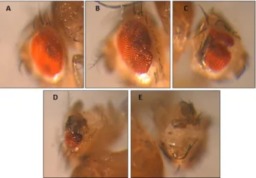
Figure 14 - Classification attributed to eye deformation in order to screen possible darhgef10 effectors. A – score 1; B – score 2; C – score 3; D – score 4; E – score 5. Score equal to 1 correspond to a normal eye without deformation and the highest score correspond to the most severely deformed eye. 35
Figure 15 - Schematic representation of Gal4 dependent UAS expression repressed by Gal80. Image adapted from (Wu and Luo, 2006). 35
Figure 16 – Boxplots representing the pupariation time of (N) larvae. The assay compared the pupariation time of darhgef10 mutant flies with the control w[1118]. Groups sharing the same letters are not statistically significant different at α = 0.017, according to the Bonferroni-Holm correction for multiple comparison. Dot blots are outliers, dark bars correspond to the medians and red dots to the averages.
Box limits indicate the lower and upper quartile and whiskers correspond to the maximum and minimum values, excluding the outliers. 37
Figure 17 – Fecundity assay. Bar-graph describing the average number of eggs laid per hour by control (w[1118]) and darhgef10 mutants females. Groups sharing the same letters are not statistically significantly different at α = 0.05, according to the Tuckey’s HSD post-hoc test. N=3 for all experiments. Error bars represent the
standard deviation of the mean (SD). 38
Figure 18 – Viability assay. Lines represent the percentages of viability through consecutive developmental stages. Total viability corresponds to the percentage of eggs that were able to reach the adult stage. All flies analyzed are isogenic. Groups sharing the same letters are not statistically significantly different at α = 0.05, according to analysis of variance (ANOVA) for multi comparisons. N=9 for w[1118] and N=6 for both darhgef10 deletions. Error bars represent the SD.
Drawings were adapted from
http://highered.mheducation.com/sites/dl/free/007352526x/873551/Reference D.pdf 39
Figure 19 – Bar graph describing the percentage of eggs that reach the adult stage, laid by w[1118] females (control), Df(1)ΔB females mated with Df(1)ΔS males and Df(1)ΔS females when mated with Df(1)ΔB males, respectively. Groups with different letters are statistically significantly different at α = 0.05, according to the Student's t-distribution. Statistical analysis was not performed to Df(1)ΔSm/Df(1)ΔBf due to insufficient set of sample elements. N=3 for all experiments except for Df(1)ΔB females mated with Df(1)ΔS males, where N=2.
Error bars represent SD. 40
Figure 20 - Bar-graph describing the percentage of laid eggs that were able to reach the adult stage. A - Viability analysis using Oregon females crossed with darhgef10 mutant males and with w[1118] males (control). B - Viability analysis using darhgef10 mutant and w[1118] (control) females crossed with Oregon males. Groups sharing the same letters are not statistically significantly different at α =
0.05, according to the Tuckey’s HSD post-hoc test. N=3 for all experiments. Error
bars represent the SD. 41
Figure 21 – Longevity of darhgef10 mutants (Df(1)ΔS and Df(1)ΔB) and w[1118] control flies: A - males; B - females. Curves with asterisks are statistically significantly different from the others at α = 0.017, according to the log rank test (http://bioinf.wehi.edu.au/software/russell/logrank/). N Df(1)ΔS = 179 (male), 183 (female); N Df(1)ΔB = 203 (male), 181 (female); N w[1118] = 203 (male), 191
(female). 42
Figure 22 - Longevity of flies expressing constitutive RNAi against darhgef10 (arm>darhgef10-IR). The ubiquitously-expressed armadillo-Gal4 (arm>) driver was used to drive UAS-darhgef10-IR thoughout the life of the flies. A - males; B - females. arm> and UAS-darhgef10-IR flies were crossed with w[1118] flies as controls. Curves with asterisks are statistically significantly different at α = 0.017, according to the log rank test (http://bioinf.wehi.edu.au/software/russell/logrank/). The color of the asterisks is relative to the color of the control curve that arm>darhgef10 longevity is statistically significant. Narm>darhgef10-IR= 61 (male), 93 (female); Narm>= 68 (male), 92(female); NUAS-darhgef10-IR= 74
(male), 76 (female). 43
Figure 23 – Negative geotaxis behavior of darhgef10 mutants and white[1118] control male adult flies aged: A - 24 h after adult eclosion (AAE), B - 20 d AAE, C - 40 d AAE and D - 60 d AAE. Graphs represent the percentage of flies (y axis) that were in the interval of height in cm (x axis), 10 s after the start of the assay (see 2.2.3). Error bars represent the standard error of the mean (SEM). Curves with an asterisk are statistically significantly different from the others at α = 0.017, according to the Fisher’s test followed by Bonferroni correction for multiple comparisons. 45 Figure 24 - Full anti-gravitational response of darhgef10 mutant male flies and w[1118] (control). Graph represents the percentage of flies at different ages that climbed more than 6 cm in the assay. This is a summary of the corresponding data presented on Figure 6. See Figure 6 for statistical analyses. The sole point that was
statistically different between the genotypes in the previous analyses is indicated
with an asterisk. Error bars represent SEM. 46
Figure 25 - Western blot analysis of darhgef10 overexpression in S2 cells transfected with a pTGw::darhgef10 plasmid for overexpression of darhgef10 marked with GFP, with a pTw::darhgef10 plasmid for overexpression of untagged darhgef10 and with a pUAST-empty plasmid to serve as a control. PAB#3 was chosen as an example, the same assay was performed with PAB#4, with similar results (see full
membrane in annexes figure III). 48
Figure 26 – S2 cells transfected with pTGw::darhgef10 (left) and pTw::darhgef10 (right), which encode GFP::dArhgef10 and untagged dArhgef10, respectively. Arrowheads indicate the transfected cells. PAB#3 was chosen as an example, but the same assay was performed with PAB#4, with similar results. Cyan, DAPI counterstain. Yellow, anti-GFP. Magenta, anti-dArhgef10 (PAB#3). Scale bar: 20
µm. 49
Figure 27 – S2 cells transfected with pTGw::darhgef10, which encodes GFP::dArhgef10. Right panel is a maximum intensity projection of all z-confocal stacks. Magenta, anti-GFP. Cyan, DAPI counterstain. Scale bar: 10 µm. 50
Figure 28 - Confocal sections of fixed S2 cells previously transfected with pTGw::darhgef10 plasmid, which encodes a GFP::dArhgef10 fusion protein. Anti-GFP, green. Phalloidin (for F-Actin), magenta. Anti-Rho1, yellow. Scale bars: 10
µm 51
Figure 29 – Confocal section of salivary glands from control (w[1118]) or ey>darhgef10 (ey-Gal4/pTw::darhgef10) L3 larvae. Salivary glands are stained with phalloidin for F-actin (magenta) and counterstained with DAPI (blue). Scale
bar: 20 µm. 53
Figure 30 – Confocal section of salivary glands from ey>darhgef10 (ey-Gal4/pTw::darhgef10) L3 larvae. Salivary glands are stained with phalloidin for F-actin (magenta) and anti-dArhgef10 (green). Scale bar: 20 µm. 53
Figure 31 – Scheme used to screen flies and S2 cells for successful homologous recombination repair. PCR product of Pair 1 is 975 bp, Pair 2 and Pair 3 is 587 bp.
54
Figure 32 - Analysis of PCR products in agarose gel of genome amplification of S2 cells transfected with: A - repair cassette into pUC57 plasmid, guideRNA into pU6-BbsI-chiRNA and pAc-sgRNA-Cas9 plasmid for cas9 expression; B - repair cassette into pUC57 plasmid, pU6-BbsI-chiRNA empty and pAc-sgRNA-Cas9 plasmid for cas9 expression. DNA volume was replaced with H20 in control
reaction. 55
Figure 33 - Analysis of PCR products amplified with primer pair 3 in agarose gel of genome amplification of S2 cells transfected with repair cassette into pUC57 plasmid, guideRNA into pU6-BbsI-chiRNA and pAc-sgRNA-Cas9 plasmid for cas9 expression - edited DNA; repair cassette into pUC57 plasmid, pU6-BbsI-chiRNA empty and pAc-sgRNA-Cas9 plasmid for cas9 expression – Control DNA. DNA volume was replaced with H20 in control reaction. 56
Figure 34 – Confocal images of Talin and βPS integrin expression detected by immunofluorescence analysis of stage 16 embryos from darhgef10 mutants and w[1118] controls. Muscle staining in w[1118] stage 16 embryo (right bottom). Arrowhead indicates muscle attachments sites. Scale bars: 20 µm. 59
Figure 35 – Live imaging of the dorsal vessel development in control (w[1118]) and darhgef10 mutant embryos. Projection of z-stacks obtained using spinning disc imaging. Arrowheads indicate the dorsal vessel defect found in darhgef10 embryos.
Scale bars: 20µm. 60
Figure 36 - Box and whiskers plot showing the interaction between dArhgef10 and candidate effectors. Plotted is the phenotypic score of the eye malformation caused by ey>darhgef10, which goes from 1 (wild-type eye) to 5 (severe malformation) - see Figure 14. Red dots represent averages, dark bars represent medians and black dots are outliers. Groups sharing the same letters are not statistically significant at α
= 0.05, according to the Tuckey’s HSD post-hoc test. N = 60, 92, 40, 88, 26, 38, 68, 100, 82 and 64, respectively, for the genotypes depicted from top to bottom. 62
Figure 37 – Confocal image showing body wall muscles stained with phalloidin in w[1118] and mef2>darhgef10 (pTW::darhgef10/mef2-Gal4) stage 16 embryos. The rounded muscle phenotype is noticeable in mef2>darhgef10 embryos (Right
List of abbreviations
aa Aminoacid
AAE After adult eclosion
Act Actin
AEL After egg laying
ANOVA Analysis of variance
Arm Armadillo
BDSC Bloomington Drosophila Stock Center. BLAST Basic Local Alignment Search Tool
bp Base pairs
BSA Bovine serum albumin
Cas9 CRISPR associated protein 9
Cdc42 Cell division cycle 42
CG Computed gene
CHD Congenital heart diseases
chiRNA Chimeric RNA
CMT Charcot-Marie-Tooth
CRISPR Clustered regularly interspaced short palindromic repeats
DAPI 4’,6-diamidino-2-phenylindole
darhgef10 Drosophila rho guanine nucleotide exchange factor 10
Df Deficiency chromosome
DH Dbl homology
dH20 Destilated Water
DHR Dock homology region
Dia Diaphanous
DNA Deoxyribonucleic acid
dNTPs Deoxynucleotide
DOCK Dedicator of cytokinesis
Dpp Decapentaplegic
DSHB Developmental Studies Hybridoma Bank EDTA Ethylenediaminetetraacetic acid
ERM Ezrin, radixin, moesin
Ey Eyeless
FBS Fetal Bovine Serum
FGF Fibroblasts growth factor
FLP Flippase
FRT Flippase recognition target
gDNA genomic DNA
GDP Guanosine diphosphate GFP Green fluorescent protein
gRNA Guide Ribonucleic Acid
GTP Guanosine triphosphate
H2O Water
HCl Chloridric ácid
Hh Hedgehod
HRP Horseradish peroxidase HSD Honest significant difference
Kb Kilo bases
kDa kilo daltons
L1 First instar
L2 Second instar
L3 Third instar
Lam A Laminin A
LIMK LIM kinase
Mef2 Myocyte enhancer factor-2
Mg2+ Magnesium
MLC Myosin light chain
MM Molecular marker
MRCK Myotonic dystrophy kinase related cdc42-binding kinase
MYPT Myosin light chain phosphatase
NaCl Sodium chloride
O.N. Overnight
PAK P21-activated kinases PAM Protospacer adjacent motif
PBAs Polyclonal antibodies
PBS Phosphate-buffered saline
PBST Phosphate-buffered saline with Triton-X PBSTw Phosphate-buffered saline with Tween20 PCR Polymerase Chain Reaction
PFA Parafolmaldehyde
PH Pleckstrin homology
Prc Pericardin
PS Penincilin-Streptomycin
qRT-PCR Quantitative reverse transcription polymerase chain reaction
Rho Ras family member
RhoGAPs RhoGTPase-activating proteins
RhoGDI Rho guanine nucleotide dissociation inhibitor RhoGEFs Rho guanine nucleotide exchange factors
RNA Ribonucleic Acid
RNAi RNA Interference
Robo Roundabout
ROCK Rho-associated protein kinase
RT Room temperature
S2 Schneider's Drosophila Line 2 cells
SD Standard deviation
SDS Sodium dodecyl sulfate
SDS-PAGE Sodium dodecyl sulfate polyacrylamide gel electrophoresis SEM Standard error of the mean
sfGFP Super folder Green fluorescence protein
sgRNA Single guide RNA
SM-RFP Somatic muscle red fluorescent protein
SNP Single nucleotide polymorphism
SP1 Specificity protein 1
TAE Tris-acetate
Tin Tinman
Tris-HCL Tris hydrochloride
Tub Tubulin promoter
UAS Upstream activating sequence
VRDC Vienna Drosophila RNAi Center
W[1118] White
WASP Wiskott-Aldrich Syndrome Protein
1.1 Congenital heart disease
Congenital heart diseases (CHD) define a group of structural and functional defects that arise during cardiac embryogenesis. These pathologies are the most common developmental birth defects, affecting about 1% of births per year worldwide. Epidemiologic studies suggest that genetic factors are the main cause of CHD (Fahed et al., 2013; Gelb and Chung, 2014; Hoffman and Kaplan, 2002). However, the genetic mechanisms of heart development continue to be poorly understood and CHD are a significant cause of infant mortality and can result in chronic disability and increased medical treatment costs (Reller et al., 2008). For instance, it is estimated that about 40 million adults live with a congenital heart defect in the world (this estimate is based on US adult CHD estimates and 2011 worldwide adult population estimates: J.I.E., 2013; Marelli et al., 2007; Central Intelligence Agency 2011, The World Factbook 2011, ISSN 1553-8133, Washington, DC, viewed 17th April, 2011). These patients with this chronic illness are in increased risk of several complications such as infective endocarditis and hemodynamic decompensation, and can face life-threatening problems during contraception and pregnancy, with cardiac disease being one of the most common causes of indirect maternal death during pregnancy.
1.2 Heart development in Drosophila
Heart formation is a critical developmental process, tightly regulated in a sequential manner by multiple signaling pathways. Gene mutations and consequently deregulation of these pathways could lead to defective morphology and function of the mature heart, compromising the adult organism.
Many genes controlling early steps of heart development are highly conserved between vertebrates and invertebrates. Drosophila has a cardiac organ, called dorsal vessel or cardiac tube that constitutes the entire cardiovascular system of the organism and acts as a muscular pump contracting automatically to distribute the hemolymph through the body. Dorsal vessel formation shares many similarities with heart
development in vertebrates providing a powerful system to study gene networks and regulatory pathways involved in heart development (Wu and Luo, 2006).
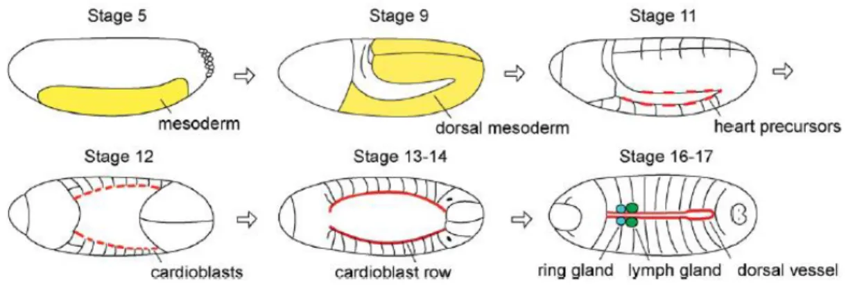
The mature dorsal vessel is formed by two major cell types: the inner contractile cardioblasts which form the lumen of the heart and the non-muscular pericardial cells which forms the outer cell layers. The pericardial cells are thought to function in detoxification of the hemolymph (Bryantsev and Cripps, 2009; Vogler and Bodmer, 2015; Zhang et al., 2013). The dorsal vessel is divided in two distinct parts: the aorta and the heart proper (Figure 1).
Figure 1 - Organization of embryonic dorsal vessel. The dorsal vessel can be divided in the
heart region (h), posteriorly, and in the aorta region, anteriorly. The heart proper contains inflow tracts termed ostia cells. Dashed arrows show the hemolymph flow. Adapted from (Tao and Schulz, 2007).
The anterior region of the tube is surrounded by two glands: the endocrine ring gland and the hematopoietic lymph gland (Bier and Bodmer, 2004; Medioni et al., 2009; Tao and Schulz, 2007). The heart is located at the posterior abdominal segments and contains inflow tracts, the ostium cells that open and close enabling entering of the hemolymph. This region, the only that shows automatically and synchronized beating, ends with a group of four cells that contribute to the major pacemaker activity of the organ. The aorta region, located anteriorly, is composed of a narrow lumen and ends in the outflow tracts. The cardiac tube is segmentally patterned, with mainly six pairs of cardioblasts per segment with a distinct genetic nature: four pairs have large nuclei and two have smaller nuclei (Medioni et al., 2009; Tao and Schulz, 2007) (Figure 1).
As in vertebrates, the Drosophila dorsal vessel arises from the mesoderm (Figure 2) being specified through similar cellular induction pathways and transcription factors (Bodmer and Venkatesh, 1998; Cripps and Olson, 2002; Zaffran and Frasch, 2002). Upon Fibroblast growth factor (FGF) signaling, mesodermal cells invaginates and spread laterally forming a layer within the embryo (Mason, 1994). In the next stage, cells of the spreading mesoderm are specified as dorsal mesoderm by Decapentaplegic (Dpp) signaling. However, Dpp signaling is not sufficient to promote cardiac fate, thus ectodermally-derived signaling molecule Wingless (Wg) is required for the development of further specialized heart precursors (Wu, Golden and Bodmer, 1995). At this stage, Notch (N) signaling is essential for distinct cardiac progenitors leading to the formation of cardioblasts and pericardial cells (Han and Bodmer, 2003). The bilateral rows of aligned cardioblasts start to migrate dorsally and meet at the dorsal midline. In order to form a functional heart, Hedgehod (Hh) signaling molecule is secreted by the ectoderm and cardioblasts start specific differentiation programs. Due to Hh signaling, cardioblasts give rise to a sub-type of cardioblasts: cells with large nuclei (Tinman-positive cells) and cells with small nuclei (Svp/Doc-positive cells). Svp (COUP-TFII class protein Seven-up) is expressed in the anterior two pairs of cardioblasts in each segment of the dorsal vessel, which differentiate into three pairs of inflow tracts, ostia cells, in the heart proper region (Molina and Cripps, 2001; Park et al., 1996; Ponzielli et al., 2002). Tinman is responsible for the contractility of the four pairs of cardioblasts in the posterior region, making these cells adopt a contractile fate instead of the ostia cell fate (Zaffran et al., 2006).
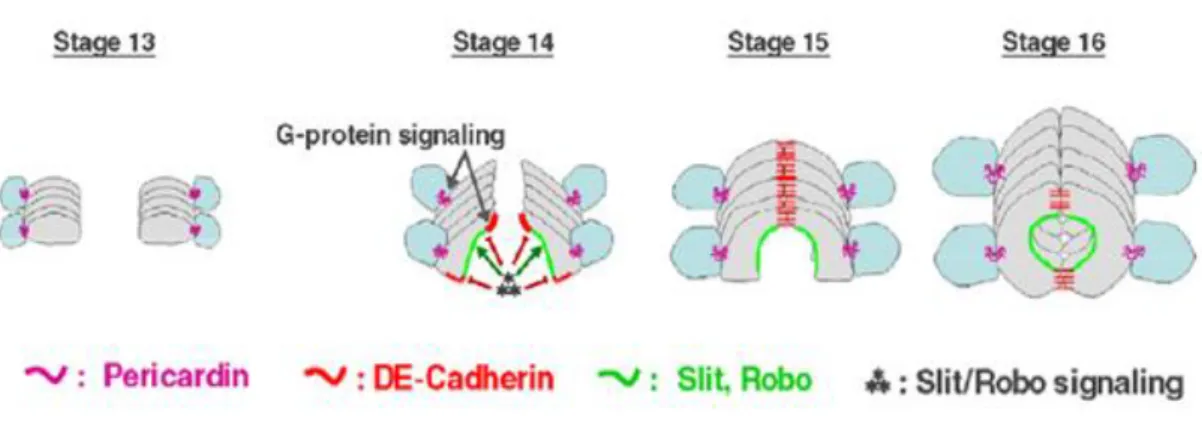
In addition to all transcription factors and signaling pathways involved in cardiac cell specification, many other genes and proteins are required for proper cardioblast alignment, migration and polarization. After specification of cardioblasts, the correct formation of the cardiac lumen is dependent on the correct polarity of cardiac cells, which is achieved by a dynamic control of cell shape changes and formation of membrane domains. In migrating cardiac cells, three distinct membrane domains can be specified due to specific cell polarity markers: L(luminal)-domain, J(junction)-domain and P(pericardin) A(adhesion)-domain. The L-domain faces the lumen of the tube. The J-domain forms adherent junctions between cardiac cells of opposite rows. The
PA-domain is the one that makes contacts between cardiac and pericardial cells (Medioni et al., 2009; Tao and Schulz, 2007; Vogler and Bodmer, 2015) (Figure 3).
Figure 2 - Cardiogenesis in the Drosophila embryo. Dorsal vessel formation starts with
mesoderm differentiation and after several specification events of heart precursors, the mature dorsal vessel is complete and functional in the stage 17. Adapted from (Tao and Schulz, 2007).
Specification and maintenance of such domains is assured by several regulators like the Slit/Robo pathway, which is required for specification of non-adherent/repulsive L-domains, DE-cadherin that establish adherent junctions and Go proteins, members of the G protein family. These are responsible for the specification of the PA domains and the localization of proteins to these membrane domains to ensure proper cardioblast and pericardial cell adhesion. At the same time that the cardioblasts migrate to the dorsal midline, the leading edge cells interact with the adjacent ectoderm layer, which is in a coincident migration towards the midline in a process called dorsal closure. These interactions contribute to the movement of the cardioblasts. Such coordinated migration between the dorsal ectoderm and the associated cardioblasts rows also requires pericardin (prc), a component of the extracellular matrix that is produced by pericardial cells (Medioni et al., 2009; Tao and Schulz, 2007) (Figure 3).
Figure 3 – Dorsal vessel formation during embryonic development. The cardioblast rows
migrate towards the dorsal midline of the embryo. At stage 15, cardioblasts of opposite rows start to adhere at their edges to form the cardiac tube. Several proteins and signaling pathways are involved in the achievement and maintenance of the three different membrane domains that mediate tube formation (see text). Adapted from (Medioni et al., 2009).
In the end of embryogenesis (stage 17), the mature dorsal vessel starts to beat as a functional cardiac organ. In the following larval stages, the cardiovascular system cells increase in size between 200-500 fold, maintaining their cellular identities. The adult heart is formed during metamorphosis by the remodeling of differentiated and already functional larval cardiomyocytes, without cell proliferation or addition of new cells (Hartenstein et al., 1992; Park, Venkatesh and Bodmer, 1998).
1.3 Rho-family GTPases
Rho GTPases are a group of evolutionarily conserved proteins that function as intracellular transducers establishing a link between the cell surface signals and multiple intracellular responses, controlling a variety of cellular signal transduction pathways (Etienne-Manneville and Hall, 2002; Murali and Rajalingam, 2014; Ridley, 2001).
These proteins belong to a main branch of the Ras superfamily of small GTPases and are ubiquitously expressed in all eukaryotic cells regulating many cellular events like actin cytoskeleton, cell-cycle progression, vesicle trafficking and gene transcription,
having an important role in cell adhesion, migration and polarity, neurite extension and retraction (Rossman, Der and Sondek, 2005; Sit and Manser, 2011).
RhoGTPases cycle between two conformational states: one GDP-bound state, and the other GTP-bound state. The GTP-bound state is able to interact and activate downstream effector proteins (Sit and Manser, 2011). Since RhoGTPases have the ability to regulate a wide variety of functions in very dynamic cellular context, they need to be tightly regulated. This regulation is achieved by three sets of proteins: Rho guanine nucleotide exchange factors (RhoGEFs), Rho GTPase activating proteins (RhoGAPs) and Rho guanine nucleotide dissociation inhibitors (RhoGDIs) (Heasman and Ridley, 2008) (Figure 4).
RhoGEFs activate RhoGTPases by catalyzing the release of GDP and the binding of GTP. In turn, RhoGAPs inactivate the proteins by enhancing the intrinsic GTPase activity to hydrolyze GTP to GDP. In order to retain the RhoGTPases in their inactive conformation, RhoGDIs bind to C-terminal prenyl groups sequestering them in the cytosol, preventing nucleotide exchange and membrane association. Altogether these proteins upregulate or downregulate the levels of membrane bound Rho proteins enabling thereby their spatio-temporal regulation (Heasman and Ridley, 2008; Pertz, 2010). Apart from these regulator proteins, RhoGTPases can also be regulated by post-translational modifications like phosphorylation and ubiquitination. Ubiquitin-dependent regulation can affect the stability of these proteins and for instance influence the plasticity of cell migration (Ekenstedt et al., 2014; Murali and Rajalingam, 2014; Pareyson, 1999).
Figure 4 - Regulation of RhoGTPases by RhoGAP, RhoGEF and RhoGDI. When active, in the
GTP bound state, RhoGTPases can interact with downstream effector proteins, regulating many cellular responses. Adapted from (Heasman and Ridley, 2008).
Until now, about 20 members of Rho GTPases have been identified in humans. Of these, RhoA, Rac1 and Cdc42 are the main and best studied groups. Over 70 GAPs and 80 GEFs have been identified, most of them showing tissue specific expression. The Drosophila genome encodes for seven Rho family members including the human homologs of Rho1, Rac1 and Cdc42 (Schmidt and Hall, 2002). The high number of functions for RhoGTPases is consistent with their large list of target proteins. This includes serine/threonine kinases, tyrosine kinases, lipases and scaffold proteins (Bishop and Hall, 2000). Despite all the functions described, RhoGTPases are best known for their function as regulators of the actin cytoskeleton and cell migration. Thus, the best studied effectors are proteins that interact with Rac1, RhoA and cdc42, which were discovered mainly for their influence in cell shape and plasticity of cell migration (Murali and Rajalingam, 2014; Sadok and Marshall, 2014).
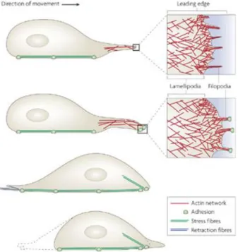
Cell migration is a dynamic process that needs to be precisely regulated in order to allow continuous remodeling of the cellular architecture. This reorganization of the actin cytoskeleton enables the cell to move and adapt to changes in the surrounding
environment (Sadok and Marshall, 2014).In figure 5, a cell migration event is depicted: highly dynamic lamellipodia and filopodia are extended at the leading edge, containing actin filaments; cell translocation occurs through actomyosin-based contraction forces; retraction fibers pull the rear of the cell forward (Mattila and Lappalainen, 2008).
Figure 5 - Cell migration. Adapted from (Mattila and Lappalainen, 2008).
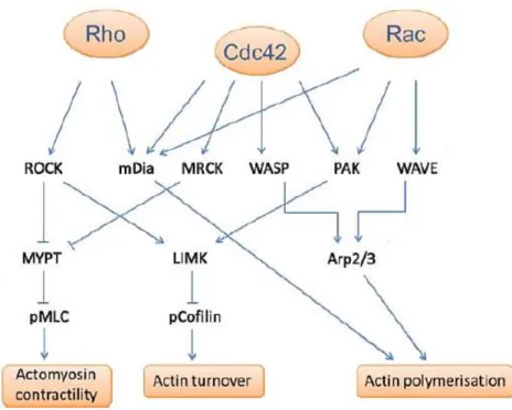
Altogether, Rac1, RhoA and Cdc42 promote a set of events that lead to new and/or reorganized actin filaments (Figure 6). Rac activates the Arp 2/3 complex through WAVE, leading to actin polymerization and lamellipodia formation. Additionally, it activates the serine/threonine kinase PAK that phosphorylates LIM kinase (LIMK), which inhibits cofilin, an actin binding protein that regulates the assembly and disassembly of actin filaments, thereby regulating actin-filament turnover. In turn, Rho activates ROCK which inactivates the myosin light chain phosphatase (MYPT) and consequently activates myosin II. Actomyosin contractility can also be promoted by Cdc42 via MRCK (myotonic dystrophy kinase related cdc42-binding kinase) because it also inactivates MYPT. Cdc42 contributes to filopodia extension through WASP and PAK, connecting with Rac and Rho. mDia (Diaphanous), a formin
protein, seems to be a common effector, leading to actin polymerization (Heasman and Ridley, 2008; Murali and Rajalingam, 2014) (Figure 6).
Figure 6 - RhoA, Rac1 and cdc42 and their effectors proteins in cell migration. Adapted from
(Sadok and Marshall, 2014).
In a simplistic way, we can summarize that Rac1 mainly regulates membrane protrusions at the leading edge, Cdc42 controls filopodia formation and cell polymerization and RhoA controls contractility at the back of the cell. Nevertheless, this is a too simple 2D model and new results have showed a more complex organization highly regulated. More and more, fluorescent probes are used to report RhoGTPase signaling in time and space, giving new insights to our knowledge in this not so simple concept (Pertz, 2010).
1.4 Rho guanine nucleotide exchange factors
Since RhoGTPases play a prominent role in many aspects of cell biology, they need to be tightly regulated. Along with the regulatory factors RhoGAP and RhoGDI, RhoGEFs provides fine control over the signaling events mediated by RhoGTPases (Cherfils and Zeghouf, 2013). RhoGEFs catalyze the displacement of the bound GDP and subsequent exchange with cytosolic GTP, generating the active form of RhoGTP that is capable of recognizing a wide variety of effector proteins (Bishop and Hall, 2000; Etienne-Manneville and Hall, 2002).
In the human genome RhoGEFs are encoded by two unrelated gene families, Dbl and DOCK, in a total of over 80 distinct proteins. The best known and major group is the Dbl family, defined by a Dbl homology (DH) domain and a pleckstrin homology (PH) domain, C-terminal to the DH domain (Rossman, Der and Sondek, 2005; Schmidt and Hall, 2002). The DH domain is necessary for GEF activity and the PH domain seems to have a role in assistance of exchange reaction (García-Mata and Burridge, 2007) and can help to target the RhoGEFs to their appropriate intracellular localization, however the precise role of this domain remains unclear (Etienne-Manneville and Hall, 2002; Schmidt and Hall, 2002). All the 23 RhoGEFs identified in Drosophila belong to the Dbl family. The DOCK (dedicator of cytokinesis) family has been defined more recently and is characterized by a conserved catalytic domain, DOCK homology region 2 (DHR2) and a phospholipid-binding domain (DHR1) that locates them to membranes (Côté et al., 2005; Meller, Merlot and Guda, 2005). DOCK proteins are specific for Rac and Cdc42 (Côté and Vuori, 2007).
Interestingly, there are about four times more RhoGEFs than their target RhoGTPases. This suggests that a specific RhoGTPase could be activated by multiple RhoGEFs. This redundancy could be explained by the tissue-specific distribution of the RhoGEFs, despite the apparent broad expression of most RhoGEFs (García-Mata and Burridge, 2007; Goicoechea et al., 2014). RhoGEFs present a huge variety of domain structures in addition to the core domains described above. These different domain structures could also contribute to the specific regulation of different signaling pathways by RhoGTPases (Goicoechea et al., 2014). RhoGEFs can be regulated in a complex manner that is not still completely understood but can include protein-protein or
protein-lipid interaction, binding of second messengers and postranslational modifications. It is thought that these interactions can lead to three major changes in GEFs: translocation to specific compartment of the cell, release from an autoinhibitory state or induction of allosteric changes in the catalytic domain (Bos, Rehmann and Wittinghofer, 2007).
1.4.1 Drosophila Rho guanine nucleotide exchange factor 10
We have previously identified a Drosophila gene, darhgef10, which is strongly expressed in the developing heart between embryonic stages 13-17 (Mantas Dias, 2012). The darhgef10 gene encodes a Drosophila Rho guanine nucleotide exchange factor (RhoGEF) homologous to a protein known as ARHGEF10 or RhoGEF10 in humans (Mantas Dias, 2012; Verhoeven et al., 2003). Interestingly, deletion of darhgef10 in Drosophila leads to subtle, yet prevalent embryonic heart abnormalities, consisting of misaligned cardioblasts in both the aorta and heart regions. Surprisingly, darhgef10 mutant flies are viable, suggesting that the presence of the cardiac defects is compatible with unchallenged life under laboratory-growth conditions.
The human Dbl family member ARHGEF10 is a specific activator of RhoA (Aoki et al., 2009; Chaya et al., 2011) and was first identified as the product of a gene associated with slowed nerve conduction velocities of peripheral nerves (Verhoeven et al., 2003). ARHGEF10 has a DH domain and a very divergent PH-like domain. Thus, it was considered to be a member of a Rho-specific GEF family with unusual protein architecture (Mohl et al., 2006). The mouse ARHGEF10 homolog, GEF10, was found to be broadly expressed with highest levels in the heart and skeletal muscle (Verhoeven et al., 2003). This expression pattern is consistent with the strong enrichment we find for darhgef10 expression in the developing Drosophila dorsal vessel (Mantas Dias, 2012). In Drosophila, dArhgef10 has an insect specific N-terminal domain, which is encode by isoform C (Figure 7).
Figure 7 - Schematic structure of the Dbl member family ARHGEF10 and dArhgef10. DH
represents the Dbl homology domain and PH, the pleckstrin homology-like domain. Although this RhoGEF possess a PH-like domain (blue region) it has a very divergent architecture when compared to other RhoGEF PH domains (Aoki et al., 2009).
1.4.1.1 ARHGEF10 in cardiovascular and neurodegenerative disorders
We identified darhgef10 as being a gene required for proper heart development in Drosophila (Mantas Dias, 2012). Interestingly, different studies have found a link between human ARHGEF10 and atherotrombotic stroke or peripheral neuropathies (Matsushita et al., 2010; Verhoeven et al., 2003; Yin et al., 2011).
In humans, a SNP in ARHGEF10 was found to be strongly associated with atherothrombotic stroke. This single point mutation may cause increased levels of
ARHGEF10 transcript due to a higher affinity for the sp1 transcription factor, enhancing the RhoA-Rho kinase activity that contributes to the development of atherothrombotic stroke (Matsushita et al., 2010). Indeed, the effector RhoA-Rho-kinase had already been implicated in the process of atherosclerotic cerebral infarction (Shimokawa and Takeshita, 2005).
Another mutation in ARHGEF10, which leads to an amino acid substitution of Threonine to Isoleucine (T332I), was identified in patients with slowed nerve conduction velocity, which was associated with thin myelination of peripheral nerves (Verhoeven et al., 2003). This was the first time that ARHGEF10 was identified as been implicated in peripheral-nerve conduction, suggesting a possible role for ARHGEF10
during the development of the peripheral nervous system in vertebrates. Cell culture experiments then suggested that the T332I change could confer constitutive GEF activity, because it maps within a large N-terminal domain that negatively affects ARHGEF10 activity. Ectopic ARHGEF10 expression induces cell contraction in rat schwann cells, mediated by Rho-ROCK activity signaling, impairing schwann cell processes and consequently proper myelination (Chaya et al., 2011).
More recently, a 10-bp deletion in the canine ARHGEF10 was associated with a severe polyneuropathy in leonberger dogs. This pathology has many clinical similarities with Charcot-Marie-Tooth (CMT) disease in humans, a genetically heterogeneous group of peripheral neuropathies sharing the same clinical phenotype (Ekenstedt et al., 2014;Pareyson, 1999). Once again, a possible role for ARHGEF10 in controlling the development and/or maintenance of peripheral nerves was suggested.
1.4.2 darhgef10 mutants
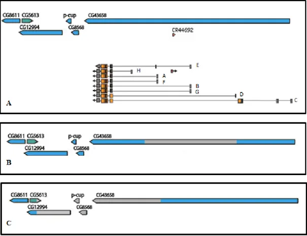
We have previously generated two darhgef10 deletion mutations (deficiencies) by the FLP-FRT method (Mantas Dias, 2012; Parks et al., 2004): a small and a large (big) deletion, hereafter termed darhgef10[Df(1)ΔS] and darhgef10[Df(1)ΔB], respectively (Figure 8). The Df(1)ΔS covers at least three transcription start sites for the darhgef10 gene and the non-coding RNA CR44692, while Df(1)ΔB covers the majority of the darhgef10 coding region, including the DH and PH-like domain coding regions, and affects at least four other genes lying 5’ of darhgef10 (CG8568, p-cup, CG5613 and CG12994). From this information alone we can predict that Df(1)ΔB is a dArhgef10 protein null allele.
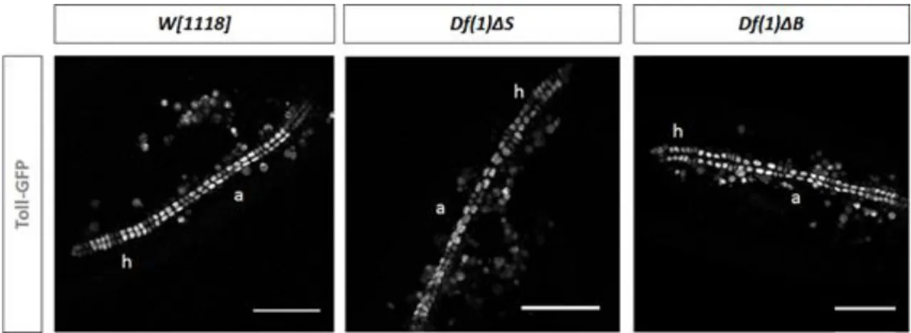
Both darhgef10 mutants are homozygous viable, suggesting that this gene is not essential for Drosophila development, however, both mutants showed a misaligned cardioblasts phenotype (Figure 9) (Mantas Dias, 2012).
Figure 8 - Scheme of the darhgef10 (aka CG43658) gene and the regions deleted in the Df(1)ΔS or Df(1)ΔB deficiency mutations (grey boxes). A - The darhgef10 gene and 5 flanking genes on the X chromosome of D. melanogaster. All darhgef10 transcripts are represented. B - The grey box depicts the region deleted in the Df(1)ΔS mutant. C - The grey box depicts the region deleted in the Df(1)ΔB mutant. Isoforms are indicated in A. Notice that four other genes are affected by Df(1)ΔB.
While Df(1)ΔB is clearly a protein null, Df(1)ΔS is not as some darhgef10 isoforms are left intact by this deficiency. However, both darhgef10 Df mutations show the same phenotype of cardioblast missalignment. This is explained because the Df(1)ΔS removes the transcriptional promoter of two of the major isoforms of darhgef10 (isoforms B and E) (Figure 8). Consequently, the total transcript levels in Df(1)ΔS are reduced by >80% in this background (annexes figure I; Heredia, Mantas Dias and Gontijo, unpublished results). In situ hybridization studies showed that no darhgef10 transcript is detectable in the developing cardioblasts in Df(1)ΔS embryos, suggesting that the isoforms B & E are responsible for most, if not all, cardioblast
expression. Thus, even though Df(1)ΔS is not a protein null, it is a strong reduction of function and could likely be a null for many cell types, including the developing cardioblasts, explaining the phenotypic similarity between Df(1)ΔS and Df(1)ΔB.
Figure 9 - Dorsal vessel visualized with a Toll-GFP reporter line showing the misaligned
cardioblasts phenotype found in darhgef10 mutants (a indicates the aorta region and h the heart region). Scale bars: 50µm. Adapted from (Mantas Dias, 2012).
1.5 Aims
Our aims were to phenotypically characterize the newly generated darhgef10 mutants by surveying their development, viability, lifespan and behavior, and in parallel to investigate the cellular basis of their misaligned cardioblasts phenotype. Since human ARHGEF10 is a susceptibility gene for cardiovascular diseases (Yin et al., 2011; Matsushita et al., 2010), we intend to explore darhgef10 mutants as potential models to study the interaction between environmental challenges and a congenitally compromised circulatory system.
2.1 Fly strains and husbandry
In this work, all fly stocks were maintained at 18ºC and raised in standard cornmeal-agar medium. When performing experiments, animals were grown at 25 ºC in appropriate humidity conditions at 75 % to achieve a controlled life cycle of about 10 days.
Virgin female collections and Drosophila stocks maintenance were performed as described by Ashburner (Ashburner and Roote, 2007).
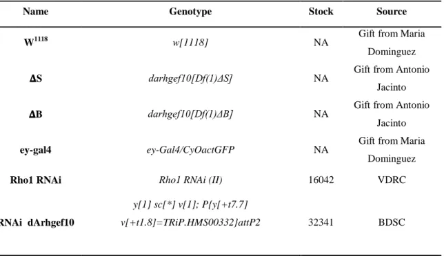
All Drosophila stocks used in this study are described in Table 1. The stocks were obtained either from the Bloomington Drosophila stock center (http://flystocks.bio.indiana.edu/), from the Vienna Drosophila Resource Center (http://stockcenter.vdrc.at/control/main), from other laboratories as a gift or were generated in our laboratory, as depicted below.
Table 1 - Drosophila melanogaster stocks used in this work.
Name Genotype Stock Source
W1118 w[1118] NA Gift from Maria Dominguez 𝚫S darhgef10[Df(1)ΔS] NA Gift from Antonio
Jacinto 𝚫B darhgef10[Df(1)ΔB] NA Gift from Antonio
Jacinto
ey-gal4 ey-Gal4/CyOactGFP NA Gift from Maria
Dominguez
Rho1 RNAi Rho1 RNAi (II) 16042 VDRC
RNAi dArhgef10
y[1] sc[*] v[1]; P{y[+t7.7]
Rho1 mutant w[*]; b[1] pr[1] cn[1] Rho1[1B] px[1]
sp[1]/CyO 9477 BDSC
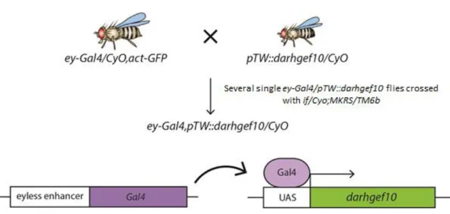
Recombinant ey-Gal4>pTW::CG43658/CyOGal80 NA Generated in our lab
Oregon Oregon-R-C 5 BDSC
FM7 Ngr[l4]/FM7c 5708 BDSC
Lifeact_𝚫S
darhgef10[Df(1)ΔS];;UAS-lifeactGFP,MefGal4/TM6b NA Generated in our lab
Lifeact_ ΔB
darhgef10[Df(1)ΔB];;UAS-lifeactGFP,Mef2Gal4/TM6b NA Generated in our lab
Rock mutant Y[1] W[1118]Rok[2]
P{ry[+t7.2]=neoFrt}19A/FM7c 6666 BDSC
Rho1 mutant W[a] N[fa-g]; Rho1 [E3.10]/CyO 3176 BDSC
Rok1 mutant W*Rok1/FM7i,P{actGFP}JMR3 6665 BDSC
Rock RNAi-TRIP y[1] v[1]; P{y[+t7.7]
v[+t1.8]=TRiP.JF03225}attP2 28797 BDSC
Rho1 RNAi Rho1 RNAi-kk (II) 109420 VDRC
Rock RNAi-kk DRHK RNAi-kk (II) 104675 VDRC
Diaphanous RNAi
y[1] v[1]; P{y[+t7.7]
v[+t1.8]=TRiP.HM05027}attP2 28541 BDSC
𝚫S_iso darhgef10[Df(1)ΔS];iso;iso NA Generated in our lab 𝚫B_iso darhgef10[Df(1)ΔB];iso;iso NA Generated in our lab
Lifeact ;;UAS-lifeactGFP,MefGal4/TM6b Gift from Antonio
Jacinto
Double balanced w; If/CyO;MKRS/Tm6b NA Gift from Antonio
2.2 Phenotypic characterization of darhgef10 mutants
darhgef10 mutations cause subtle heart abnormalities that arise during embryonic development. To learn more about the possible effects of these abnormalities on other developmental and life history traits and/or to identify novel darhgef10-dependent phenotypes we characterized a series of parameters of darhgef10 animals, including larval development and adult viability, fecundity, longevity and negative geotaxis behavior of adult flies.
2.2.1 Viability and fecundity assays
To study the fertility of darhgef10 mutant flies, we performed fecundity and viability assays. The fecundity consists in the number of eggs that a single female laid per hour and viability is based on the number of eggs delivered by a single female per hour that hatch to larvae.
For these experiments, the adult flies were raised in optimal developmental conditions (with a controlled number of eggs per vial to avoid overcrowding, and controlled temperature at 25 ºC) in order to minimize any nutritional and/or environmental negative effect on their fecundity (such as a reduction on ovariole number). A day before (24 h) each fecundity/viability experiment, 5-10 virgin females and an equal number of males all aged between 3 to 7 days old were crossed. The flies were kept in laying pots for 6 h and apple plates were replaced every 2 h. After 6 h, the eggs laid during the day were transferred to a strip of paper impregnated in 1x PBS and counted. The strip of paper was transferred to a new vial and viability analysis was performed 24 h later by scoring the number of eggs that hatched. This procedure was repeated for 3 consecutive days and each fly that died or escaped was discounted in the egg/female ratio. The fraction of larvae that pupariated and then eclosed into adults were registered for each genotype as further indicators of viability. The fecundity and viability measurements between genotypes were compared using analysis of variance (ANOVA) followed by post-hoc Tukey’s HSD (honest significant difference) test, using α = 0.05.
2.2.2 Developmental time assay
In order to verify if darhgef10 larvae had any major problem developing as larvae, we measured the time they took to pupariation. If darhgef10 affected heart physiology, maybe the larvae had difficulties in any of the behaviors required to reassure the rapid growth necessary to achieve a typical timing of the onset of metamorphosis. We therefore performed pupariation assays following the development of synchronously growing larvae until reaching the pupal stage.
As in the fertility assay, flies were crossed 24 h before starting the assay and kept at 25 ºC in a laying pot. In each day of the assay, flies laid eggs during 9 h in a laying pot with an apple plate that was switched every 3 hours. 48 h AEL (after egg laying), the larvae were transferred to vials, usually 3 vials of 10 larvae each, to avoid overcrowding. This was repeated in 3 consecutive days. Two days following transfer of the larvae, we started scoring time of pupariation for each larvae by counting pupae at 10 am, 3 pm and 7 pm, until all larvae had pupariated. Statistical analyses was performed using the Wilcoxon rank sum test followed by bonferroni–holm method for p-value adjustment to multiple comparisons, using α = 0.05.
2.2.3 Negative geotaxis behavior assay
In Drosophila, negative geotaxis is an innate behavior where flies preferentially move against the gravitational force when agitated. This can be observed by their tendency to climb up a tube after the tube with the flies is tapped on a table top. To perform this behavior the flies require an intact locomotor capacity. Not surprisingly, this behavior declines with the age of the animals and is affected by mutations in genes that affect locomotor activity. Our aim with this experiment was to use the negative geotaxis behavior assay as an indicator of the overall locomotor capacity of the darhgef10 mutant lines as compared to their controls (w[1118]background). In other words, we wanted to see if the congenital heart defect in the mutant lines leads to a reduced locomotor behavior capacity of the flies (Nichols, Becnel and Pandey, 2012).
For this assay, newly emerged adult male flies were collected in a total of 5 sets of 10 male flies of each genotype, and kept at 25 ºC during the experiment. In order to follow the height climbed for each flies, we used a tube with a 2 cm, 4 cm and 6 cm mark. To perform the assay, the tubes were sharply tapped down three times, ensuring that each tap is hard enough to knock down all the flies to the bottom of the tube. The behavior of the flies was followed by a video camera during 10 s. After this time, the number of flies that passed 2 cm, 4 cm and 6 cm was registered. This procedure was repeated 10 times for each set of flies analyzed, with a 30 s interval between each assay to allow the flies to recover. The assay was performed with male flies aged 1, 20, 40 and 60 days after eclosion. Statistical analysis was performed using Fisher’s test followed by Bonferroni Correction (α/n) for multiple comparisons, using α=0.05.
2.2.4 Longevity assays
In order to study the influence of darhgef10 on total lifespan, we performed a longevity assay, following the flies from adult eclosion until death. Newly emerged adult flies, grown in optimal developmental conditions (with a controlled number of eggs per vial to avoid overcrowding, and controlled temperature at 25 ºC) were collected. The collection day was established as day 0. The vials were changed twice a week, allowing optimal growth conditions (food supply and media texture) in order to guarantee more robust data. Thereby, the possibility of the recorded deaths happening because of environmental conditions instead of the age factor in study is reduced. Data was analyzed statistically using the log rank test
(http://bioinf.wehi.edu.au/software/russell/logrank/)followed by Bonferroni Correction (α/n) for multiple comparisons, using α=0.05.
2.3 Targeted genome editing using CRISPR-Cas 9
2.3.1 Guide RNA and repair cassette designIn order to access the localization of dArhgef10 protein, we used a CRISPR-Cas9 mediated homologous repair-based technique(Jinek et al., 2012; Port et al., 2014)to tag this protein with a green fluorescent protein (sfGFP) and OLLAS (E.coli OmpF Linker and mouse Langerin fusion Sequence) at its Amino (C)-terminus, separated by two flexible linker spacer sequences GSGSGS.
The guide RNA (Figure 10) was designed with http://tools.flycrispr.molbio.wisc.edu/targetFinder, synthetized by Sigma and cloned into pU6-BbsI-chiRNA.
Figure 10- GuideRNA used in the CRISPR-Cas9 strategy with PAM sequence. Nucleotides represented
in red are only used to clone into pU6-BbsI-chiRNA.
The repair cassette was synthesized de novo and cloned into a pUC57 plasmid by GeneScript and co-injected, by BestGene, with the gRNA into w1118; PBac{y[+mDint2]=vas-Cas9}VK00037/CyO,P{w[+mC]=Tb[1]}Cpr[CyO-V] embryos, which expresses Cas9 under control of Vas regulatory sequences. The following sequence corresponds to the repair cassette, where dark blue corresponds to the homology region after darhgef10 end, dark red to the homology region up to darhgef10 end, green to the sfGFP, blue to the spacers (GSGSGS), grey to the OLLAS epitope, brown to the restriction site for BglII and orange to the restriction site for AgeI:
GCCAGCCGCCGCGGTGGCCACTTTAAAGCGAAAACAGAAGCAGAACTCCAAGCAGCAGCAGCAACAGAACGCCGACG GACCAAGGACAGTGATCACCCTGATGGGCGGACGCGGCTACTGGCGCCAGATGTGGTACAACGGGGCGGGTGGTTCG CCCAGCCATAAGAACAGCAGCTCCGGCGGCGGCGGAGGCAGTGGCTCCGGATTCAGTGGCCAAACAATGCAGTCGGG
TAATCCCAGCTGCTCACCGCTGACGGCCAACTCCAATGACGCCCACATCGTTGTCTGGGAGAAGAAGTTAGGCTCCG
GTAGTGGTTCCAGATCTCGTAAGGGCGAGGAGTTGTTCACGGGAGTTGTGCCCATATTGGTTGAGCTGGATGGAGAT GTGAATGGCCACAAGTTCAGTGTGCGGGGTGAGGGAGAAGGAGACGCAACAAACGGTAAGCTGACACTGAAGTTCAT TTGTACTACGGGCAAGCTCCCGGTGCCATGGCCCACATTGGTCACCACCCTGACCTATGGCGTGCAATGCTTCGCCC GATATCCAGATCATATGAAGCAGCATGATTTCTTTAAGTCGGCCATGCCCGAGGGTTACGTACAAGAGCGCACTATT AGCTTTAAGGACGACGGTACGTATAAAACCAGGGCTGAGGTGAAGTTTGAGGGTGATACCCTGGTGAACCGCATTGA ATTGAAGGGCATCGATTTTAAGGAGGACGGCAACATCCTGGGCCACAAGCTCGAATATAATTTTAATAGCCATAATG TTTACATTACCGCGGACAAGCAGAAGAATGGAATTAAGGCTAATTTCAAGATCCGACATAATGTGGAGGACGGATCC GTTCAGTTGGCCGATCACTACCAGCAAAACACCCCCATCGGAGATGGCCCCGTCCTGCTGCCCGATAACCACTACCT GAGTACCCAGTCCGTCCTGTCGAAGGATCCTAATGAGAAGCGGGATCATATGGTGCTGCTGGAGTTTGTGACTGCCG
CCGGCATAACGCATGGAATGGACGAGCTGTATAAAGGCTCCGGTAGTGGTTCCTCCGGCTTCGCCAACGAGCTGGGC
CCCCGCCTGATGGGCAAGACCGGTTAATCGCAGGAGCTGGTGTGCTGCTGCACCCTTCAGCAGCATCCCCACCAGCA
CCATCCACCGCTAACCGAGCTGTGGCGCCGCGTGGGCGAGCAGCATCAGCAGCACCAGCAGCTGCAGCAGGCGCAGC GTCGTGGCCGTGGACGGGTCCGGTTCCGGGGATCCTTTATTCAGCGGGATCGCAGTGCCGGCAGCGATGGAGGAAGC GGTTACGGATCAGGCGGATTGTCAGGTGCTGGCGGATCGGGGGGCATGTTGGGTGCCAGTCTGAAGCGACTGACCAA GCTAAAGCGCGGCGGCA
The adults originating from the injected embryos, hereafter refered to as founder flies, were separately crossed with Ngrl4/FM7c flies. Since darhgef10 is on the X chromosome, we followed two different strategies depending on whether the founder flies were male or female: 1) For female founders, either male or female F1 progeny were separately crossed again with Ngrl4/FM7c; 2) For male founders, only the F1 female flies were crossed again with Ngrl4/FM7c. After eclosion of the F2, we extracted genomic DNA (gDNA) from single F2 male flies and performed PCR to search for insertion of the repair cassette in the genome.
2.3.2 Screen for mutants
For gDNA extractions, we isolated a single male fly, which was macerated with a pestle homogenizer in 100 μl of DNA Extraction Buffer (1 M Tris-HCl, pH 8.2, 0.5 M EDTA, 5 M NaCl). When properly homogenized, we added 1 μl of Protease K 50 ng/μl (Roche) followed by 1h incubation at 37°C. To inactivate the protease, gDNA was incubated at 95 ºC for 5 min. gDNA was stored at -20ºC.
We used two sets of primers to look for mutants, designed and tested for specificity using Primer – BLAST (http://www.ncbi.nlm.nih.gov/tools/primer-blast/) and synthetized by Sigma (USA). Primers were designed to check for the presence of the repair cassette from the right and left side of the target region (Table 2 and Figure 11).
Table 2 - Primers used for gDNA PCR amplification in CRISPR-Cas9 screen.
Primer name Primer sequence (5’→3’) Fragment size (bp)
Pair 1: CG43658_Guide1_check_R SuperfolderGFP_F CCAAGCCAGATGAGATGAGC CAGCACCATATGATCCCGCT 975 Pair 2: CG43658_Guide1_check_F SuperfolderGFP_R CCGCCGGTTTTTGAGCCTAA CCTATGGCGTGCAATGCTTC 1034 Pair 3: CG43658_Guide1_check_R SuperfolderGFP_RC_F CCAAGCCAGATGAGATGAGC GAAGCATTGCACGCCATAGG 587
![Figure 16 – Boxplots representing the pupariation time of (N) larvae. The assay compared the pupariation time of darhgef10 mutant flies with the control w[1118]](https://thumb-eu.123doks.com/thumbv2/123dok_br/15822087.1081900/58.893.156.770.481.692/figure-boxplots-representing-pupariation-compared-pupariation-darhgef-control.webp)
