Submitted4 December 2015 Accepted 24 February 2016 Published28 March 2016
Corresponding author Oren Levy, [email protected]
Academic editor
Mauricio Rodriguez-Lanetty
Additional Information and Declarations can be found on page 15
DOI10.7717/peerj.1814
Copyright
2016 Maor-Landaw and Levy
Distributed under
Creative Commons CC-BY 4.0
OPEN ACCESS
Gene expression profiles during
short-term heat stress; branching
vs.
massive Scleractinian corals of the Red
Sea
Keren Maor-Landaw and Oren Levy
The Mina and Everard Goodman Faculty of Life Sciences, Bar-Ilan University, Ramat Gan, Israel
ABSTRACT
It is well-established that there is a hierarchy of susceptibilities amongst coral genera during heat-stress. However, molecular mechanisms governing these differences are still poorly understood. Here we explored if specific corals possessing different morphologies and different susceptibilities to heat stress may manifest varied gene expression patterns. We examined expression patterns of seven genes in the branching corals Stylophora pistillata and Acropora eurystoma and additionally in the massive robust coral,Poritessp. The tested genes are representatives of key cellular processes occurring during heat-stress in Cnidaria: oxidative stress, ER stress, energy metabolism, DNA repair and apoptosis. Varied response to the heat-stress, in terms of visual coral paling, algal maximum quantum yield and host gene expression was evident in the different growth forms. The two branching corals exhibited similar overall responses that differed from that of the massive coral. A. eurystoma that is considered as a susceptible species did not bleach in our experiment, but tissue sloughing was evident at 34◦C. Interestingly, in this species redox regulation genes were up-regulated at the very onset of the thermal challenge. InS. pistillata, bleaching was evident at 34◦C and most of the stress markers were already up-regulated at 32◦C, either remaining highly expressed or decreasing when temperatures reached 34◦C. The massivePoritesspecies displayed severe bleaching at 32◦C but stress marker genes were only significantly elevated at 34◦C. We postulate that by expelling the algal symbionts fromPoritestissues, oxidation damages are reduced and stress genes are activated only at a progressed stage. The differential gene expression responses exhibited here can be correlated with the literature well-documented hierarchy of susceptibilities amongst coral morphologies and genera in Eilat’s coral reef.
SubjectsEcology, Marine Biology
Keywords Coral, Gene expression, Heat stress, Branching coral, Massive coral, Coral morphology, Hsps, Oxidative stress, ER stress
INTRODUCTION
(Weis, 2008) of the unicellular photosynthetic dinoflagellate symbionts. Frequent episodes of high SSTs induce severe coral bleaching events, which typically lead to coral death (Brown, 1997a;Hoegh-Guldberg, 2010). Even though the forecast of continuing coral bleaching and mortality is grim, coral susceptibility to heat stress is highly variable and there are indications that some corals can thrive even at temperature extremes (Coles & Brown, 2003;Weis, 2010).
Many mechanisms have been suggested to explain differential bleaching susceptibilities; these hypotheses can be grouped into three categories. The first explanation involves the possibility that the genotype of the algal endosymbiont (Symbiodinium) affects the holobiont’s thermal tolerance via effects on photosynthetic dysfunction (Rowan et al., 1997;Baker, 2001;Berkelmans & Van Oppen, 2006;Hawkins et al., 2014). Recent work by
Hume et al. (2015)supports the genotypic difference hypothesis as they discovered a new Symbiodinium species,Symbiodinium thermophilum, prevalent in corals in the world’s hottest sea; southern Persian/Arabian Gulf. The symbiotic dinoflagellate may be able to utilize the xanthophyll cycle as a photoprotective mechanism by dissipation of excess excitation energy (Brown et al., 1999). An additional explanation is that it is the dynamic physiological characteristics of the host which are including its photoprotective mechanisms (MAAs) (Shick & Dunlap, 2002;Lesser, 2004), changes in fluorescent pigments (FP) (Salih et al., 2000), regulation of heat-shock proteins (Black, Voellmy & Szmant, 1995;Hayes & King, 1995;Downs et al., 2000;Richier et al., 2006) and antioxidant enzymes, that mitigate oxidation damage (Baird et al., 2009), differential regulation of host apoptosis (Tchernov et al., 2011) and/or generation of nitric oxide (NO) (Hawkins et al., 2014) in reaction to stress. The third hypothesis also includes the importance of the coral’s thermal history aiding in acclimation and increasing its capacity for mitigating cellular stress (Brown et al., 2002;Barshis et al., 2010;Weis, 2010).
There is a hierarchy of susceptibilities amongst coral genera during heat stress (Harriott, 1985;Glynn, 1988;Cook et al., 1990). Branching species, especially acroporids, are generally known to be more susceptible to bleaching when compared to massive corals, such asPorites (Jokiel & Coles, 1974;Brown & Suharsono, 1990;Loya et al., 2001). This phenomenon is consistent over wide geographic ranges and was documented in Hawaii (Jokiel & Coles, 1974), Java Sea (Brown & Suharsono, 1990), Japan (Loya et al., 2001) and the Great Barrier Reef (Marshall & Baird, 2000). However, an exception from that pattern was observed in juvenileAcroporacolonies. This finding was explained by the colony size:Acroporacolonies <5 cm are often flat prior to branching and forming 3-dimensional structures, hence will survive better than larger colonies (Loya et al., 2001;Van Woesik et al., 2011). Moreover, short-term response will not necessarily apply to the long-term and recovery behavior that is depended on number of variables (Van Woesik et al., 2011).
To date there have been few studies aimed at explaining the discrepancy in bleaching susceptibilities between massive and branching forms in physiological terms. One possible explanation is that massive corals, on average, have thicker tissues than branching species (Loya et al., 2001). These thick tissues may posses more photoprotective abilities through self-shading properties, especially when the polyps are retracted (Hoegh-Guldberg, 1999;
in corals, and documented in poritids and other less-susceptible taxa, may be an additional explanation (Salih, Hoegh-guldberg & Cox, 1998;Baird et al., 2009). It is also possible that colony morphology influences flow regimes and the differences in boundary layers affect differences in mass transfer at the tissue water interface (Nakamura & Van Woesik, 2001;
Loya et al., 2001;DeSalvo et al., 2010b). The light-absorbing properties of the zooxanthellae symbiont were suggested to be effected by colony morphology, shape and size (Enríquez, Méndez & Iglesias-Prieto, 2005;Stambler & Dubinsky, 2005). Furthermore, differences in morphological and physiological features between massive corals and branching species were correlated to more effective acclimatization abilities in these species (Gates & Edmunds, 1999). Coral ‘‘losers’’ i.e., those with branching morphologies (Loya et al., 2001), exhibit high symbiont flexibility (generalist), while massive ‘‘winner’’ corals are often symbiont specific (Putnam et al., 2012). Host flexibility to symbionts under environmental stress, may drive competitive interactions and impair the overall function of the symbiotic interactions resulting in damaged holobiont fitness (Putnam et al., 2012). Furthermore, massive corals resilience can also be explained by a compensating mechanism of increasing heterotrophic feeding and decreasing energy allocated to calcification (Grottoli, Rodrigues & Palardy, 2006;Levas et al., 2013). Recently,Wooldridge (2014)argued that the differences in susceptibilities are due to different strategies of ensuring a continuity of CO2 for
photosynthesis. Only one attempt was made (DeSalvo et al., 2010b) to compare gene expression profiles (using microarray) of massive and branching corals following similar thermal stress experiment. The authors found that in the massiveOrbicella faveolataand the branchingAcropora palmatadespite the small gene overlap between the two microarrays (10%), and despite the fact that the percentage of annotated differentially expressed genes was different, there were similar core responses for the two species including an increase of multiple heat shock protein and antioxidant transcripts, a decrease in expression of calcium homeostasis proteins and ribosomal proteins, and changes in the extracellular matrix and in actin cytoskeleton (DeSalvo et al., 2010b). In addition,DeSalvo et al. (2010b)identified expression of markers inA. palmatathat did not appear inO. faveolataincluding markers for osmotic stress, p53 and NF-κB signaling, sensory perception, the glyoxylate cycle, and nitric oxide signaling.
In this research we attempt to ascertain if specific corals with different morphologies would have diverse responses to heat stress, in terms of gene expression. We hypothesized that these will have varied gene expression patterns occurring following controlled short-term heat stress. We expect that corals known as relatively sensitive would have a stronger reaction of heat stress markers. So, we tested this in three selected highly abundant coral species of the Gulf of Eilat, the Red Sea (Shaked & Genin, 2015), in which we had also permits to collect, the branchingStylophora pistillataandAcropora eurystomaand massive robust coralPorites sp.These corals grow in relatively shallow waters and are therefore subjected to daily and seasonal water temperature changes (Shaked & Genin, 2015). We explored the expression patterns of seven genes, representatives of key cellular processes occurring during stress in Cnidaria including those in charge of redox regulation, heat shock, energy metabolism, DNA repair, and apoptosis (Maor-Landaw & Levy, in press). These processes (excluding the apoptotic caspase 3) were previously defined as a part of a minimal cellular stress proteome that is highly conserved throughout the metazoan (Kültz, 2005).
MATERIAL AND METHODS
Sample collection and experimental design
During November 2013 and January 2014 single colonies of the branching coralsStylophora pistillataandAcropora eurystoma, as well as a colony of the massivePorites sp.were collected using SCUBA from a depth of 10 m in the Gulf of Eilat (Red Sea) (the Israeli Nature and National Parks Protection Authority approved the collection of corals in this study, permit No. 2013/40159). The upper branches of each theS. pistillataandA. eurystoma colonies were cut providing 32 fragments each, those were approximately 5 cm in length. A 25 cm-sizedPorites sp.was fragmented using a core-forming drill into 32 fragments. By fragmenting single colonies, we established duplicate ‘micro-colonies,’ eliminating unwanted sources of genetic and biological variability (Granados-Cifuentes et al., 2013;
Hemond, Kaluziak & Vollmer, 2014;Parkinson et al., 2015) derived from colony size, shapes and thermal/light life histories (Tambutté et al., 1995;Brown et al., 2002). In April 2014, following an acclimation period of at least two months at 24 ◦C in an indoor aquarium at Bar-Ilan University, the fragments were placed randomly in six indoor aquaria (see
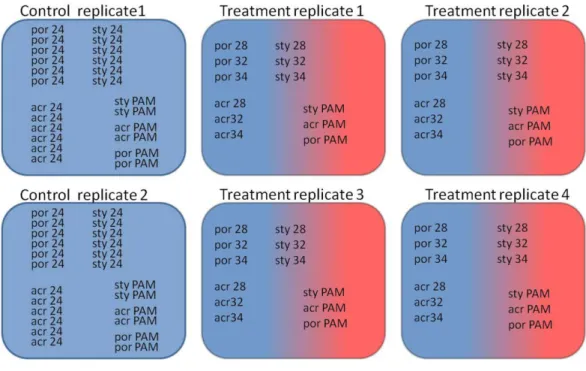
Fig. 1): two control aquaria (8 fragments from each coral species at each aquarium) were maintained at 24◦C and four experimental aquaria (4 fragments from each coral species at each aquarium) were subjected to a temperature increase of 1◦C per day from 24◦C to 34 ◦C. The aquaria were maintained with continuous water flow (artificial seawater (Brightwell Aquatics, Pennsylvania, USA)) using a computer-controlled closed circulation system, which compensates for salinity fluctuations and water level changes (constant salinity level of 35h). Light periodicity was achieved using an Advanced Control Lighting System (ACLS, Sfiligoi, Italy) with HQI (hydrargyrum quartz iodide) light bulbs (400 w, 14,000 Kelvin) configured to simulate a year-long diurnal-dimming light regime (PAR (photosynthetically active radiation) of 150µmol quanta m−2s−1). Four fragments, were
Figure 1 Experimental design of the experiment.Two control aquaria and four experimental aquaria: Coral fragments (por;Porites sp., sty;S. pistillata, acr;A. eurystoma) were sampled at the time points cor-responding to 24 h incubation at the following temperatures (28, 32 and 34; 28◦C, 32◦C and 34◦C) and
concurrent PAM measurements of fragments was used for evaluating the maximum quantum yield (PAM; fragments used for evaluating the maximum quantum yield).
experiment), 32◦C (day 10) and 34◦C (day 13) (seeFig. 1). The four treatment fragments were sampled from four independent aquaria. In contrast, the four control fragments were sampled from only two aquaria. Our long previous experience with our controlled system suggests that (exampleMaor-Landaw et al., 2014) there is no difference between the independent aquaria. Therefore, in this experiment the four control fragments sampled from two aquaria were considered as four replicates.
PAM flourometry
RNA extractions
Extraction protocols changed according to coral species and growth form. Total RNA was extracted from each fragment ofS. pistillataandA. eurystomausing Trizol (Invitrogen Life Technologies, Carlsbad, CA, USA) according to the methods presented in (Levy et al., 2011), and the samples were further purified using a RNA Clean and Concentrator kit (Zymo Research Corp., Irvine, CA, USA). Total RNA was extracted fromPoritesfragments using RNAqueous 4-PCR kit (Ambion), as described by (Kenkel et al., 2011). RNA quantity was assessed using a NanoDrop spectrophotometer (ND-1000). RNA integrity was checked via a Bioanalyzer (Agilent) or alternatively through agarose gel electrophoresis and evaluated based on clear 28S and 18S ribosomal RNA bands.
Primer design
We examined the expression of six genes: thioredoxin, peroxiredoxin, DNAJ, heat shock protein 70, enolase and Rad51, which were up-regulated following heat-stress in our previousS. pistillatastudy (Maor-Landaw et al., 2014) and also examined the expression of caspase 3. With the exception ofS. pistillatacaspase 3 that was adapted from (Kvitt et al., 2011) the primers for amplifyingS. pistillatatarget genes of interest (GOI) were designed based uponS. pistillataEST libraries previously constructed in our lab (Karako-Lampert et al., 2014). Degenerate primers were designed forPorites sp.and A. eurystomaGOI using Porites astroides(Kenkel, Meyer & Matz, 2013),Acropora tenuis(Matz lab website) transcriptome databases and the Cnidarian Database of Centre Scientifique de Monaco (http://data.centrescientifique.mc/CSMdata-home.html). The sequences were aligned using ClustalW and degenerate primers were designed based on conserved regions. Gradient rtPCR was applied for each pair of degenerate primers using Ready Mix RedTaq reaction mix (SIGMA) or with DreamTaq Green DNA Polymerase (Thermo Scientific). Each 50µl reaction contained 25µl polymerase, 4µl of each of the forward and reverse
primers, 1µl of cDNA and 16µl ddH2O (nuclease free water). PCR temperature profiles
were as manufacturer’s instructions. PCR products with the most stringent temperature that yielded a band of the suitable size upon 1% agarose gel were sent for sequencing in Hylabs or Macrogene. Resulting sequences were assembled and sequence identity was confirmed using BLAST search through the NCBI server on the GenBank database. Sequences generated in this study were deposited in GenBank under accession numbers
KT957160– KT957173. These partial sequences then served as a template for specific primers design for real-time PCR primers (seeTable S1), using Primer Quest design tool.
Real-Time Polymerase chain reaction
Complementary DNAs were synthesized from 1µg of total RNA with 1µl Solaris RNA
with 0.5µl mix of forward and reverse primers, 5µl of GoTaq qPCR Master Mix (Promega)
and 0.5µl of RNAse free water, for 45 cycles. A melt curve analysis was performed for each
pair of primers, to test for nonspecific amplification products by incubating the reactions for 10 s at 0.5◦C increments between 60◦C and 90◦C. Primer efficiencies were determined using a standard curve analysis with a 4-fold dilution series and according to the formula: % Efficiency =(E−1)×100% (E is calculated from the slope of the standard curve: E=10−1/slope).
The comparative11CTs method was applied, and fold changes were calculated using the 2−11Ctformula to estimate the relative amounts of transcripts in each sample (Livak
& Schmittgen, 2001). Ct refers to the cycle at which the fluorescence signal crosses the threshold and by using the solaris spike control (Mayfield, Hirst & Gates, 2009;Mayfield et al., 2012;Putnam et al., 2013) we normalized the expression to RNA loading. The MIQE guidelines were taken into account in designing real time profiles and analyzing their results (Bustin et al., 2009).
Statistical analysis
Results fromFv/Fmdata and gene expression were tested for normality and equal variances. In order to distinguish significant results we used the One-way ANOVA analysis followed by post hoc LSD/Bonferroni multiple-comparisons test (p<0.05). All statistical analyses were performed using SPSS software (Version 20.0. Armonk, NY, IBM Corp).
Protein oxidation assay
Protein oxidation was determined in extracts of corals fragments by measuring the degree of protein carbonylation present using Oxyblot protein oxidation kit (Millipore, Billerica, MA, USA) (seeSupplemental Information 1).
RESULTS
PAM fluorometer
Figure 2 Maximum quantum yield of coral fragment symbionts throughout the experiment. Maximum quantum yield (Fv/Fm) values for heat-stressed (light grey) and control (dark grey) coral fragments; (A)S. pistillata, (B)A. eurystomaand (C)Porites. Asterisk represents a significant difference between control and treatment (p<0.05). (D) Percentage ofFv/Fmrelative to control for the three studied coral species, as indicated in the legend. The table in the upper right hand corner represents the significantly different treatments (using post hoc LSD multiple-comparisons) (por; Porites sp., sty; S. pistillata, acr;A. eurystoma24, 28, 32 and 34; fragments sampled at the time points corresponding to 28◦C, 32◦C, 34◦C).
Gene expression
With the purpose of evaluating gene expression, Real-Time PCR was used to quantify seven genes of interest (GOI) in the three coral species. Utilized PCR primers (Table S1) were based upon partial sequences achieved using degenerate primers. No correlation was found in all control11CTs (from fragments of two aquaria and three sampling points) between aquariums and also between sampling times in One-way ANOVA (p>0.05). Since control11CTs were not different from one another, the replicates were considered to be independent and an arithmetic mean was calculated for all control values. Average values of comparative11CT are presented inFig. 4for heat-stress treatments showing significant gene expression differences in comparison to the average of control samples and considered to be up-regulated values (One-way ANOVA followed by post hoc multiple comparisons analyses,p<0.05).
In general, GOI expression profiles were similar betweenS. pistillataandA. eurystoma, while for most of the cases,Poritesexhibited a different gene expression response (Fig. 4
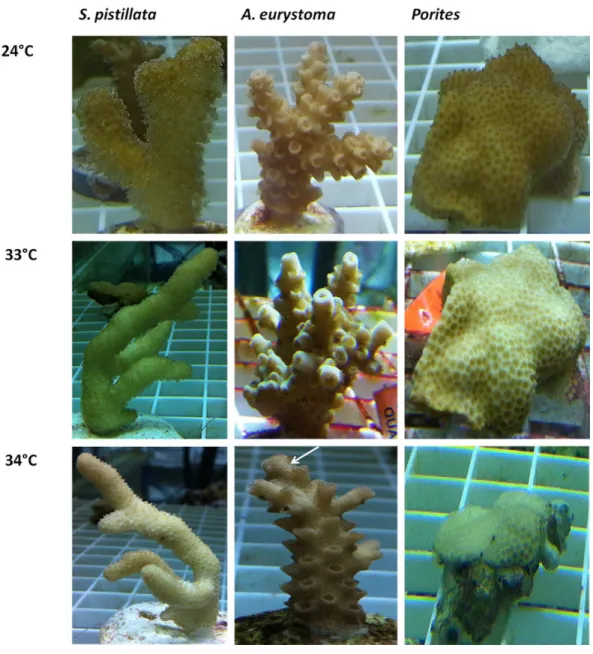
Figure 3 Visual appearance of coral fragments at ambient 240◦C and following thermal stress of 33◦C
and 34◦C.Acropora eurystoma t
stissue sloughing is indicated in the figure with a white arrow.
Figure 4 Gene expression following treatment of 28◦C, 32◦C, 34◦C and in 24◦C control treatment.
Gene expression (represented as average11CT) of thioredoxin, peroxiredoxin-6, Dnaj C3, Hsp70, cas-pase 3, enolase and Rad51 inS. pistillata,A. eurystomaandPoritesfollowing treatment of 28◦C, 32◦C,
34◦C and in 24◦C control treatment. Results were subjected to One-way ANOVA followed by post-hoc
LSD/bonferroni multiple-comparisons test (p<0.05). Treatments significantly different from control were considered as up-regulated and indicate with an asterisk. The table in the right hand upper corner contains additional significantly different treatments.
Table 1 Up-regulated genes inStylophora pistillata,Acropora eurystomaandPoritesfollowing treat-ment of 28◦C, 32◦C and 34◦C, based onFig. 4.
S. pistillata A. eurystoma Porites
28◦C 32◦C 34◦C 28◦C 32◦C 34◦C 28◦C 32◦C 34◦C
Thioredoxin ↑ ↑ ↑ ↑
peroxiredoxin-6 ↑ ↑ ↑
Dnaj C3 ↑ ↑ ↑ ↑
Hsp70 ↑ ↑ ↑ ↑
Caspase 3 ↑ ↑ ↑ ↑
Enolase ↑ ↑
Rad51 ↑
by post hoc multiple comparisons analyses,p<0.05) but not at other temperatures or in the other corals sampled at these temperatures.
DISCUSSION
To ascertain if specific corals possessing different morphologies may manifest varied gene expression patterns, we studied the expression of seven key representative genes of cellular processes known to occur during heat-stress in Cnidaria: two redox regulation agents: thioredoxin and peroxiredoxin, heat shock protein 70, Dnaj which is involved in the unfolded protein response (UPR) in ER stress, energy metabolism agent enolase, DNA repair mediator rad51, and apoptosis executioner caspase 3. The three studied coral species showed a variety of cellular responses that were correlated to their morphology as well as to their taxonomic classification.
A varied response to the heat stress, in terms of visual coral paling, algal maximum quantum yield and host gene expression was evident following heat stress on the coral. Overall, the two branching corals exhibited a more similar response to each other, than to the massive coral. Fv/Fmvalues under elevated temperatures decreased quickly in Porites, while in S. pistillatathis occurred only in the severe temperatures treatment of 34◦C while inA. eurystomathey remained high throughout the experiment. The visual appearance of coral bleaching corresponded with this pattern. Bearing in mind some of our gene expression results, it seems likely thatPoritesexhibited a delay in the stress response. Compared toS. pistillataandA. eurystomagenes that in most cases were up-regulated as the temperatures reached 28◦C or 32◦C, in
Poritesthese genes were elevated mostly only at 34◦C, or not at all. The relative resilience ofPoritesand other massive corals to heat stress is well known in coral literature (Jokiel & Coles, 1974;Brown & Suharsono, 1990;
Figure 5 Illustration of coral visual appearance and up-regulated genes throughout the heat stress ex-periment inAcroporaandPoritesfragments. Acropora eurystomafragments didn’t exhibit bleaching, but at 34◦C live tissue started to peel off the skeleton, whilePoritesfragments were paler at 28◦C and
bleach-ing was maximized at 34◦C. Below coral figures, up-regulated genes are indicated, corresponding to the
temperature they were elevated at.
beginning of the experiment, at temperatures as low as 28◦C (seeFig. 5). Interestingly, this species began losing its tissues at the elevated temperatures, perhaps as a response to accumulations of free radicals in their tissues.S. pistillatamay represent an intermediate version of the two, with bleaching occurring only in extreme temperatures and the redox regulation thioredoxin being up-regulated not early as inA. eurystomabut sooner than inPorites. Coral bleaching was previously suggested in the literature as a host resort for survival; expelling or degrading the compromised ROS-causing symbionts and breakdown of the symbiosis (Downs et al., 2002;Downs et al., 2009). This study demonstrates how gene expression may reflect this characteristic.
The two relatively heat-stress sensitive coral species of this study showed elevated levels of caspase 3 at 32 ◦C. Members of the family of caspases—cysteine-dependent aspartate specific proteases—are the core effectors of the apoptotic cascade (Nicholson & Thornberry, 1997) that cleave a variety of cellular subtracts resulting in programmed cell death (Chowdhury, Tharakan & Bhat, 2008). Caspase 3 is an executioner caspase that was studied in corals with regards to gene expression (Kvitt et al., 2011;Tchernov et al., 2011;
Kaniewska et al., 2012;Shearer et al., 2012) and enzyme specific activity (Pernice et al., 2011;
(Kvitt et al., 2011). In that study the decrease in caspase-3 was attributed to acclimatization of the coral to the chronic heat stress together with the completion of symbiosis breakdown (Kvitt et al., 2011). InA. eurystoma, caspase-3 levels remained elevated at 34◦C, suggesting an inability to acclimatize. This response is also reflected in the peeling off of the live tissue at this temperature. These results rank A. eurystomaas the most susceptible species to heat-stress in this experiment. As opposed to caspase 3 inS. pistillataandA. eurystoma, in Poritescaspase-3 was elevated only when the heat-stress was most severe, at 34◦C, which may explain the higher resilience observed in this coral.
The gene expression patterns of enolase, a representative of energy regulation process, and rad51, an agent of DNA repair process, differed greatly between the coral species. There are only few reports in the literature indicating the role of these genes following environmental stress in corals. These include the association of enolase with heat stress, or with macroalgal exposure (Maor-Landaw et al., 2014;Shearer et al., 2012). The association of rad51 expression with UV radiation exposure in coral larvae was also reported (Aranda et al., 2011). There are several possible explanations to these results; (a) the sampling points may have missed the maximal gene expression point (e.g., gene expression peaks at 30◦C); (b) Enolase and rad51 genes may not be suitable or prominent representatives of these stress processes in coral cells; or (c) The genes are good representative of stress response in these pathways, but the processes themselves of higher energy demands and DNA repair are not occurring in this experiment. These possible interpretations can be relevant to one of the species or common to all. A fundamental issue is whether the genes that govern these cellular processes in the different coral species are the same and only the timing varies, or alternatively, the key players in mitigating stress are different. The above-mentioned discrepancies still need to be resolved.
In contrast, the two-redox regulation agents and the two heat shock proteins studied here, were all up-regulated at some point in all the three coral species. Therefore, these may provide suitable candidates as markers of redox regulation and heat shock processes in the three corals. Thioredoxin, an enzyme that detoxifies oxidized molecules, was previously reported to be up-regulated in corals following thermal stress (DeSalvo et al., 2010a;
Maor-Landaw et al., 2014), high irradiance (Starcevic et al., 2010), macroalgal exposure (Shearer et al., 2012), and elevated salinity (Edge et al., 2005). Peroxiredoxin elevation was also previously documented after heat-stress (Maor-Landaw et al., 2014), and also was related to white band disease inAcropora (Libro, Kaluziak & Vollmer, 2013). Hsp70 is known to be an important factor in protein folding and repair of stress-induced protein damage (Tavaria et al., 1996) and is well documented during coral stress (Brown et al., 2002;
(UPR) during ER stress and also serves as a co-chaperone to hsp70 (Cyr, Langer & Douglas, 1994) indicating that it may be a suitable marker for heat stress. In the EilatS. pistillata,32◦C was previously suggested to be the temperature of initial stress reaction (Maor-Landaw et al., 2014) and 34◦C as the upper thermal limit (Shaish, Abelson & Rinkevich, 2007;Kvitt
et al., 2011). This may differ with ambient temperature regime of these populations as colonies of this species are known to flourish in much warmer waters (Bauman, Baird & Cavalcante, 2011). Indeed DNAJ is elevated at 32◦C inS. pistillatabefore bleaching occurs, before tissue peeling off inA. eurystomaand before most of the gene expression heat-stress response inPorites.
Protein carbolyation a common marker of protein oxidation of stress-induced damage (Murik & Kaplan, 2009) was used to estimate protein oxidation levels (Supplemental Information 1). The results indicated that protein oxidation differed between species. In Porites spthe profile of protein oxidation following 34◦C treatment did not significantly differ from the 24◦C control. InS. pistillatamaximum protein oxidation was at 34◦C. In A. eurystomaprotein oxidation peaked at 32◦C (seeFigs. S1andS2). This pattern is comparable with our previous results and with the hierarchy documented in the literature (Jokiel & Coles, 1974;Brown & Suharsono, 1990;Loya et al., 2001). It also provides additional explanation for the resilience of ‘‘winner’’ corals with massive-morphology (Porites sp.), when compared to that of ‘‘looser’’ branching coralsS. pistillataand especially A. eurystoma, that are more sensitive to heat stress.
The corals studied here representing different growth forms, S. pistillata,A. eurystoma andPorites sp., demonstrated different physiological response to short-term heat stress. These responses included visual coral paling and algal maximum quantum yield, and varied host gene-expression reactions to elevated temperature.
We acknowledge the possible role of zooxanthellae in the thermal tolerance of corals (Berkelmans & Van Oppen, 2006); however, this was not the scope of our research. Most of the corals of the Gulf of Eilat hostSymbiodiniumclade A or C that are both known to be relatively sensitive to heat stress (Karako-Lampert et al., 2004;Lampert-Karako et al., 2008;Fine, Gildor & Genin, 2013). Lately, symbiont enzymatic antioxidant activity was found to be independent of thermal sensitivity (Krueger et al., 2015), so the dispute over the potential coupling of symbiont antioxidant capacity and bleaching outcome (Hawkins et al., 2015) is still ongoing.
coral in expelling symbiont process as an approach to elevate heat stress resistance, which was not the main scope of this research.
ACKNOWLEDGEMENTS
We thank Mr. M Samuelson, Mr. G Luria and Dr. G Miller of the Faculty of Life Sciences, BIU, Israel for their help during this study. This study represents partial fulfillment of the requirements for a PhD thesis for K Maor-Landaw at Faculty of Life Sciences Bar-Ilan University, Israel. We also thank The Interuniversity Institute for Marine Sciences in Eilat (IUI) for the support in this research.
ADDITIONAL INFORMATION AND DECLARATIONS
Funding
The authors received no funding for this work.
Competing Interests
The authors declare there are no competing interests.
Author Contributions
• Keren Maor-Landaw conceived and designed the experiments, performed the experiments, analyzed the data, wrote the paper, prepared figures and/or tables, reviewed drafts of the paper.
• Oren Levy conceived and designed the experiments, analyzed the data, contributed reagents/materials/analysis tools, wrote the paper, prepared figures and/or tables, reviewed drafts of the paper.
Field Study Permissions
The following information was supplied relating to field study approvals (i.e., approving body and any reference numbers):
The Israeli Nature and National Parks Protection Authority approved the collection of corals in this study, permit No. 2013/40159.
DNA Deposition
The following information was supplied regarding the deposition of DNA sequences: Sequences generated in this study were deposited in GenBank NCBI under accession numbersKT957160–KT957173.
Data Availability
The following information was supplied regarding data availability: The research in this article did not generate any raw data.
Supplemental Information
REFERENCES
Ainsworth TD, Hoegh-Guldberg O, Heron SF, Skirving WJ, Leggat W. 2008.
Early cellular changes are indicators of pre-bleaching thermal stress in the coral host.Journal of Experimental Marine Biology and Ecology364:63–71
DOI 10.1016/j.jembe.2008.06.032.
Aranda M, Banaszak AT, Bayer T, Luyten JR, Medina M, Voolstra CR. 2011.
Differential sensitivity of coral larvae to natural levels of ultraviolet radiation during the onset of larval competence.Molecular Ecology20:2955–2972
DOI 10.1111/j.1365-294X.2011.05153.x.
Baird AH, Bhagooli R, Ralph PJ, Takahashi S. 2009.Coral bleaching: the role of the host.
Trends in Ecology & Evolution24:16–20DOI 10.1016/j.tree.2008.09.005.
Baker AC. 2001.Reef corals bleach to survive change.Nature411:765–766
DOI 10.1038/35081151.
Barshis DJ, Ladner JT, Oliver TA, Seneca FO, Traylor-Knowles N, Palumbi SR.
2013.Genomic basis for coral resilience to climate change.Proceedings of the
National Academy of Sciences of the United States of America110:1387–1392
DOI 10.1073/pnas.1210224110.
Barshis DJ, Stillman JH, Gates RD, Toonen RJ, Smith LW, Birkeland C. 2010.Protein
expression and genetic structure of the coral Porites lobata in an environmentally extreme Samoan back reef: does host genotype limit phenotypic plasticity?Molecular Ecology 19:1705–1720DOI 10.1111/j.1365-294X.2010.04574.x.
Bauman AG, Baird AH, Cavalcante GH. 2011.Coral reproduction in the world’s
warmest reefs: southern Persian Gulf (Dubai, United Arab Emirates).Coral Reefs
30:405–413DOI 10.1007/s00338-010-0711-5.
Berkelmans R, Van Oppen MJH. 2006.The role of zooxanthellae in the thermal
tolerance of corals: a ‘‘nugget of hope’’ for coral reefs in an era of climate change.Proceedings of the Royal Society B: Biological Sciences273:2305–2312
DOI 10.1098/rspb.2006.3567.
Black NA, Voellmy R, Szmant AM. 1995.Heat shock protein induction in montastraea
faveolata and aiptasia pallida exposed to elevated temperatures.Biological Bulletin
188:234–240DOI 10.2307/1542301.
Brown BE. 1997a.Coral bleaching: causes and consequences.Coral Reefs16:s129–s138
DOI 10.1007/s003380050249.
Brown BE. 1997b.Adaptations of reef corals to physical environmental stress.Advances
in Marine Biology31:222–301 DOI 10.1016/S0065-2881(08)60224-2.
Brown BE, Ambarsari I, Warner ME, Fitt WK, Dunne RP, Gibb SW, Cummings DG.
1999.Diurnal changes in photochemical efficiency and xanthophyll concentrations
in shallow water reef corals: evidence for photoinhibition and photoprotection.Coral Reefs18:99–105DOI 10.1007/s003380050163.
Brown BE, Downs C, Dunne R, Gibb S. 2002.Exploring the basis of thermotolerance
in the reef coral Goniastrea aspera.Marine Ecology Progress Series242:119–129
Brown BE, Suharsono. 1990.Coral reefs damage and recovery of coral reefs affected by El Nifio related seawater warming in the thousand islands, Indonesia.Coral Reefs
8:163–170DOI 10.1007/BF00265007.
Bustin SA, Benes V, Garson JA, Hellemans J, Huggett J, Kubista M, Mueller R, Nolan T,
Pfaffl MW, Shipley GL, Vandesompele J, Wittwer CT. 2009.The MIQE guidelines:
minimum information for publication of quantitative real-time PCR experiments. Clinical Chemistry55:611–622DOI 10.1373/clinchem.2008.112797.
Carpenter LW, Patterson MR, Bromage ES. 2010.Water flow influences the
spatiotem-poral distribution of heat shock protein 70 within colonies of the scleractinian coral Montastrea annularis (Ellis and Solander, 1786) following heat stress: implications for coral bleaching.Journal of Experimental Marine Biology and Ecology387:52–59
DOI 10.1016/j.jembe.2010.02.019.
Chowdhury I, Tharakan B, Bhat GK. 2008.Caspases—an update.Comparative
Biochemistry and Physiology. Part B, Biochemistry & Molecular Biology 151:10–27
DOI 10.1016/j.cbpb.2008.05.010.
Coles SL, Brown BE. 2003.Coral bleaching—capacity for acclimatization and
adapta-tion.Advances in Marine Biology46:183–223DOI 10.1016/S0065-2881(03)46004-5.
Cook CB, Logan A, Ward J, Luckhurst B, Berg Jr CJ. 1990.Coral reefs elevated
tempera-tures and bleaching on a high latitude coral reef: the 1988 Bermuda event.Coral Reefs
9:45–49DOI 10.1007/BF00686721.
Cyr DM, Langer T, Douglas MG. 1994.DnaJ-like proteins: molecular chaperones
and specific regulators of Hsp70.Trends in Biochemical Sciences19:176–181
DOI 10.1016/0968-0004(94)90281-X.
DeSalvo MK, Sunagawa S, Fisher PL, Voolstra CR, Iglesias-Prieto R, Medina M. 2010a.
Coral host transcriptomic states are correlated with Symbiodinium genotypes. Molecular Ecology 19:1174–1186DOI 10.1111/j.1365-294X.2010.04534.x.
DeSalvo MK, Sunagawa S, Voolstra C, Medina M. 2010b.Transcriptomic responses
to heat stress and bleaching in the elkhorn coralAcroporapalmata.Marine Ecology Progress Series402:97–113DOI 10.3354/meps08372.
DeSalvo MK, Voolstra CR, Sunagawa S, Schwarz JA, Stillman JH, Coffroth MA,
Szmant AM, Medina M. 2008.Differential gene expression during thermal stress
and bleaching in the Caribbean coral Montastraea faveolata.Molecular Ecology
17:3952–3971DOI 10.1111/j.1365-294X.2008.03879.x.
Downs CA, Fauth JE, Halas JC, Dustan P, Bemiss J, Woodley CM. 2002.Oxidative
stress and seasonal coral bleaching.Free Radical Biology & Medicine33:533–543
DOI 10.1016/S0891-5849(02)00907-3.
Downs CA, Kramarsky-winter E, Martinez J, Kushmaro A, Woodley CM, Loya Y. 2009.
Symbiophagy as a cellular mechanism for coral bleaching.Autopjagy5:211–216.
Downs CA, Mueller E, Phillips S, Fauth JE, Woodley CM. 2000.A molecular biomarker
Edge SE, Morgan MB, Gleason DF, Snell TW. 2005.Development of a coral cDNA array to examine gene expression profiles in Montastraea faveolata exposed to environmental stress.Marine Pollution Bulletin51:507–523
DOI 10.1016/j.marpolbul.2005.07.007.
Enríquez S, Méndez ER, Iglesias-Prieto R. 2005.Multiple scattering on coral skeletons
enhances light absorption by symbiotic algae.Limnology and Oceanography
50:1025–1032DOI 10.4319/lo.2005.50.4.1025.
Fine M, Gildor H, Genin A. 2013.A coral reef refuge in the Red Sea.Global Change
Biology19:3640–3647DOI 10.1111/gcb.12356.
Fitt W, Brown B, Warner M, Dunne R. 2001.Coral bleaching: interpretation of thermal
tolerance limits and thermal thresholds in tropical corals.Coral Reefs20:51–65
DOI 10.1007/s003380100146.
Gates RD, Edmunds PJ. 1999.The physiological mechanisms of acclimatization in
tropical reef corals.American Zoologist39:30–43DOI 10.1093/icb/39.1.30.
Glynn PW. 1988.El Niño warming, coral mortality and reef framework destruction by
Echinoid bioerosion in the eastern pacific.Galaxea7:129–160.
Granados-Cifuentes C, Bellantuono AJ, Ridgway T, Hoegh-guldberg O,
Rodriguez-Lanetty M. 2013.High natural gene expression variation in the reef-building coral
Acroporamillepora: potential for acclimative and adaptive plasticity.BMC Genomics
14:228 DOI 10.1186/1471-2164-14-228.
Grottoli AG, Eakin CM. 2007.A review of modern coralδ18O and114C proxy records.
Earth-Science Reviews81:67–91DOI 10.1016/j.earscirev.2006.10.001.
Grottoli AG, Rodrigues LJ, Palardy JE. 2006.Heterotrophic plasticity and resilience in
bleached corals.Nature440:1186–1189DOI 10.1038/nature04565.
Harriott VJ. 1985.Mortality rates of scleractinian corals before and during a mass
bleaching event.Marine Ecology Progress Series21:81–88DOI 10.3354/meps021081.
Hawkins TD, Krueger T, Becker S, Fisher PL, Davy SK. 2014.Differential nitric oxide
synthesis and host apoptotic events correlate with bleaching susceptibility in reef corals.Coral Reefs33:141–153 DOI 10.1007/s00338-013-1103-4.
Hawkins TD, Krueger T, Wilkinson SP, Fisher PL, Davy SK. 2015.Antioxidant
responses to heat and light stress differ with habitat in a common reef coral.Coral Reefs34:1229–1241DOI 10.1007/s00338-015-1345-4.
Hayes RL, King CM. 1995.Induction of 70-kD heat shock protein in scleractinian corals
by elevated temperature: significance for coral bleaching.Molecular Marine Biology and Biotechnology4:36–42.
Hemond EM, Kaluziak ST, Vollmer SV. 2014.The genetics of colony form and function
in CaribbeanAcroporacorals.BMC Genomics15:1133
DOI 10.1186/1471-2164-15-1133.
Hoegh-Guldberg O. 1999.Climate change, coral bleaching and the future of the world’s
coral reefs.Marine Freshwater Research50:839–866DOI 10.1071/MF99078.
Hoegh-Guldberg O. 2010.Coral reef ecosystems and anthropogenic climate change.
Hoegh-Guldberg O, Mumby PJ, Hooten AJ, Steneck RS, Greenfield P, Gomez E, Harvell CD, Sale PF, Edwards AJ, Caldeira K, Knowlton N, Eakin CM,
Iglesias-Prieto R, Muthiga N, Bradbury RH, Dubi A, Hatziolos ME. 2007.Coral reefs under
rapid climate change and ocean acidification.Science318:1737–1742
DOI 10.1126/science.1152509.
Hume BCC, D’Angelo C, Smith EG, Stevens JR, Burt J, Wiedenmann J. 2015.
Symbio-dinium thermophilumsp. nov., a thermotolerant symbiotic alga prevalent in corals of the world’s hottest sea, the Persian/Arabian Gulf.Scientific Reports5:8562.
Jokiel PL, Coles SL. 1974.Effects of heated effluent on hermatypic corals at Kahe Point,
Oahu.Pacific Science28:1–18.
Kaniewska P, Campbell PR, Kline DI, Rodriguez-Lanetty M, Miller DJ, Dove S,
Hoegh-Guldberg O. 2012.Major cellular and physiological impacts of ocean acidification
on a reef building coral.PLoS ONE7:e34659DOI 10.1371/journal.pone.0034659.
Karako-Lampert S, Katcoff DJ, Achituv Y, Dubinsky Z, Stambler N. 2004.Do clades of
symbiotic dinoflagellates in scleractinian corals of the Gulf of Eilat (Red Sea) differ from those of other coral reefs?Journal of Experimental Marine Biology and Ecology
311:301–314DOI 10.1016/j.jembe.2004.05.015.
Karako-Lampert S, Zoccola D, Salmon-Divon M, Katzenellenbogen, Mark Tambuttéb S, Bertucci A, Hoegh-Guldberg, Ove Deleuryd E, Allemandb D, Levy O. 2014.
Transcriptome analysis of the scleractinian coralStylophora pistillata.PLoS ONE
9:e88615DOI 10.1371/journal.pone.0088615.
Kenkel CD, Aglyamova G, Alamaru A, Bhagooli R, Capper R, Cunning R, DeVillers A, Haslun JA, Hédouin L, Keshavmurthy S, Kuehl KA, Mahmoud H, McGinty ES, Montoya-Maya PH, Palmer CV, Pantile R, Sánchez JA, Schils T, Silverstein RN,
Squiers LB, Tang P-C, Goulet TL, Matz MV. 2011.Development of gene expression
markers of acute heat-light stress in reef-building corals of the genus Porites.PLoS ONE6:e26914DOI 10.1371/journal.pone.0026914.
Kenkel CD, Meyer E, Matz MV. 2013.Gene expression under chronic heat stress in
populations of the mustard hill coral (Porites astreoides) from different thermal environments.Molecular Ecology22:4322–4334DOI 10.1111/mec.12390.
Krueger T, Hawkins TD, Becker S, Pontasch S, Dove S, Hoegh-Guldberg O, Leggat W,
Fisher PL, Davy SK. 2015.Differential coral bleaching—Contrasting the activity
and response of enzymatic antioxidants in symbiotic partners under thermal stress. Comparative Biochemistry and Physiology Part A: Molecular & Integrative Physiology
190:15–25DOI 10.1016/j.cbpa.2015.08.012.
Kültz D. 2005.Molecular and evolutionary basis of the cellular stress response.Annual
Review of Physiology67:225–257DOI 10.1146/annurev.physiol.67.040403.103635.
Kvitt H, Rosenfeld H, Zandbank K, Tchernov D. 2011.Regulation of apoptotic
Lampert-Karako S, Stambler N, Katcoff DJ, Achituv Y, Dubinsky Z, Simon-Blecher N.
2008.Effects of depth and eutrophication on the zooxanthella clades ofStylophora
pistillatafrom the Gulf of Eilat (Red Sea).Aquatic Conservation: Marine and Freshwater Ecosystems1045:1039–1045.
Lesser MP. 2004.Experimental biology of coral reef ecosystems.Journal of Experimental
Marine Biology and Ecology300:217–252DOI 10.1016/j.jembe.2003.12.027.
Levas SJ, Grottoli AG, Hughes A, Osburn CL, Matsui Y. 2013.Physiological and
biogeochemical traits of bleaching and recovery in the mounding species of coral Porites lobata: implications for resilience in mounding corals.PLoS ONE8:e63267
DOI 10.1371/journal.pone.0063267.
Levy O, Kaniewska P, Alon S, Eisenberg E, Karako-Lampert S, Bay LK, Reef R,
Rodriguez-Lanetty M, Miller DJ, Hoegh-Guldberg O. 2011.Complex diel cycles
of gene expression in coral-algal symbiosis.Science331:175
DOI 10.1126/science.1196419.
Libro S, Kaluziak ST, Vollmer SV. 2013.RNA-seq profiles of immune related genes in
the staghorn coralAcroporacervicornis infected with white band disease.PLoS ONE
8:e81821DOI 10.1371/journal.pone.0081821.
Livak KJ, Schmittgen TD. 2001.Analysis of relative gene expression data using
real-time quantitative PCR and the 2-ddCT method.Methods25:402–408
DOI 10.1006/meth.2001.1262.
Loya Y, Sakai K, Nakano Y, Woesik R Van. 2001.Coral bleaching: the winners and the
losers.Ecology Letters4:122–131DOI 10.1046/j.1461-0248.2001.00203.x.
Maor-Landaw K, Karako-Lampert S, Ben-Asher HW, Goffredo S, Falini G,
Du-binsky Z, Levy O. 2014.Gene expression profiles during short-term heat stress
in the red sea coralStylophora pistillata.Global Change Biology20:3026–3035
DOI 10.1111/gcb.12592.
Maor-Landaw K, Levy O. Survey of cnidarian gene expression profiles in response to
environmental stressors; Summarizing 20 years of research, what are we heading for? In: Goffredo S, Dubinsky Z, eds.The Cnidaria, past, present and future: the world of Medusa and her sisters. Dordrecht: Springer In Press.
Marshall PA, Baird AH. 2000.Bleaching of corals on the great barrier reef: differential
susceptibilities among taxa.Coral Reefs19:155–163DOI 10.1007/s003380000086.
Mayfield AB, Chan P-H, Putnam HM, Chen C-S, Fan T-Y. 2012.The effects of a variable
temperature regime on the physiology of the reef-building coral Seriatopora hystrix: results from a laboratory-based reciprocal transplant.The Journal of Experimental Biology215:4183–4195DOI 10.1242/jeb.071688.
Mayfield AB, Hirst MB, Gates RD. 2009.Gene expression normalization in a
dual-compartment system: a real-time quantitative polymerase chain reaction protocol for symbiotic anthozoans.Molecular Ecology Resources9:462–470
DOI 10.1111/j.1755-0998.2008.02349.x.
Murik O, Kaplan A. 2009.Paradoxically, prior acquisition of antioxidant
activ-ity enhances oxidative stress-induced cell death.Environmental Microbiology
Nakamura T, Van Woesik R. 2001.Water-flow rates and passive diffusion partially explain differential survival of corals during the 1998 bleaching event.Marine Ecology Progress Series212:301–304DOI 10.3354/meps212301.
Nicholson DW, Thornberry NA. 1997.Caspases: killer proteases.Trends in Biochemical
Sciences22:299–306 DOI 10.1016/S0968-0004(97)01085-2.
Parkinson JE, Banaszak AT, Altman NS, LaJeunesse TC, Baums IB. 2015.Intraspecific
diversity among partners drives functional variation in coral symbioses.Scientific Reports5: 15667.
Pernice M, Dunn SR, Miard T, Dufour S, Dove S, Hoegh-Guldberg O. 2011.
Regulation of apoptotic mediators reveals dynamic responses to thermal stress in the reef building coralAcroporamillepora.PLoS ONE6:e16095
DOI 10.1371/journal.pone.0016095.
Polato NR, Voolstra CR, Schnetzer J, DeSalvo MK, Randall CJ, Szmant AM,
Med-ina M, Baums IB. 2010.Location-specific responses to thermal stress in larvae
of the reef-building coral Montastraea faveolata.PLoS ONE5:e11221
DOI 10.1371/journal.pone.0011221.
Putnam HM, Mayfield AB, Fan TY, Chen CS, Gates RD. 2013.The physiological and
molecular responses of larvae from the reef-building coral Pocillopora damicornis exposed to near-future increases in temperature and pCO2.Marine Biology
160:2157–2173DOI 10.1007/s00227-012-2129-9.
Putnam HM, Stat M, Pochon X, Gates RD. 2012.Endosymbiotic flexibility associates
with environmental sensitivity in scleractinian corals.Proceedings of the Royal Society B: Biological Sciences279:4352–4361DOI 10.1098/rspb.2012.1454.
Richier S, Sabourault C, Courtiade J, Zucchini N, Allemand D, Furla P. 2006.Oxidative
stress and apoptotic events during thermal stress in the symbiotic sea anemone, Anemonia viridis.The FEBS Journal273:4186–4198
DOI 10.1111/j.1742-4658.2006.05414.x.
Rodriguez-Lanetty M, Harii S, Hoegh-Guldberg O. 2009.Early molecular responses
of coral larvae to hyperthermal stress.Molecular Ecology18:5101–5114
DOI 10.1111/j.1365-294X.2009.04419.x.
Rowan R, Knowlton N, Baker A, Jara J. 1997.Landscape ecology of algal symbionts
creates variation in episodes of coral bleaching.Nature388:265–269
DOI 10.1038/40843.
Salih A, Hoegh-guldberg O, Cox G. 1998.Photoprotection of symbiotic dinoflagellates
by fluorescent pigments in reef corals. In:ACRS Proceedings - 75th Anniversary Conference, 217–230.
Salih A, Larkum A, Cox G, Kühl M, Hoegh-Guldberg O. 2000.Fluorescent pigments in
corals are photoprotective.Nature408:850–853DOI 10.1038/35048564.
Shaish L, Abelson A, Rinkevich B. 2007.How plastic can phenotypic plasticity be?
The branching coralStylophora pistillataas a model system.PLoS ONE 2:e644
Shaked Y, Genin A. 2015.The Israel National Monitoring Program in the Northern Gulf of Aqaba. Scientific report 2014. Jerusalem: Israel Ministry of Environmental Protection.
Shearer T, Rasher D, Snell T, Hay M. 2012.Gene expression patterns of the coral
Acroporamillepora in response to contact with macroalgae.Coral Reefs31:1177–1192
DOI 10.1007/s00338-012-0943-7.
Shick JM, Dunlap WC. 2002.Mycosporine-like amino acids and related Gadusols:
biosynthesis, acumulation, and UV-protective functions in aquatic organisms. Annual Review of Physiology 64:223–262
DOI 10.1146/annurev.physiol.64.081501.155802.
Stambler N, Dubinsky Z. 2005.Corals as light collectors: an integrating sphere approach.
Coral Reefs24:1–9DOI 10.1007/s00338-004-0452-4.
Starcevic A, Dunlap WC, Cullum J, Shick JM, Hranueli D, Long PF. 2010.Gene
expression in the scleractinianAcroporamicrophthalma exposed to high solar irradiance reveals elements of photoprotection and coral bleaching.PLoS ONE
5:e13975DOI 10.1371/journal.pone.0013975.
Tambutté É, Allemand D, Bourge I, Gattuso J, Jaubert J. 1995.An improved 45 Ca
protocol for investigating physiological mechanisms in coral calcification.Marine Biology122:453–459.
Tavaria M, Gabriele T, Kola I, Anderson RL. 1996.A hitchhiker’s guide to the human
Hsp70 family.Cell Stress & Chaperones1:23–28
DOI 10.1379/1466-1268(1996)001<0023:AHSGTT>2.3.CO;2.
Tchernov D, Kvitt H, Haramaty L, Bibby TS, Gorbunov MY, Rosenfeld H, Falkowski
PG. 2011.Apoptosis and the selective survival of host animals following thermal
bleaching in zooxanthellate corals.Proceedings of the National Academy of Sciences of the United States of America108:9905–9909DOI 10.1073/pnas.1106924108.
Van Woesik R, Sakai K, Ganase A, Loya Y. 2011.Revisiting the winners and the
losers a decade after coral bleaching.Marine Ecology Progress Series434:67–76
DOI 10.3354/meps09203.
Weis VM. 2008.Cellular mechanisms of Cnidarian bleaching: stress causes the
collapse of symbiosis.The Journal of Experimental Biology211:3059–3066
DOI 10.1242/jeb.009597.
Weis VM. 2010.The susceptibility and resilience of corals to thermal stress: adaptation,
acclimatization or both?Molecular Ecology 19:1515–1517
DOI 10.1111/j.1365-294X.2010.04575.x.
Wooldridge SA. 2014.Differential thermal bleaching susceptibilities amongst coral taxa:
re-posing the role of the host.Coral Reefs33:15–27DOI 10.1007/s00338-013-1111-4.
Yuyama I, Ito Y, Watanabe T, Hidaka M, Suzuki Y, Nishida M. 2012.Differential