doi:10.3897/compcytogen.v4i2.32
Histone H3 gene in the Pacific oyster,
Crassostrea gigas Thunberg, 1793: molecular and
cytogenetic characterisations
K. Bouilly
1, R. Chaves
2, M. Fernandes
3, H. Guedes-Pinto
4Institute for Biotechnology and Bioengineering, Centre of Genomics and Biotechno- logy, University of Trás-os-Montes and Alto Douro, (IBB/CGB-UTAD), Apartado
1013, 5001-801 Vila Real, Portugal. E-mails: 1
kbouilly@yahoo.fr, 2
rchaves@utad.pt, 3
marymgf@gmail.com, 4
hgp@hotmail.com
Abstract. The Pacific oyster, Crassostrea gigas Thunberg, 1793 (2n = 20) is an economically important mollusc species cultured throughout the world. The most frequently used technique for molecular cytogenetic studies is fluorescence in situ hybridisation which offers new opportunities for the identification of oyster chromo- somes. In oysters, it has been used to locate telomeric sequences, satellite DNA, sim- ple sequence repeats, ribosomal RNA genes, and bacteriophage P1 clones. However, regarding chromosome identification, no study has been done with histone H3 gene. Histone H3 is among the most conserved eukaryotic proteins. Most histone H3 genes are repeatedly organised into clusters, which make them an ideal chromosomal mark- er. In bivalves, some data exist concerning sequence information but little knowledge is available concerning the physical mapping of histone genes. The histone H3 gene was sequenced in C. gigas and phylogenetic analysis revealed that C. gigas was more closely related to Ostrea edulis Linnaeus, 1758 and species of the genus Mytilus Lin- naeus, 1758. In C. gigas, the histone H3 gene was mapped on two different pairs of chromosomes, one at an interstitial site on the long arm of chromosome pair 4, and the other on the telomeres of the smaller chromosome pair (pair 10). Polymorphism was detected on the telomeres of pair 10, once it was possible to observe single or double signals. Comparative chromosomal mapping should improve our understanding of bivalve genome organisation.
Key words: bivalves, Crassostrea gigas, fluorescence in situ hybridisation, histone H3 gene, phylogenetics, physical mapping.
I
NTRODUCTIONThe Pacific oyster, Crassostrea gigas Thunberg, 1793, is an economically
important oyster species cultured
throughout the world. This species has a diploid chromosome number of 2n = 20, and all chromosomes are metacentric. Several banding tehniques
were already applied to oyster
chromosomes. They are very useful for
the identification of individual
chromosomes and also of particular regions of chromosomes. “Classical” cytogenetic banding (G-, C- and NOR (nucleolus organiser regions) - banding techniques) was already performed in several species
of the genus Crassostrea Sacco, 1897
(review in Leitão, Chaves, 2008).
Molecular cytogenetic banding was also applied to several species of the genus Crassostrea (review in Leitão, Chaves, 2008). RE (restriction endonucleases) - banding (Leitão et al., 2004, 2007; Bouilly et al., 2005; Cross et al., 2005) and FISH
(fluorescence in situ hybridisation)
technique with different kinds of labelled probes were already produced in different species of the genus Crassostrea. FISH is a rapid and reliable technique for
chromosome identification, gene
mapping, localisation of gene expression,
and analysis of chromosome
genomes. Different probes have already been applied successfully to species of the genus Crassostrea: telomeric sequences (Guo, Allen, 1997; Wang, Guo, 2001; Cross et al., 2005), satellite DNA (Clabby et al., 1996; Wang et al., 2001), simple sequence repeats (Cross et al., 2005; Bouilly et al., 2008), ribosomal RNA genes (Zhang et al., 1999; Xu et al., 2001; Cross et al., 2003, 2005; Wang et al., 2004, 2005a), and bacteriophage P1 clones (Wang et al., 2005b).
DNA sequences of nuclear and
mitochondrial genes (Boudry et al., 2003; Reese et al., 2008), including repetitive satellite DNA sequences
(López-Flores et al., 2004) have been variously
used to examine the phylogenetic
relationships between oysters.
Histone proteins are the major
constituents of the eukaryotic chromatin. The family of histone proteins has been subdivided into the core histones (H2A, H2B, H3, and H4), which form the core particle of the nucleosome, and the linker histones (H1), which are involved in the generation of the higher-order chromatin structure (Thoma et al., 1979). Histone H3 is one of the most conserved eukaryotic proteins (Miller et al., 1993) and is an excellent probe for chromosome mapping as histone genes are usually organised in clusters (Maxson et al., 1983). Histone
gene mapping has beenachieved in a few
species. In bivalves, little knowledge is available concerning the physical mapping of histone genes. Only five species were studied until now: a mussel (Eirín-López et al., 2002, 2004) and four scallops (Zhang
et al., 2007). In Mytilidae, FISH
experiments on Mytilus galloprovincialis Lamarck, 1819 chromosomes revealed the presence of three pairs of signals in three chromosome pairs with the linker histone H1 probe (Eirín-López et al., 2002), and two pairs of signals in two chromosome pairs with a core histone gene probe
(Eirín-López et al., 2004). In Pectinidae, histone H3 gene was clustered at two
different loci in the genome of
Patinopecten yessoensis Jay, 1857 or a single locus in the genome of Chlamys farreri Jones et Preston 1904, Chlamys nobilis Reeve, 1852, and Argopecten irradians Lamarck, 1819 (Zhang et al., 2007). Regarding mollusc molecular phylogenetic studies using histone genes, little information is available. No studies were realised in Ostreidae, but some studies have already been performed in Pectinidae (Puslednik, Serb, 2008), Mytilidae (Eirín-López et al., 2004), Veneridae (Kappner, Bieler, 2006) and in gastropods (Colgan et al., 2000).
In the present work, we partially sequenced the histone H3 gene in C. gigas and used that data to ascertain the phylogenetic relationships. We also applied FISH technique with histone H3 gene probe in order to determine its location and distribution in the genome of C. gigas.
M
ATERIAL AND METHODSAnimal material and genomic DNA extraction. DNA was extracted from muscle tissue preserved in 70% ethanol using the Quickgene DNA tissue kit S (Fujifilm Life Science). Tissue was sampled from adult oysters
bred at the IFREMER
(Institut Français de Recherche pour l’Exploitation de la Mer) hatchery in
La Tremblade (Charente- Maritime,
France).
For chromosome preparations,
embryos
were used. Gametes were collected by strip- spawning sexually mature animals. Fertilized gametes were cultured at 23°C in 150-L fibreglass larval tanks of seawater.
PCR. PCR amplification of the histone H3
(forward: 5’- CGTAAATCCACTGGA
GGCAAGG-3’; reverse:
5’GGATGGCGCA CAGGTTGGTGTC-3’) designed from the H3 nucleotide sequences of Pecten jacobaeus Linnaeus, 1758 and Mimachlamys varia Linnaeus, 1758 retrieved from GenBank (AY070153 and AY070154 respectively). The PCR reactions used were the standard ones, except that in the labelling PCR we also used digoxigenin-11- dUTP (Roche
Molecular Biochemicals). PCR was
performed under the following conditions: 5
min denaturation at 94°C, 30 cycles of 1 min at 94°C, 40 s at 66°C and 1 min at 72°C, and a final extension of 5 min at 72°C. PCR amplification products were
subjected to 1.5% agarose gel
electrophoresis and stained with ethidium bromide.
Sequencing of histone H3 gene in Crassostrea gigas, sequence and phylogenetic analyses. The PCR amplification product was excised from a 1.5% agarose gel and purified using the Geneclean II Kit (QBioGene MP
Biomedicals). A band of ~ 300 bp
was obtained. The purified PCR product was sequenced in both directions using the primers H3 forward (5’-CGT AAATCCACTGGAGGCAAGG-3’) and
H3 reverse
(5’-GGATGGCGCACAGGTTGG TGTC-3’).
The DNA sequence of the histone H3 gene from C. gigas was deposited in the GenBank Database with the following accession number: HQ009488. The histone H3 gene sequence in C. gigas was
analysedfor similarity with other known
sequences using the National Centre for Biotechnology Information Blast database
tool (http://www. ncbi.nlm.gov/Blast/).
CLC Sequence Viewer version 6.2
(http://www.clcbio.com) was used to aligne the sequence with other histone H3 gene sequences and for phylogenetic
reconstruction. A total of 42 histone H3 gene sequences were used for phylogenetic analysis (Table 1). Distance-tree was constructed with the Neighbour-Joining (NJ) algorithm (Saitou, Nei, 1987), 100 bootstrap replicates. Bootstrap values lower than 50% were not shown.
Chromosome preparations.
Chromosome preparations were made as described in Bouilly et al. (2008). Briefly, 6 hours old embryos were treated with 0.005% colchicine in seawater for 25 min. A hypotonic shock was applied for 10 min in 0.9% sodium citrate. Then, embryos were fixed in 3:1 absolute ethanol-acetic
acid solution. Embryos cells were
dissociated in 50% acetic acid and the
suspension obtained was dropped onto slides heated at 42°C and air-dried.
Fluorescence in situ hybridisation (FISH).
Chromosome spreads were pretreated with 0.005% pepsin in 10 mM HCl at 37°C for 5 min and air-dried. Slides were then fixed with formaldehyde as described in Chaves et al. (2002). Briefly, slides were rinsed twice in phosphate-buffered saline (PBS) for 5 min, and after incubation in 1% formaldehyde in PBS, for 20 min at room temperature, they were washed twice in PBS for 5 min. The slides were then dehydrated in a chilled ethanol series (70%, 90%, and 100%; 5 min each) and air-dried. Slides were aged overnight at 65°C, and then, dehydrated in 100% ethanol at –
20°C for 3 min. Chromosomes
preparations were denaturated at 72°C in 70% formamide in 2 × SSC for 2 min,
followed by dehydratation in 70%, 90%,
and 100% chilled ethanol, for 5 min each. The probe was denaturated at 80°C for 10 min and cooled immediately. Hybridisation solution containing the denaturated probe was dropped onto the denaturated spreads, the slides were covered and kept overnight in a humid chamber at 37°C.
After hybridisation, the slides were washed at 37°C once in 2 × SSC for 5 min, then twice in 50% formamide in 2 × SSC for 5 min, and once in 2 × SSC for 5 min. The blocking agent was 3% BSA, for 10 min at room temperature.
The digoxigenin-labelled-probe was
detected with anti-digoxigenin-rhodamine
fab fragments (Roche Molecular
Biochemicals). The slides were
counterstained with DAPI and mounted in Vectashield (Vector Laboratories).
Chromosome observation. Chromosomes
were observed with a Zeiss Imager.Z1 microscope coupled with an Axiocam digital camera and a Zeiss LSM Image Browser software. Digitised photos were prepared for printing in Adobe Photoshop (Version 9.0); contrast and color optimisation were the functions used and all affected the whole of the image equally.
R
ESULTS AND DISCUSSIONMolecular characterisation. A histone H3 gene sequence in C. gigas with a size of approximately 300 base pairs was isolated and sequenced. A contig was produced using Vector NTI Advance 10. The length of this contig was 326 bp. Primers were aligned with the contig and unalignable regions were excluded from phylogenetic analysis. These included positions 1 to 3 and 320 to 326. The sequence, after analysis, was 316 base pairs long (Fig.1). The analysis of C. gigas H3 gene sequence using NCBI Blast database tool showed a high similarity with H3 gene sequence in different mollusc species. For example, C. gigas H3 gene had 87%
similarity with O. edulis H3 gene
(AY070151). The GC content (56.3%) was higher than the AT content (43.7%). The histone H3 gene sequence from C. gigas and other histone H3 gene sequences were reduced to 305 bp for phylogenetic analysis to have a good alignment with the
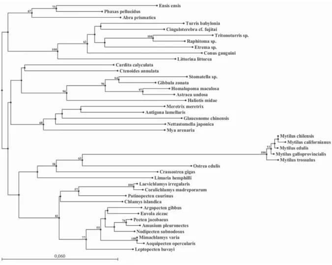
41 histone H3 gene sequences selected from GenBank Database. The NJ tree separated the species into two clades (Fig. 2).The first clade was subdivided into two groups, one constituted of 20 Bivalvia (12 Pectinidae, 5 Mytilidae, 2 Ostreidae and 1 Limidae) all from the subclass Pteriomorphia, and the other one of 5
Gastropoda (corresponding to the
superorder of Vetigastropoda), and 7 Bivalvia (all Heteroconchia: 4 Veneroida, 2
Myoida, except one which was
Pteriomorphia: 1 Limoida).
In bivalves, phylogenetic
relationships with histone H3 gene
sequences have already been studied in Pectinidae (Puslednik, Serb, 2008), in Veneridae (Kappner, Bieler, 2006), and in Mytilidae (Eirín-López et al., 2004).
Unfortunately, little knowledge was
available concerning Ostreidae as until now only two sequences of histone H3 gene were available in oysters, the one from O. edulis and the one from C. gigas (this study).
Ostreida, Mytiloida, Pectinoida and
Limoida are from the same subclass: Pteriomorphia, therefore it was expected to find them in the same clade. The other Limoida (Ctenoides annulata Lamarck, 1819) was found in another clade which was unexpected, but it was also not supported by a significative bootstrap value. The closest relative of this Limoida was a Veneroida. This Limoida
was also closest to a group of
Vetigastropoda species (representatives of another class, Gastropoda). The bootstrap values were not significative, thus, no relationships between these different species can be established. C. gigas was more closely related to O. edulis which is a predictable result as they both belong to the same family Ostreidae, and also to
different Mytilidae species: Mytilus
californianus Conrad, 1837, M. chilensis Hupé, 1854, M. edulis Linnaeus, 1758, M.
galloprovincialis, and M. trossulus Gould, 1850. However, O. edulis seems to be more closely related to species of the genus Mytilus than to C. gigas which is a species from the same family. The reason for this peculiar result could be the short length of the sequence (only around 300 bp) used for the reconstruction. Eirín-López et al. (2004) studied 5 Mytilidae, the same sequences that we selected in GenBank Database. The results observed by Eirín-López et al. (2004) were quite similar to the ones observed in this study as they showed that the 5 Mytilus species: M. californianus, M. edulis, M. chilensis, M. galloprovincialis and M. trossulus were clustered together.
According to the phylogenetic tree some closely related species were not clustered together. The partial sequence of histone H3 gene seems to be too short to give a good resolution in phylogenetic reconstructions. Indeed, some groups were not supported by a significative bootstrap value, and thus, H3 gene did not allow reconstructing an accurate phylogeny as the different clusters not always correspond to the taxonomical (family level or class level) groups.
Cytogenetic characterisation. We examined 80 good chromosome spreads from oyster embryos. Our results showed that the histone H3 gene was mapped at two different loci in the genome of C. gigas (Fig. 3). The histone H3 gene was located at an interstitial site (about half-way from the centromere to the telomeres) on the long arm of chromosome pair 4, and on the telomeres of the smaller chromosome pair (pair 10). Polymorphism was detected on the telomeres of pair 10. On the 80
metaphases that were examined, 16
presented single signals in both
chromosomes (20%) (Fig. 3, A-B), 29 showed double signals (36%) (Fig. 3, C-D), and 35 had a single signal on one
chromosome and a double signal on the homologue (44%) (Fig. 3, E-F). The
histone H3 gene is a qualified chromosomal
marker.
In other bivalve species, histone genes were also localised in one, two or three loci. In Mytilidae, FISH results on Mytilus galloprovincialis chromosomes located core histone genes at two loci in two different chromosome pairs
(Eirín-López et al., 2004), and the linker
histone H1 probe at three loci in three chromosome pairs (Eirín- López et al.,
2002). In both experiments, the
hybridisation signals mapped the histone genes at telomeric chromosomal
positions or interstitial positions. In
Pectinidae, histone H3 gene was mapped at two loci in the genome of Patinopecten yessoensis or a single locus in the genome of Chlamys farreri, Chlamys nobilis, and Argopecten irradians (Zhang et al., 2007).
Positions were also interstitial or
telomeric as in our experiment. In Mytilidae and Pectinidae studies, no polymorphism was reported. Histone gene clusters were rather conservative in chromosome location in most cases where
closed species have been analysed
(Hankeln et al., 1993; Ranz et al., 2003; Cabrero et al., 2009). Chromosomal localisation of histone H3 gene should
be determined in other Crassostrea
species for oyster comparative studies. Histone H3 gene in O. edulis was characterised molecularly (Giribet, Distel, 2003), but no cytogenetic studies were realised with this sequence.
Major ribosomal DNA genes (Xu et
al., 2001; Wang et al., 2004), NORs
(Thiriot- Quiévreux, Insua, 1992) and C-bands (Bouilly et al., 2008) were also localised on the telomeres of long arm of pair 10 in C. gigas. During mitosis, the major ribosomal genes are the nucleotidic component of the NORs (Goessens, 1984),
therefore, the chromosomal sites of major ribosomal gene clusters correspond to NOR sites. NOR sites were already detected to colocalise with C-banded
heterochromatic region in an insect
Bradysia hygida Sauaia et Alves, 1968
(Gaspar et al., 2002). In salmonids, the
chromosomal location of the major histone cluster was adjacent or close to regions with C+ heterochromatin (Pendás et al., 1994). Histone genes were already found to be associated with 5S rDNA in two Crustacean species, Artemia salina Linnaeus, 1758 (Andrews et al., 1987) and Asellus aquaticus Linnaeus, 1758 (Barzotti et al., 2000). Multicolour-FISH will be of great interest to check if the
major and minor rDNA genes are
colocalised or not with the histone H3 genes in C. gigas.
In conclusion, histone H3 gene is a useful karyotypic marker which can be used in oyster cytotaxonomy. Our data support that molecular and cytogenetic characterisations of histone H3 gene in other oyster and bivalve species will be useful for comparative studies, evolutionary and phylogenetic studies, and for understanding genome organisation in oysters.
A
CKNOWLEDGEMENTSThis work was supported by the project PTDC/MAR/66143/2006 and a post-doc grant SFRH/BPD/20538/2004 of the Science and Technology Foundation (FCT) from Portu- gal and also by the project FCOMP-01-0124- FEDER-007377 of the European Fund of Re- gional Development (FEDER). We are deeply grateful to A. Benabdelmouna for providing the fixed embryos of C. gigas.
R
EFERENCESAndrews M.T., Vaughn J.C., Perry B.A.,
Bagshaw J.C. 1987. Interspersion of histone and 5S genes in Artemia // Gene. 51(1): 61-67. http://dx.doi. org/10.1016/0378-1119(87)90474-4
Barzotti R., Pelliccia F., Bucciarelli E., Rocchi A. 2000. Organization, nucleotide sequence, and chromosomal mapping of a tandemly repeated unit containing the four core histone genes and a 5S rRNA gene in an isopod crustacean species // Genome. 43(2): 341-345. http://dx.doi.org/10.1139/ gen-43-2-341
Boudry P., Heurtebise S., Lapègue S. 2003.Mitochondrial and nuclear DNA sequence variation of presumed Crassostrea gigas and
Crassostrea angulata specimens: a new oyster
species in Hong Kong? // Aquaculture. 228(1-4): 15-25. http:// dx.doi.org/10.1016/S0044-8486(03)00443-5
Bouilly K., Leitão A., Chaves R., Guedes-Pinto H. 2005. Endonuclease banding reveals that atrazine- induced aneuploidy resembles spontaneous chromosome loss in Crassostrea
gigas // Genome. 48(1): 177-180. http://dx.doi.org/10.1139/g04-087
Bouilly K., Chaves R., Leitão A., Benabdelmouna A., Guedes-Pinto H. 2008. Chromosomal organization of simple sequence repeats in the Pacific oyster (Crassostrea gigas): (GGAT)4, (GT)7 and (TA)10 chromosome patterns // J. Genet. 87(2): 119-125.
http://dx.doi.org/10.1007/s12041-008-0018-2 Cabrero J., López-León M.D., Teruel M.,
Camacho J.P.M. 2009. Chromsome mapping of H3 and H4 histone gene clusters in 35 species of acridid grasshoppers // Chromosome
Res. 17(3): 397-404. http://dx.doi.org/10.1007/s10577-009-9030-5 Chaves R., Adega F., Santos S., Guedes-Pinto
H., Heslop-Harrison J.S. 2002. In situ hybridization and chromosome banding in mammalian species // Cytogenet. Genome Res. 96(1-4): 113-116. http:// dx.doi.org/10.1159/000063020
Clabby C., Goswami U., Flavin F., Wilkins N.P., Houghton J.A., Powell R. 1996. Cloning, characterization and chromosomal location of a satellite DNA from the Pacific oyster,
Crassostrea gigas // Gene. 168(2): 205-209.
http://dx.doi. org/10.1016/0378-1119(95)00749-0 Colgan D.J., Ponder W.F., Eggler P.E. 2000. Gastropod evolutionary rates and phylogenetic relationships assessed using partial rDNA and histone H3 sequences // Zool. Scr. 29(1): 29-63. http://dx.doi.
org/10.1046/j.1463-6409.2000.00021.x
Cross I., Vega L., Rebordinos L. 2003. Nucleolar organizing regions in Crassostrea
angulata: chromosomal location and polymorphism // Genetica. 119(1): 65-74. http://dx.doi.org/10.1023/ A:1024478407781 Cross I., Díaz E., Sánchez I., Rebordinos L.
2005. Molecular and cytogenetic characterization of Crassostrea angulata
chromosomes // Aquaculture. 247(1-4): 135-144. http://dx.doi.org/10.1016/j.aquaculture.2005.02. 039
Eirín-López J.M., González-Tizón A.M., Martínez A., Méndez J. 2002. Molecular and evolutionary analysis of mussel histone genes (Mytilus spp.): possible evidence of an “orphon origin” for H1 histone genes // J. Mol. Evol. 55(3): 272-283. http:// dx.doi.org/10.1007/s00239-002-2325-1
Eirín-López J.M., Ruiz M.F., González-Tizón A.M., Martínez A., Sánchez L., Méndez J. 2004. Molecular evolutionary characterization of the mussel Mytilus histone multigene family: first record of a tandemly repeated unit of five histone genes containing an H1 subtype with “orphon” features // J. Mol.
Evol. 58(2): 131-144. http:// dx.doi.org/10.1007/s00239-003-2531-5
Gaspar V.P., Borges A.R., Fernandez M.A. 2002. NOR sites detected by Ag-DAPI staining of an unusual autosome chromosome of Bradysia hygida (Diptera: Sciaridae) colocalize with C-banded heterochromatic region // Genetica. 114(1): 57-61. http://dx.doi.org/10.1023/A:1014698401988 Giribet G., Distel D.L. 2003. Bivalve
phylogeny and molecular data, (pp. 45-90) // Lydeard C., Lindberg D.R. (Eds). Molecular
systematics and phylogeography of mollusks.
Washington: Smithsonian Books. 328p.
Goessens G. 1984. Nucleolar structure // Int.
Rev. Cytol. 87: 107-158. http://dx.doi.org/10.1016/
S0074-7696(08)62441-9
Guo X., Allen S.K. Jr 1997. Fluorescence in
situ hybridization of vertebrate telomere sequence to chromosome ends of the Pacific oyster, Crassostrea gigas Thunberg // J. Shellfish
Res. 16(1): 87-89.
Hankeln T., Keyl H.G., Ross R., Schmidt E.R. 1993. Evolution of histone gene loci in chironomid midges // Genome. 36(5): 852-862. http://dx.doi. org/10.1139/g93-113
Kappner I., Bieler R. 2006. Phylogeny of venus clams (Bivalvia: Venerinae) as inferred from
nuclear and mitochondrial gene sequences //
Mol. Phylogenet. Evol. 40(2): 317-331. http://dx.doi.org/10.1016/j. ympev.2006.02.006 Leitão A., Chaves R. 2008. Banding for
chromosomal identification in bivalves: A 20-year history // Dyn. Biochem. Process Biotechnol. Mol. Biol. 2 (special issue 1): 44-49.
http://www.globalsciencebooks.info/JournalsSup /08DBPBMB_2_SI1.html
Leitão A., Chaves R., Santos S., Guedes-Pinto H., Boudry P. 2004. Restriction enzyme digestion chromosome banding in Crassostrea and Ostrea species: comparative karyological analysis within Ostreidae // Genome. 47(5): 781-788. http://dx.doi. org/10.1139/g04-035
Leitão A., Chaves R., Santos S., Guedes-Pinto H., Boudry P. 2007. Interspecific hybridization in oysters: restriction enzyme digestion
chromosome banding confirms Crassostra angulata x López-Flores I., de la Herrán R., Garrido-Ramos
M.A., Boudry P., Rejón C., Ruiz-Rejón M. 2004. The molecular phylogeny of oysters based on a satellite DNA related to transposons //Expression and organization of histone genes // Annu. Rev. Genet. 17: 239-277. http://dx.doi.org/10.1146/annurev.ge.17.120183. 001323
Maxson R., Cohn R., Kedes L., Mohun T. 1983. A new method for reconstructiong phylogenetic bivalve molluscs by fluorescence
in situ hybridization // J. Shellfish Res. 20(3):
1187-1190.
Miller D.J., Harrison P.L., Mahony T.J., McMillan J.P., Miles A., Odorico D.M., ten Lohuis M.R.1993. Nucleotide sequence of the histone gene cluster in the coral Acropora
formosa (Cnidaria; Scleractinia): Features of
histone gene structure and organization are common to diploblastic and triploblastic metazoans // J. Mol. Evol. 37(3):
245-253.
http://dx.doi.org/10.1007/BF00175501
Pendás A.M., Morán P., García-Vázquez E. 1994.
Organization and chromosomal location of the major histone cluster in brown trout, Atlantic salmon and rainbow trout // Chromosoma.
103(2): 147-152.
http://dx.doi.org/10.1007/BF00352324
Puslednik L., Serb J.M. 2008. Molecular phylogenetics of the Pectinidae (Mollusca: Bivalvia) and effect of increased taxon sampling and outgroup selection on tree topology // Mol. Phylogenet. Evol.
48(3):1178-1188. http://dx.doi.org/10.1016/j. ympev.2008.05.006
Ranz J.M., González J., Casals F., Ruiz A. 2003. Low occurrence of gene transposition events during the evolution of the genus
Drosophila // Evolution. 57(6): 1325-1335.
http://dx.doi.org/10.1111/j.0014-3820.2003.tb00340.x
Reese K.S., Cordes J.F., Stubbs J.B., Hudson K.L., Francis E.A. 2008. Molecular phylogenies help resolve taxonomic confusion with Asian Crassostrea oyster species // Mar.
Biol. 153(4): 709-721. http:// dx.doi.org/10.1007/s00227-007-0846-2
Saitou N., Nei M. 1987. The neighbor-joining method: trees // Mol. Biol. Evol. 4(4): 406-425. http://mbe.oxfordjournals.org/cgi/content/abstrac t/4/4/406
Thiriot-Quiévreux C., Insua A. 1992. Nucleolar organizer region variation in the chromosomes of 3 oyster species // J. Exp.
Mar. Biol. Ecol. 157(1): 33-40. http://dx.doi.org/10.1016/0022-0981(92)90072-I Thoma F., Koller T., Klug A. 1979. Involvement
of histone H1 in the organization of the nucleosome and of the salt-dependent superstructures of chromatin // J. Cell Biol. 83(2): 403-427. http://www.ncbi.nlm. nih.gov/pmc/articles/PMC2111545/
Wang Y., Guo X. 2001. Chromosomal mapping of the Gene. 339: 181-188. http://dx.doi.org/10.1016/j.vertebrate telomeric sequence (TTAGGG)n in fourgene.2004.06.049 Wang Y., Xu Z., Guo X. 2001. A centromeric
satellite sequence in the Pacific oyster (Crassostrea gigas Thunberg) identified by fluorescence in situ hybridization // Mar.
Biotechnol. 3(5): 486-492. http://dx.doi.org/10.1007/s10126-001-0063-3
Wang Y., Xu Z., Guo X. 2004. Differences in the rDNA- bearing chromosome divide the Asian Pacific and Atlantic species of Crassostrea (Bivalvia, Mollusca) // Biol. Bull. 206: 46-54. http://www.biolbull.org/cgi/content/abstract/206/ 1/46
Wang Y., Xu Z., Guo X. 2005a. Chromosomal mapping of 5S ribosomal RNA genes in the Eastern oyster, Crassostrea virginica Gmelin by fluorescence in situ hybridization // J. Shellfish
Res. 24(4): 959-964.
Wang Y., Xu Z., Pierce J.C., Guo X. 2005b. Characterization of Eastern oyster (Crassostrea virginica Gmelin) chromosomes by fluoresence in situ hybridization with bacteriophage P1 clones // Mar. Biotechnol. 7(3): 207-214. http://dx.doi. org/10.1007/s10126-004-0051-y
Xu Z., Guo X., Gaffney P.M., Pierce J.C. 2001. Chromosomal location of the major ribosomal RNA genes in Crassostrea virginica and Crassostrea gigas // Veliger. 44(1): 79-83. Zhang L., Bao Z., Wang S., Huang X., Hu
J. 2007. Chromosome rearrangements in Pectinidae (Bivalvia: Pteriomorphia) implied based on chromozomal localization of histone H3 gene in four scallops // Genetica. 130(2): 193-198. http:// dx.doi.org/10.1007/s10709-006-9006-8
Zhang Q., Yu G., Cooper R.K., Tiersch T.R. 1999. Chromosomal location by fluorescence in
situ hybridization of the 28S ribosomal RNA
gene of the Eastern oyster // J. Shellfish Res. 18(2): 431-435.
Table 1. List of histone H3 gene sequences in different molluscan species used for the phylogenetic analysis with GenBank accession numbers.
Phylum Class Superorder Order Family Species Accession
number
Crassostrea gigas Thunberg, 1793 HQ009488
Ostreoida Ostreidae
Ostrea edulis Linnaeus, 1758 AY070151
Mytilus californianus Conrad, 1837 AY267745
Mytilus chilensis Hupé, 1854 AY267746
Mytilus edulis Linnaeus, 1758 AY267749
Mytilus galloprovincialis Lamarck,
1819
AY267748
Mytiloida Mytilidae
Mytilus trossulus Gould, 1850 AY267747
Limaria hemphilli Hertlein & Strong,
1946
EU379502
Limoida Limidae
Ctenoides annulata Lamarck, 1819 EU379493
Laevichlamys irregularis Sowerby,
1842
EU379537
Coralichlamys madreporarum Sowerby
II, 1842
EU379505
Patinopecten caurinus Gould, 1850 FJ263662
Chlamys islandica Müller, 1776 FJ263666
Argopecten gibbus Linnaeus, 1758 EU379496
Euvola ziczac Linnaeus, 1758 EU379538
Pecten jacobaeus Linnaeus, 1758 AY070153
Amusium pleuronectes Linnaeus, 1758 EU379523
Nodipecten subnodosus Sowerby, 1835 EU379535
Mimachlamys varia Linnaeus, 1758 AY070154
Aequipecten opercularis Linnaeus, 1758EU379516
Pectinoida Pectinidae
Leptopecten bavayi Dautzenberg, 1900 EU379487
Antigona lamellaris Schumacher, 1817 DQ184882
Veneridae
Meretrix meretrix Linnaeus, 1758 FJ429106
Glaucono- midae
Glauconome chinensis Gray, 1828 DQ184899
Ensis ensis Linnaeus, 1758 AY070159
Pharidae
Phaxas pellucidus Pennant, 1777 DQ280006
Semelidae Abra prismatica Montagu, 1808 AY070160
Veneroida
Carditidae Cardita calyculata Linnaeus, 1758 AY070156
Pholadidae Nettastomella japonica Yokoyama,
1920
AB439267
Bivalvia
-Myoida
Myidae Mya arenaria Linnaeus, 1758 AY070164
Turridae Turris babylonia Linnaeus, 1758 EU015786
Terebridae Cinguloterebra cf. fujitai Kuroda &
Habe, 1952 EU015832 Etrema sp. EU015800 Tritonoturris sp. EU015823 Raphitoma sp. EU015813 Conidae
Conus gauguini Richard & Salvat, 1973 EU015856
Caenogastropoda Sorbeoconcha
Littorinidae Littorina littorea Linnaeus, 1758 DQ093507
Stomate- llidae
Stomatella sp. AY923978
Trochidae Gibbula zonata Wood, 1828 AY923977
Astraea undosa Wood, 1829 AY923980
Turbinidae
Homalopoma maculosa Pease, 1868 AY923982
Mollusca
Gastropoda
Vetigastropoda
Fig. 1. Histone H3 gene sequence from Crassostrea gigas.
Fig. 2. The phylogenetic relationships among different molluscan species, as analysed by the Neighbour- Joining method. Bootstrap values over 50% are written above the branches. Bar = Substitutions/Site.
Fig. 3, A-F. FISH with histone H3 gene probe applied on chromosomes of Crassostrea gigas (in red). The chromosomes were contrasted with DAPI (blue). A - a metaphase cell with single/single signals on telomeres of pair 10, and B - its corresponding karyotype. C - a metaphase cell with double/double signals on telomeres of pair 10, and D - its corresponding karyotype. E - a metaphase cell with single/double signals on telomeres of pair 10, and F - its corresponding karyotype. Bars = 10 µm.