Universidade de Trás-os-Montes e Alto Douro
Jejunal Diverticulum in the Dog
Dissertação de Mestrado Integrado em Medicina Veterinária
Raquel Seabra Lima Reis
Orientador:
Professor Doutor Luís Miguel Viana Maltez da Costa
Co-orientador:
Drª Maria Emanuel Dias Barbosa Frada
iii
Universidade de Trás-os-Montes e Alto Douro
Jejunal Diverticulum in the Dog
Dissertação de Mestrado Integrado em Medicina Veterinária
Raquel Seabra Lima Reis
Orientador:
Professor Doutor Luís Miguel Viana Maltez da Costa
Co-orientador:
Drª Maria Emanuel Dias Barbosa Frada
Composição do Júri:
Doutor Carlos Alberto e Silva Venâncio
Doutora Maria Isabel Ribeiro Dias
Doutor Luís Miguel Viana Maltez da Costa
v
.
Dissertação apresentada à Escola de Ciências Agrárias e Veterinárias - Departamento de Ciências Veterinárias - da Universidade de Trás-os-Montes e Alto Douro, como requisito para a obtenção do grau de Mestre em Medicina Veterinária O conteúdo do presente trabalho é da inteira responsabilidade da autora.
vii
Acknowledgements
I would like to thanks to University of Trás-os-Montes e Alto Douro, its director and all the teachers.
To Veterinary Hospital of University of Trás-os-Montes e Alto Douro, as well as, to Veterinary Hospital of Trás-os-Monte for the opportunity to learn during my internship time.
To my mentors, Professor Luís Maltez da Costa and Doctor Maria Frada for accepting this dissertation’s orientation and for all the dedication and availability shown during its elaboration. To my parents and family, for always supporting me and making this dream come true.
To Sara Gonçalves, Melissa Silva, Ana Catarina Silva, Rui Figueiredo and all the other friends that I made during these past six years, who stood by my side, making it a truly unforgettable travel through college life.
viii
Abstract
A diverticulum is a bulging sack that occurs in any portion of the gastrointestinal tract and may be either congenital or acquired. In small animals, intestinal diverticula are rarely reported. Recent literature reports four cases of intestinal diverticula in dog, where two are duodenal diverticula and two are jejunal diverticula. The etiology of intestinal diverticula is unknow. Diagnosis is made by ultrasonography and confirmed during surgery. The treatment for intestinal diverticula is surgical resection. The prognosis is excellent.
The clinical case reported in this dissertation was attended at Veterinary Hospital of University of Trás-os-Montes e Alto Douro. A 6-month-old female Beagle was presented with the main complaint of diarrhea with melena and hyporexia over the preceding 4 days. Complementary diagnostic tests showed leukocytosis and hypoproteinemia, presence of foreign bodies in the stomach and small intestine and a highly distended intestinal segment compatible with intestinal obstruction.
Medical treatment made followed the standard indications. Surgical treatment consisted on gastrotomy and duodenotomy to remove foreign bodies. During surgery, a jejunal diverticulum was discovered and removed by partial enterectomy with end-to-end anastomosis. Biopsies to the duodenum, jejunum and jejunal lymph nodes were made, as well as a serosal patch before the abdomen was closed.
Histologically the diverticulum showed normal appearance and no signs of gastric-type mucosa or pancreatic tissue.
The dog was clinically discharged six days after the surgery with no signs of gastrointestinal problems. Seven months after surgery the patient did not present clinical signs of gastrointestinal pathology and recovered corporal condition.
ix
Resumo
Um divertículo é uma protuberância (bolsa) que ocorre em qualquer porção do trato gastrointestinal e pode ser classificado como congénito ou adquirido. Em animais de companhia, os divertículos jejunais são raramente reportados. Na literatura recente são relatados quatro casos de divertículos intestinais em cães, sendo dois casos divertículos duodenais e dois divertículos jejunais. A etiologia desta patologia é desconhecida. O diagnóstico é alcançado usando a ultrassonografia e confirmado durante a cirurgia. O tratamento para divertículos intestinais é a resseção cirúrgica. O prognóstico é excelente.
O caso clínico descrito nesta dissertação foi acompanhado no Hospital Veterinário da Universidade de Trás-os-Montes e Alto Douro. Uma cadela de 6 meses de idade da raça Beagle deu entrada por apresentar diarreia com melena e hiporexia há 4 dias. Os exames de diagnóstico complementares mostraram uma leucocitose e hipoproteinemia, presença de corpos estranhos no estômago e intestino delgado, bem como, um segmento intestinal muito dilatado compatível com imagem de obstrução intestinal.
O tratamento médico administrado vai de acordo com as indicações padrão. O tratamento cirúrgico consistiu numa gastrotomia e duodenotomia para remoção de corpos estranhos. Durante a cirurgia, um divertículo jejunal foi descoberto e removido por enterectomia parcial com anastomose topo-a-topo. Foram executadas, também, algumas biopsias de duodeno, jejuno e linfonodos jejunais, bem como, um patch de serosa antes de se encerrar o abdómen.
Histologicamente, o divertículo tem uma aparência normal, sem presença de tecido gástrico ou pancreático.
O animal teve alta seis dias após a cirurgia sem sinais de problemas gastrointestinais. Sete meses após a cirurgia o paciente não mostrou qualquer sinal de patologia gastrointestinal e recuperou a sua condição corporal.
x
Table of Contents
Acknowledgements ... vii
Abstract ... viii
Table of Contents ... x
List of Figures ... xiii
List of Tables ... xiii
List of abbreviations and acronyms ... xv
Introduction ... 1
Chapter I – Literature Review ... 3
1.1. Pathophysiology of intestinal obstruction ... 3
1.2 Intestinal obstruction in small animals ... 6
1.2.1 Foreign bodies ... 6
1.2.2 Intussusception ... 10
1.2.3 Neoplasia ... 11
1.2.4 Intestinal torsion ... 13
1.2.5 Abscessation, granulomas and strictures formation ... 14
1.3 Intestinal diverticula in humans ... 16
1.3.1 Meckel diverticula ... 17
1.3.2 Acquired diverticula ... 19
1.4 Intestinal diverticula in small animals ... 20
1.5 Small intestine surgery ... 22
1.5.1 Gastrotomy ... 22
1.5.2 Enterotomy ... 23
1.5.3 Intestinal resection and anastomosis ... 25
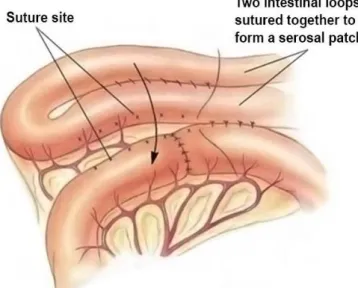
1.5.4 Omental patch and serosal patch ... 27
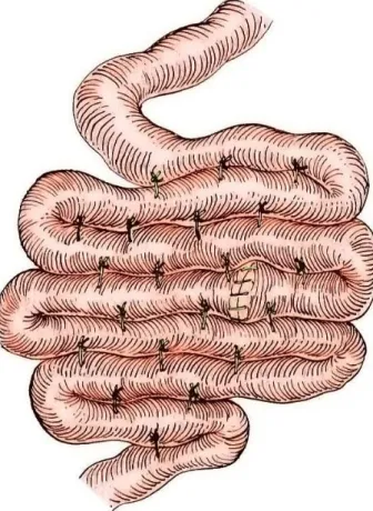
1.5.5 Enteroplication ... 28
Chapter II – Case Report ... 29
2.1 Signalment and presenting complaint... 29
2.2 Past pertinent history ... 29
2.3 Physical exam ... 29
2.4 Complementary diagnostic tests ... 29
2.4.1 Hematology and serum biochemical profile ... 29
2.4.2 Radiographic exam ... 31
xi 2.5 Problem List ... 32 2.6 Definitive diagnosis ... 32 2.7 Treatment decisions ... 32 2.7.1 Medical management ... 32 2.7.2 Surgical management ... 34 2.8 Histopathology results ... 40 2.9 Outcome... 40
Chapter III - Discussion ... 43
Chapter IV - Conclusion ... 46
xiii
List of Figures
Figure 1.: Pathophysiology of intestinal obstruction. Figure 2.: Gastrotomy.
Figure 3.: Enterotomy. Figure 4.: Enterectomy.
Figure 5.: Serosal patch technique. Figure 6.: Bowel plication.
Figure 7.: Day 1 lateral and ventrodorsal radiographic view. Figure 8.: Day 2 lateral and ventrodorsal radiographic exam. Figure 9.: Jejunal Diverticulum at surgery.
Figure 10.: Jejunal Diverticulum.
Figure 11.: Histologic image of the jejunal diverticulum. List of Tables
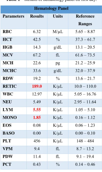
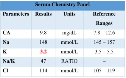
Table 1 – Results of hematology panel on first day. Table 2 – Results of Serum Chemistry on first day.
Table 3 – Results of serum chemistry panel on second day. Table 4 – Results of Serum Chemistry Panel on third day. Table 5 - Results of Hematology Panel on fifth day. Table 6 – Results of Serum Chemistry on fifth day.
xv
List of abbreviations and acronyms % – percent
µg/kg – microgram per kilogram
µg/kg/min – microgram per kilogram per minute cm – centimeter
CRI – constant rate infusion fL – femtoliter
g/dL – grams per deciliter HTC – hematocrit
K/µL – thousands per microliter kg – kilogram
M/µL – million per microliter mg/dL – milligrams per deciliter mg/kg – milligram per kilogram ml – milliliter
mmol/L – millimoles per liter
ml/Kg/L – milliliter per kilogram per hour mEq/L – milliequivalents per liter
ºC – degrees Celsius pg – picogram U/L – units per liter º – degrees
bpm – beats per minute BID – twice a day TID – three times a day IV – Intravenously PO – per os
1 Introduction
Gastrointestinal obstruction is a mechanical or functional blockage of the stomach, or small or large intestine (Rozanski & Rush, 2007). Intestinal torsion (Patsikas et al., 2003), intra-luminal foreign body, intestinal intussusception, neoplasia, abscessation, granulomas and strictures formation are included in reported cases of gastrointestinal obstruction (Nordquist & Culp, 2013).
Intestinal diverticula are rarely reported in small animals (Van Klaveren et al., 2008), although can be a cause of intestinal obstruction (Polf & Poteet, 2010). In recent literature, only 4 cases reports addressed intestinal diverticula, two duodenal diverticula (Van Klaveren et al., 2008; Polf & Poteet, 2010;) and two jejunal diverticula (Castaneda, n.d.; Mills et al., 2017).
Diverticular disease includes diverticulosis characterized by the presence of diverticula protruding the colonic wall and diverticulitis that is an acute inflammation of diverticula (Kozak
et al., 2006). In humans, diverticula in the duodenum and colon are common but they are rare
in the remaining small bowel, with reported rate of 0.06% to 1.3% (comparing with all intestinal diverticula) on autopsy predominantly involving jejunum (Kassahun et al., 2007). Intestinal obstruction, hemorrhage and perforation are some of the complications of intestinal diverticula (Elsayes et al., 2007; Tavakkoli et al., 2015).
Due to its rarity and similar complications, both in small animals and humans, it is possible to make an analogy between the two species. The lack of information in small animals’ bibliography, and the possible similarity between intestinal diverticula in humans and small animals, lead to a deeper research on human literature.
This dissertation intends to report an additional case of jejunal diverticula in the dog. Despite the rarity of this pathology, intestinal diverticula should be considered as cause of intestinal obstruction.
3 Chapter I – Literature Review
1.1. Pathophysiology of intestinal obstruction
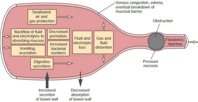
Gastrointestinal obstruction is a mechanical or functional blockage (partial or complete) of the stomach, or small or large intestine. Acid-base and electrolyte status, hydration and gastrointestinal functions (like peristalsis and secretion or absorption of ions) can be affected whatever the area of the obstruction (Rozanski & Rush, 2007).
Causes of gastrointestinal obstruction include foreign bodies (linear and nonlinear), neoplasia (intra- and extraluminal), inflammatory disease, hypertrophy/hyperplasia, strangulation, volvulus, intussusception and stenosis. Obstruction can also be iatrogenic like post-surgical stenosis, strangulation, or excessive angulation post plication. Diseases that decrease peristalsis (ileus) can easily be misattributed to obstruction (Rozanski & Rush, 2007). Gastrointestinal obstruction can be classified as acute or chronic. The degree of obstruction and subsequent duration, timing, and severity of clinical signs are determined by the size of the remaining luminal opening. Chronic signs are more likely to appear on partial gastrointestinal obstruction (Rozanski & Rush, 2007).
Intestinal obstruction includes a complex interaction of local and systemic factors (Jones & Blivslager, 2002) which is still not fully understood. Fluid, acid-base and electrolyte imbalances, hypovolemia and toxemia lead to life-threatening complications (Guilford & Strombeck, 1996). Secondary to a complete intestinal obstruction, a luminal distention is formed proximal to the obstruction due to accumulation of gas and fluid (Lantz, 1981). Most of the gas results from swallowed air, carbon dioxide formation due to bicarbonate neutralization in the intestinal lumen and organic gases resulting from bacterial fermentation (Lantz, 1981). Nitrogen is not absorbed by the intestinal mucosa which leads to gaseous luminal distention from bacterial fermentation (Walshaw, 1985).
Accumulating fluids result from two different sources: an increased amount of secretion in the upper gastrointestinal tract (saliva and bile; gastric, pancreatic and small intestine secretions) and retention of ingested fluids (Ellison, 1993). Lymphatic and venous congestion, increased intraluminal osmolality and decreased enterocyte turnover rate cause solute absorption reduction (Guilford & Strombeck, 1996). The distended bowel may lose its ability to absorb fluids, and local hypersecretion is observed, after 24 hours of obstruction (Ellison, 1993); as the obstruction becomes prolonged, intraluminal fluid volume increases (Mishra et
4 ingested fluids cannot be reabsorbed because fluids will not contact with jejunal or ileal mucosa. In distal intestinal obstructions, some fluids, by reverse peristalsis, move proximally and reach a nondistended intestinal loop, where normal absorption occurs (Ellison, 1993). The pathophysiology of these events has yet to be completely defined, despite that, four major mechanisms of hypersecretion and decreased absorption may co-exist (Papazoglou et al., 2003):
➢ Enteric bacterial toxins secreted by noninvasive pathogenic bacteria that bind specific enterocyte receptors and stimulate salt and water production may be responsible for hypersecretion (Jones & Blivslager, 2002);
➢ In the obstruction site, there is an increased concentration of bile and fatty acids and tissue ischemia products (Guilford & Strombeck, 1996);
➢ In the proximal part of intestinal obstruction there is an increased blood flow that may stimulate secretory activity (Enochsson et al., 1982);
➢ Increased luminal distention may stimulate enteroendocrine cells to release serotonin (5-hydroxytryptamine) which activates reflex pathways that increase chloride ion secretion (Cooke & Reddix, 1994).
Additionally, acetylcholine, vasoactive intestinal polypeptide and substance P, that are chemical mediators of the enteric nervous system, active chloride ion-rich fluid secretion by several mechanisms (Jones & Blivslager, 2002).
The increasing intraluminal pressure generated proximal to the intestinal obstruction and the duration of the obstruction are related to the effects of luminal distention (Lantz, 1981). Fluid and gas accumulation increases gradually the intraluminal pressure, proximal to the obstruction. The arterial circulation is not affected, but intestinal wall edema may occur. This happens when capillary bed congestion leads to elevated hydrostatic pressure that produces wall edema (Walshaw, 1985). Moreover, fluid can be extravasated through the serosal surface to the peritoneal cavity (Ellison, 1993).
In intestinal luminal distention, there is an increased myoelectric activity proximal to the obstruction and a simultaneous decrease distally. As time passes and the obstruction is prolonged, clusters of intense myoelectric activity that migrate distally are interrupted by periods of absent motor activity. Uninhibited hyperperistalsis may result in elevated intraluminal pressure that can lead to ischemia and rupture, so these periods of inactivation may represent a protective mechanism (Summers et al., 1983). Proximal to an intestinal obstruction increased myoelectric activity was thought to have cholinergically mediated, when in fact
5 noncholinergic nonadrenergic pathways may mediate distal inhibition of spike bursts (Prihoda
et al., 1984).
Due to stasis or loss of the migratory myoelectric complex, which helps on moving intestinal contents distally and keeping bacterial numbers low, simple intestinal obstruction may cause an increase in the intraluminal pathogenic bacterial population (Guilford & Strombeck, 1996). When the luminal distention is prolonged and severe, the enteric mucosal barrier can be damage and result in increased permeability and entry of bacteria and toxins into the systemic circulation. This can cause endotoxic shock and septic peritonitis if bacteria and toxins reach the peritoneal cavity (Lantz, 1981). An efficient decompression of the obstruction is advisable because it allows quicker mucosal regeneration (Shikata et al., 1983). In simple obstruction, a significant amount of fluids and electrolytes can be lost, resulting in hypovolemia and electrolyte and acid-base imbalances (Lantz, 1981). The degree, location, and duration of the intestinal obstruction correlates with the volume of fluid lost and its metabolic effects. Vomiting, sequestration in the intestinal lumen, diarrhea, intestinal wall edema and extravasation into the peritoneal cavity are responsible for fluid and electrolyte losses (Guilford & Strombeck, 1996).
Clinical signs of nutrients maldigestion and malabsorption (stagnant loop syndrome) can be associated with partial intestinal obstruction. Diarrhea can be related to secretory activity of enterocytes and combined osmotic effects of unabsorbed substances in intestinal lumen (Guilford & Strombeck, 1996). Colonic obstruction induced by foreign bodies is generally and partially due to distensibility of the colon and its slightly rhythmic segmentation (Walshaw, 1985). In result of the obstruction, large quantities of feces accumulate in the colon, where water and electrolyte absorption to take place as well, often prolonging the course of the disease (Pass, 1985).
6
Figure 1.: Pathophysiology of intestinal obstruction (Adapted from Radlinsky, 2013).
1.2 Intestinal obstruction in small animals
Gastrointestinal obstruction (focal and linear) in dogs and cats is a common condition which frequently requires emergency surgical intervention and attentive medical management (Nordquist & Culp, 2013). Intestinal torsion (Patsikas et al., 2003), intra-luminal foreign body, intestinal intussusception, neoplasia, abscessation, granulomas and strictures formation are included in reported cases of gastrointestinal obstruction. Depending on the underlying cause and location of the obstruction, clinical signs, physical examination findings, and diagnostic results will be different (Nordquist & Culp, 2013).
1.2.1 Foreign bodies
Intestinal foreign bodies commonly cause intestinal obstruction in dogs and cats, and may lodge in any part of the intestinal tract. The size of the foreign body dictates the degree of obstruction. Partial obstruction can be caused by small irregular or linear foreign bodies whereas large circular foreign bodies usually cause complete obstruction. The location of the obstruction can be classified as high intestinal obstruction (involves the duodenum or upper jejunum), middle intestinal obstruction (middle jejunal region), low intestinal obstruction (distal small intestine) and colonic. Foreign bodies that don’t compromise the intestinal wall blood supply induce a simple (or mechanical) intestinal obstruction. When a strangulating obstruction is present (usually complete), it involves impairment of the blood supply of the
7 involved intestinal segment. In clinical situations, obstructions are rarely purely simple because there is almost always some compromise of the wall blood supply (Walshaw, 1985).
In dogs, the most commonly reported foreign body are latex, rubber or plastic objects, stones, balls, underwear, pantyhose, towel or fabric, carpet, and corn cobs. Cats commonly ingest linear foreign bodies like string or thread with or without a needle attached; yet focal obstructions, have been reported after ingestion of almonds, plastic and rubber ingestion (Hayes, 2009) as well as derived from trichobezoars (Barrs et al., 1999).
Nonlinear foreign bodies that are round and smooth may cause complete intestinal obstruction and pressure necrosis of the intestinal wall and sometimes if they move down, intestinal edema occurs (Walshaw, 1985). Septic peritonitis and adynamic ileus are caused by sharp foreign bodies that penetrate the intestinal wall (Papazoglou et al., 2003). Proximal obstructions are more acute than distal obstructions, complete obstructions are frequently more acute than partial obstructions, and strangulating obstructions are more severe than simple obstructions (Pass, 1985). Anorexia, dehydration, depression, abdominal pain or discomfort and vomiting are the most common clinical signs. Abdominal palpation may be painful and can occasionally reveal the presence of a foreign body and intestinal fluid and gas accumulating proximally (Capak et al., 2001). Laboratory findings will be different depending on the nature of the intestinal obstruction. Serum albumin concentration can be low and packed cell volume and total protein levels may be high. Leukocytosis with shift to the left may be present in intestinal perforation. Vomiting of intestinal contents containing hydrochloric acid and pancreatic secretions rich in bicarbonate results in mild metabolic acidosis and dehydration in case of duodenal and proximal jejunal obstruction (Ellison, 1993). Bicarbonate loss, dehydration and starvation contribute to the development of metabolic acidosis in distal intestinal obstruction because proximal intestinal secretions rich in bicarbonate are sequestered at the obstruction site (Lantz, 1981). A small increase in the activity of alanine aminotransferase, alkaline phosphatase, lipase, blood urea nitrogen and creatinine concentrations can be noted in cases of chronic intestinal obstruction (Papazoglou et al., 2003). Intestinal foreign bodies can easily be seen in plain radiographs if they are radiopaque, whereas nonopaque foreign bodies can be identified based on a typical shape and contained gas lucencies (Lamb & Hansson, 1994). Radiographs signs of intestinal obstruction is the basis for the diagnosis of radiolucent intestinal foreign bodies. These signs may include localized intestinal distention related to gas or fluid accumulation, distended intestinal loops that lie in layers parallel to each other and connected with sharp hairpin turns, and unequal gas-fluid interface
8 seen in a standing lateral projection (McNeel, 1994). Examination of intestinal tract with contrast improvement is necessary to make a diagnosis in some questionable cases, where may be visualized a radiolucent area surrounded by contrast material outlining the foreign body (Gomez, 1974). A valuable addition to radiography for detection of foreign bodies is ultrasonography. The acoustic patterns may vary depending on the physical properties of foreign body and the interaction with the ultrasound beam. Some foreign bodies are able to transmit ultrasounds and can be accurately detected, and some strongly attenuating foreign bodies produce acoustic shadows that if seen with in the intestinal lumen, can be a useful indicator of their presence (Tidwell & Penninck, 1992). Asymptomatic animals with some small, sharp foreign bodies (such as pins, sewing needles and fish hooks) may be treated conservatively. The foreign body may pass uneventfully, possibly because of the generation of local intestinal dilation in response to contact between the mucosa and this object (Guilford & Strombeck, 1996). Colonic and rectal perforation is still possible even when the foreign body passes through the ileocolic junction (Walshaw, 1985). To ensure uncomplicated foreign body passage the animal should be evaluated radiographically and clinically on a regular basis (Orsher & Rosin, 1993). Nonlinear foreign body that causes a large intestinal obstruction may, occasionally, be dislodged endoscopically. Hairballs treatment include administration of semisolid petrolatum-based laxatives for lubrication and introduction of a commercial diet to facilitate passage of ingested hair (Barrs et al., 1999). In animals with intestinal obstruction prophylactic antibiotics are indicated for two reasons: (1) surgeries that involve access the intestinal lumen are considered clean-contaminated, and (2) the high risk of contamination due to bacterial overgrowth (Bright, 2001). Enterotomy may be sufficient to remove most foreign bodies rather than resection and anastomosis unless intestinal necrosis or perforation is present (Radlinsky, 2013). Some complications like intestinal necrosis, perforation, leakage, dehiscence, peritonitis, endotoxic shock, stenosis and short bowel syndrome (if resected large segments of intestines) may occur (Ralphs et al., 2003). Prognosis without surgery is guarded mainly because hypovolemic or endotoxic shock, septicemia, peritonitis or starvation can kill the animal (Radlinsky, 2013). Prognosis is good when surgical treatment is performed and if peritonitis and extensive resections are avoided (Hayes, 2009).
Linear foreign bodies are frequently reported in cats and produce a specific type of intestinal obstruction because they can cause serious and extensive damage to the intestinal tract. This type of foreign bodies causes a partial intestinal obstruction and a chronic problem will result in large portions of the small intestines being damaged and nonfunctional that result
9 in clinical signs similar to complete intestinal obstruction. Linear foreign bodies can wrap around the base of the tongue or entrap in the pylorus. Pleating or gathering of the intestine around the foreign body is caused by peristaltic waves continuing to move the free end of the object in a distal direction, and may occasionally occur intussusception. Foreign body may cut through the intestinal wall at the mesenteric border causing perforation of the intestinal loops, followed by local or generalized peritonitis. Common clinical signs are vomiting, anorexia and bloody diarrhea. Depression, fever, dehydration, abdominal pain are include in physical examination findings. Additionally, a thread looped around the tongue, a foreign body hanging from the mouth or protruding from the anus, and palpable intestinal plication may be present (Felts et al., 1984). A detailed oral examination is essential for animals with a suspected linear foreign body intestinal obstruction. Hematologic and biochemical findings are similar from those in an animal with a nonlinear foreign body (Papazoglou et al., 2003). Radiograph examination may show the intestines bunched or pleated together, with small gas bubbles in the lumen (comma shaped) and without gas-distended intestinal loops (plication), although, some patients (especially cats) show no obvious lesions on plain radiographs. Contrast studies are infrequently done because the contrast may or may not be able to reach the lesion site (Radlinsky, 2013). An ultrasonography study can detect intestinal plication, facilitating this way do the diagnosis of linear foreign body, nevertheless it’s not always possible to visualize the foreign body during the examination (Tidwell & Penninck, 1992). Endoscopy sometimes is helpful to remove linear foreign body that are lodge at the pylorus. A cat that is presented 1 to 3 days after ingestion of a linear foreign body lodged at the base of the tongue, can be treat conservatively, depending on how ill the cat appears. The linear foreign body can be cut and then monitored for passage. The animal should be taken to surgery if clinical signs persist within 12 hours after cutting the foreign body off the tongue. Surgical treatment to remove linear foreign bodies requires frequently multiple enterotomies (two to four). Intestinal resection is a possibility if the linear object has been present for a long time and it became embedded in the mucosa (Radlinsky, 2013). Complications and postoperative care are identical to those for animals with nonlinear foreign body (Orsher & Rosin, 1993). Animals with longer duration of clinical signs, linear foreign bodies or that need more than one gastrointestinal incision present higher risk of death. The prognosis is also poor in situations of chronic obstructions, greater bowel compromise, longer surgery time, and increased risk of contamination. On the other hand, the location of the foreign body and degree of obstruction are not related with the prognosis (Hayes, 2009).
10 1.2.2 Intussusception
Intussusception of the intestine is the telescoping or invagination of a portion of the intestinal tract (intussusceptum) into the lumen of an adjacent intestinal segment (intussuscipiens) in the direction of normal peristalsis or rarely in a retrograde direction (Rallis
et al., 2000). Ileocolic intussusception is the most common condition although gastroduodenal,
duodenojejunal, jejunojejunal, ileoileal and colocolic intussusceptions have been described in young dogs (Patsikas et al., 2008). Motility dysfunction, enteritis, infection (viral, bacterial or parasitic), foreign bodies, previous intestinal surgery, adhesions and neoplasia are conditions associated with intussusception (Brown, 2011). Mechanical obstruction of intestines in dogs is commonly caused by intussusception. Clinical presentation, radiographic and ultrasonographic examination are necessary to achieve a diagnosis (Holt & Brockman, 2003).
In dogs and cats, the major clinical signs associated with intussusception are anorexia, lethargy and vomiting. Less commonly animals may present diarrhea with or without blood and weight loss (Atray et al., 2012). The severity and type of clinical signs are partly determined by patient factors, duration of the intussusception and if the obstruction is complete or incomplete (Kovak & Buriko, 2016).
When intestinal obstruction is suspected, careful abdominal palpation may reveal a tubular structure within the cranial or mid-abdomen. Frequently reported bloodwork abnormalities are hyponatremia, hypochloremia and hypokalemia. Other clinicopathologic abnormalities relevant in dogs consist in hemoconcentration, hyperlactatemia, hypoalbuminemia and leukocytosis with neutrophilia (Atray et al., 2012). In radiograph examination, lateral and dorsoventral projections can reveal a mass effect and evidence of obstruction. Additionally, barium contrast material (per os (PO) or via enema) may confirm the diagnosis (Wilson & Burt, 1974). Ultrasonographic exam provides a sensitive and specific method for accurate diagnosis, by revealing concentric rings, hyperechoic or anechoic center surrounded by multiple hyper- and hypoechoic concentric rings, in transverse section and multiple hyper- and hypoechoic parallel lines in longitudinal section (Patsikas et al., 2003).
Exploratory laparotomy is done when appropriate stabilization is guaranteed. To evaluate for any underlying causes, such as foreign bodies or neoplasia, a full assessment of the intestinal tract should be performed (Sridhar et al, 1994). Manual reduction in some cases can be attempted with very gentle traction on the intussusceptum, but usually resection and anastomosis is recommended as portions of the intestines are probably necrotic or nonviable
11 (Applewhite et al., 2001). To reduce the incidence of future occurrence, enteroplication may be performed after reduction or resection of the intussusception (Oakes, 1998). Hydration, perfusion, comfort level and electrolyte and acid-base status should be closely monitored on the postoperative period (Klinger et al., 1990). Major surgical complications can occur including dehiscence, peritonitis, short bowel syndrome if a large portion of intestine is resected, conjointly with reoccurrence or obstruction (Radlinsky, 2013).
Prognosis is good after an easy reduction or resection (Oakes, 1998). None specific risk factors for reoccurrence have been identified apart from the fact that in patients undergoing manual reduction, reoccurrence is more frequent comparatively with patients where resection and anastomosis was performed (Applewhite et al., 2001). Treatment for any identified underlying disease needs to be done to minimize the reoccurrence of intussusception. This risk for reoccurrence can be diminished by enteroplication, but is not without the potential for serious complications (Kovak & Buriko, 2016).
1.2.3 Neoplasia
In intestinal neoplasia, tumors arise from one of the layers of the intestinal wall, its glands, or associated cells or lymphatics (Radlinsky, 2013).
Gastrointestinal neoplasia is rare in dogs and cats (Eisele et al., 2010). Lymphoma is the most common intestinal neoplasia occurring in cats, while lymphomas or adenocarcinomas are the more common intestinal tumors in dogs (Rissetto et al., 2011). Leiomyosarcoma, gastrointestinal stromal tumor, mast cell tumor, carcinoids and several others can also occur in the small intestines (Worley, 2013).
Tumors most commonly invade the muscular layer of the intestinal wall where they compromise the lumen diameter and reduce distensibility causing intramural or intraluminal mechanical obstruction (Radlinsky, 2013).
Initially, patients manifest indistinct clinical signs of depression, anorexia and lethargy, that can progress to diarrhea and/or vomiting, with progressive weight loss. Dehydration, melena, hematemesis, anemia, fever, icterus, polyuria, polydipsia, and/or abdominal effusion are other clinical signs that can occur. Signs of intestinal obstruction, abscessation, and malabsorption may be present. Clincal signs secondary to metastasis in other organs may develop. Abdominal palpation can reveal a solid abdominal mass, thickned intestinal loops or mesenteric lymphadenomegaly. Body condition is poor in some animals (Cohen et al., 2003). Hematologic and biochemical profiles may reveal blood loss anemia, neutrophilic leukocytosis
12 with left shift, hypoalbuminemia, hypoglycemia and/or elevated serum hepatic enzyme concentrations, but these analytic profiles are frequently normal. Cytology or histopathology are the accurate methods to achieve a definitive diagnosis of intestinal neoplasia (Radlinsky, 2013).
Diagnosis of stomach and intestinal tumors using noninvasive methods can be challenging. Transcutaneous fine-needle aspiration, transcutaneous needle-core biopsies for solid masses and endoscopic biopsies are some of the tools available for diagnosis (Bonfanti et
al., 2006). Abdominal radiographs may reveal abnormal masses, abnormal gas and fluid
patterns, visceral displacement and abdominal fluid. The use of contrast agents can be helpful to delineate regions of mucosal irregularity, luminal narrowing and intraluminal infiltration, thickening or nodularity. Thoracic radiographs should be obtained if neoplasia is suspected (Radlinsky, 2013). Ultrasonographic findings may include intestinal wall thickening and loss of discrete wall layers, even though a normal-appearing intestine may be seen in a presence of lymphosarcoma. Intestinal tumors can manifest extensive ultrasonic patterns but abdominal ultrasonography frequently allows mass delimitation, finds evidence of metastasis and may facilitate percutaneous fine needle aspiration (Bonfanti et al., 2006). Endoscopy can reveal mucosal irregularities, ulceration and a narrowed duodenal or high jejunal lumen, or on the contrary, the intestines may appear normal. Evaluation of intestinal tumors beyond the duodenum may be difficult. In tumors involving the mucosa, mucosal biopsies can be diagnostic (Radlinsky, 2013).
Medical management like chemotherapy can be use in lymphosarcoma, and small cell lymphoma in cat, but the response of other tumor types is unknow or poor. Surgical resection is the treatment of choice, although many tumors are too advanced to allow complete resection by the time they are diagnosed. This resection is palliative if metastasis has occurred, yet if it is attempted, is important to ensure the presence of healthy tissue in both margins of the anastomosis (Radlinsky, 2013). Recommended resection margins are 5 cm in the intestines, and greater than 1 cm (preferably 2-6 cm) in the stomach and rectum. Due to anatomic limitations, wider margins in stomach and rectum could be difficult to achieve (Morello et al., 2008). Metastatic lesions can hide within the liver and regional lymph nodes, thus is essential to perform a full abdominal exploration (Worley, 2013). Dehiscence can be a complication if tumor containing tissue is sutured to healthy tissue. Leakage and peritonitis are other possible complications more frequent in debilitated patients, and if large portions of bowel are resected,
13 short bowel syndrome is possible. Recurrent signs of obstruction can be caused by stenosis after surgery or tumor recurrence (Radlinsky, 2013).
The presence of a solitary mass, surgical margins completely free of any tumor cell, and (for cats) response to chemotherapy for gastrointestinal lymphoma are positive prognostic indicators. In opposition, the presence of metastasis in regional lymph nodes, lung and peritoneum are poor prognostic indicators (Crawshaw et al., 1998). After a curative-intent therapy of gastrointestinal neoplasia, additional monitoring is recommended with reexamination every 3 months for the first year and every 6 months thereafter (Worley, 2013).
1.2.4 Intestinal torsion
Twisting of the intestines that causes obstruction is called intestinal volvulus, twisting of the intestines about the root of the mesentery is called intestinal torsion, but usually the terms are used interchangeably (Radlinsky, 2013). These pathologies are uncommon in small animals because of their short mesenteric attachments (Knell et al., 2010). The intestinal segment most frequently involved is the jejunum. When the intestinal volvulus causes strangulation and mechanical obstructions, it is considered a medical and surgical emergency. Physical activity and normal peristalsis provoke movement and physiologic twisting or turning of the suspended intestine around the mesenteric axis or root. However, when the attachments fail to prevent excessive rotation, the result is vascular compromise, tissue ischemia and luminal obstruction. Rotation, clockwise or counterclockwise, can exceed 360º. If the condition is not immediately corrected, the animal can die as a consequence of rapid sequence of vascular obstruction, intestinal anoxia, circulatory shock, endotoxemia and cardiovascular failure (MacPhail, 2013). Another contribution to mortality is reperfusion injury caused by the oxygen-derived free radicals after intestinal derotation and tissue reoxygenation (Radlinsky, 2013).
Many factors can be related with intestinal volvulus, some examples are: vigorous activity, dietary indiscretion, trauma, recent gastrointestinal surgery, enteritis, parvoviral infection, parasitism, foreign bodies, intussusceptions, obstructive masses, exocrine pancreatic insufficiency and concurrent gastric dilatation-volvulus. Clinical signs range from hyperacute to acute, because the animal can be ill for many days and suddenly deteriorate or appear normal and be near death in less than 6 hours. Clinical signs are frequently associated with partial obstruction and ischemia (Bentley et al., 2005). Some clinical signs include acute pain, shock, mild abdominal enlargement, nausea, retching, vomiting, hematochezia, depression, weakness or recumbency. Abdominal palpation is painful and may detect dilated loops of intestine and
14 abdominal fluid. This abdominal fluid can be collected and the quantity varies with the duration of the clinical signs (MacPhail, 2013).
Normal packed cell volume, leukocytosis, hypoproteinemia, hypoalbuminemia and hypokalemia are common laboratory findings. Abdominal fluid is initially a transudate from serosal vessel leakage secondary to lymphatic and venous congestion, but when mucosal barrier becomes incompetent, bacteria and degenerative neutrophils can be found (Radlinsky, 2013). Radiographic examination shows the entire intestinal tract uniformly distended with gas, the loops lie parallel to each other and are expected to appear intestinal fluid, free abdominal fluid and generalized loss of serosal detail (MacPhail, 2013). These results linked with the physical presentation and history are highly suggestive of intestinal volvulus but the definitive diagnosis is made at surgery or necropsy (Radlinsky, 2013).
Medical management is essential. Aggressive fluid therapy, using crystalloid and/or colloid fluids, should be instituted. Administration of a broad-spectrum antibiotic is important as well. Immediate surgery, after diagnosis, is crucial for the patient to survive (MacPhail, 2013). During the surgery, the intestine will appear dilated, edematous and discolored, with the serosal surface going from red to black. To allow derotation and reposition of the intestines to the correct anatomic site is necessary to decompress the intestine. Then, after evaluating intestinal viability, it is necessary to resect devitalized tissue (Radlinsky, 2013). Assessment of intestinal viability may be difficult, some subjective criteria include: intestinal color, intestinal wall texture, arterial pulsations and peristalsis. Intravenous injection of fluorescein dye is an objective way to evaluate intestinal viability, but is not often used clinically (MacPhail, 2013). Right-side gastropexy, gastrocolopexy and colopexy are recommended by some surgeons (Radlinsky, 2013).
After surgery, the presence of vomiting, diarrhea, shock, intestinal necrosis, dehiscence and peritonitis is common. After a massive intestinal resection, some patients may develop short bowel syndrome (Tobias, 2010). Many of the animals that survived after an intestinal volvulus were accidently diagnosed during celiotomy for another problem, had a limited rotation of 180 degrees and were operated on within a few hours of occurrence (Radlinsky, 2013).
1.2.5 Abscessation, granulomas and strictures formation
Abscessation, granulomas and strictures formation are included in reported cases of gastrointestinal obstruction (Nordquist & Culp, 2013). In dogs, mechanical obstruction, is commonly caused by intestinal wall neoplasm (Ellison, 1993) and fungal granulomas such
15 those associated with Pythium species (Fisher et al., 1994). Less commonly, intestinal obstruction is caused by intestinal haematomas (Moore & Carpenter, 1984), parasitic granulomas associated with migrating larvae (Hayden & Van Kruiningen, 1973), idiopathic eosinophilic granulomas (Guilford, 1996; Lyles et al., 2009) and abscessation (Wolfe & Meyer, 1975). Intestinal pyogranulomas in cats (Harvey et al., 1996), an uncommon manifestation of feline infectious peritonitis, have been described as a cause of intestinal obstruction, as well as an intestinal pyogranuloma in dogs associated with foreign body (Papazoglou et al., 2010). Lecoindre et al (2016), reported cases of dogs with focal lipogranulomatous lymphangitis with signs of gastrointestinal obstruction.
In human beings, non-specific intestinal granulomas resulting in intestinal obstruction or perforation and granulomas associated with tuberculosis, cholesterol crystals, vegetable fibers or parasites have been described (Bapat et al., 1995).
Stenosis is an incomplete occlusion of the intestinal lumen. Atresia denotes complete intrinsic occlusion of the intestinal lumen as a result of anomalous development of its walls (Bockus, 1976). Stenosis and atresia of small bowel and colon have three suggested causes: development anomalies (primary hypoplasia, abnormal influence of the ductus omphalomesentericus, congenital absence of arteries), inflammatory causes (meconium peritonitis and enteritis) and mechanical lesions of the blood supply (volvulus, intussusception, omphacele malrotation, strangulation) (Tibboel et al., 1980). Stenosis can be caused by iatrogenic causes as well. Some examples are intestinal surgery (Radlinsky, 2013) or gastrointestinal chronic ulceration due to long-term or incorrect use of nonsteroidal anti-inflammatory drugs or corticosteroids (KuKanich et al., 2012).
Van Der Gaag & Tibboel (1980), reported cases of atresia of the small intestine in several animals such as pups, calves, lambs and piglets; and stenosis of the small intestines in foal, calf, pup and cat. In these reported cases, the etiology was a congenital. Nel et al (2015), reported a case of a duodenal stenosis, due to intestinal ulceration caused by nonsteroidal anti-inflammatory drugs and corticosteroids, in a dog.
All the pathologies referred above must be considered a differential diagnosis to intestinal obstruction.
16 1.3 Intestinal diverticula in humans
Diverticular disease includes diverticulosis characterized by the presence of diverticula protruding the colonic wall and diverticulitis that is an acute inflammation of diverticula that can lead to fever, leukocytosis and pain (Kozak et al., 2006).
A diverticulum is a bulging sack that appears in any portion of the gastrointestinal tract. The large intestine is the most common site for the formation of diverticula. The site at which an artery enters the muscle layer, is where normally the diverticulum forms because is a weakness area of the intestine. It is believed that intestinal spams can create more diverticula and enlarging existing ones because they increase pressure in the intestine. Diverticula can be real or false: if diverticulum is composed of all the intestine layers it is real, if it consists of the uncus of the mucosa and the submucosa it is classified as a false diverticulum (Chow et al., 1997).
Prior to the age of 40 years old in human’s, diverticula are rare and after that age they become increasingly common. And, in theory, everyone at age of 90 years old has many diverticula. This situation does not appear to have symptoms; but, when present, there can be abdominal cramping of unexplained cause, diarrhea and other symptoms common on intestinal disorders. Stool trapped in the diverticula can cause bleeding, inflammation and infection, originating diverticulitis (Rubesin, 2003)
Intra- and extraluminal is another classification for diverticula. Intraluminal and Meckel diverticula are congenital, while extraluminal can be found in any part of the intestine and are denominated depending on the site they appear, duodenal, jejunal, ileal or jejunoileal (Clarke
et al., 2010).
Diverticula in the duodenum and colon are common but they are rare in the remaining small bowel, with reported rate of 0.06% to 1.3% on autopsy predominantly involving jejunum (Kassahun et al., 2007). It is twice frequently to occur diverticula of small bowel in males than in females. In proximal jejunum, the acquired diverticula tend to be larger and higher in number, and when progressing distally in the small bowel diverticula are smaller and fewer, with exception of jejunoileal diverticula that are often multiple (Bree et al., 1998). Almost all small bowel diverticula are asymptomatic and are usually discovered during laparotomy, barium studies, or autopsy. When they are symptomatic they may cause complications such as dyspepsia, mild abdominal discomfort, malabsorption, obstruction, volvulus, and bleeding (Grana et al., 2009). The initial treatment for patients with non-acute symptoms is a
17 conservative management, however when in the presence of complications, surgery is the preferred treatment (Park & Lee, 2009).
Colon diverticular disease is much common then small intestine diverticular disease. Abnormalities in intestinal peristalsis, intestinal dyskinesis and very high intraluminal pressure can be involved on the diverticula development, although their etiology is unknown. Where the mesenteric vessels penetrate the muscular layer of the small intestine is where the diverticulum emerges. In oppositon to Meckel diverticula, the remainder of small intestine diverticula are acquired and the incidence is very related with age. Colon and small intestine diverticula are usually asymptomatic, being the Meckel diverticula the exception (Clarke et al., 2010).
1.3.1 Meckel diverticula
The onfalomesenteric duct is a embryonic structure that connects the primitive medium intestine with the yolk sac until the 7th week in intrauterine life. After, it changes to a thin fibrous
band that disappears in the 10th week. The incomplete disintegration of the onfalomesenteric
duct will result in some abnormalities and one of them is a Meckel diverticulum, that represents 98% of all abnormalities of onfalomesenteric duct (Jenkins & Sylvester, 2004).This abnormality appears in ileum on the anti-mesenteric side, 40-100 cm proximal to the ileocecal valve. Meckel’s diverticulum is a true diverticulum because it contains all three layers of the bowel wall, mucosa, muscularis and serosa. Ectopic mucosa can be found on the Meckel diverticula, with gastric mucosa being the most common (50-60%). Pancreatic, duodenal or rarely colonic or hepatobiliary mucosa can be found too (St-Vil et al., 1991)
Haemorrhage, perforation, volvulus, intussusception, enterolith formation, intestinal obstruction and neoplasm development are complications of the Meckel’s diverticulum and are associated to his morbidity and clinical symptoms. Haemorrhage is the most common complication (Elsayes et al., 2007). Detection of heterotopic mucosa is important. Gastric or pancreatic mucosa acid-secreting can lead to painless gastrointestinal bleeding that is the major complication of Meckel’s diverticulum. Predisposing factors to complications are length and base diameter of the diverticulum. Long- and narrow-based diverticula can lead to obstruction or inflammation (Choi et al., 2016). Especially in pediatric populations, bleeding is the most common complication and is associated with peptic ulceration. This ulceration is due to heterotopic or ectopic gastric mucosa that are found in 20-55% of patients. This ectopic mucosa manifests with bleeding in approximately 90% of Meckel’s diverticula (Elsayes et al., 2007). Intestinal obstruction is the second most common complication. It presents as abdominal
18 distension, bilious vomiting, constipation and abdominal pain; it usually occurs in older children or adults. Adhesion, intussusception, inverted diverticulum can cause intestinal obstruction by Meckel’s diverticulum. Diverticulitis, volvulus or internal hernia can cause intestinal obstruction too due to persistent attachment of the diverticulum to the umbilicus by an obliterated mesodiverticular band, and neoplasm. Enterolith, fecalith, foreign body, parasite, neoplasm or fibrosis from recurrent peptic ulcer cause inflammation of diverticular orifice and is narrowing. This is the mechanism of Meckel’s diverticulitis. Abdominal pain and fever are symptoms of acute Meckel’s diverticulitis that frequently mimics acute appendicitis (Levy & Hobbs, 2004). Inflammation, gangrene, peptic ulceration, ingested foreign body and intestinal obstruction can lead to perforation. Although it is a rare complication of Meckel’s diverticulum, perforation can be a serious problem (Schmidt et al., 2001). Chronic and intermittent abdominal pain or gastrointestinal bleeding are clinical symptoms of enterolith formation. Enterolithiasis is a rare complication of Meckel’s diverticula, evident in 3-10% of cases (Kusumoto et al., 1992). When Meckel’s diverticulum invert or invaginates into the small bowel lumen, the mesenteric fat surrounding it is pulled into the center and can progress to intestinal obstruction or intussusceptions. If intussusception or inversion are not detected, they can progress to bowel necrosis, perforation and sepsis. Therefore, these cases can be very dangerous situations (Levy & Hobbs, 2004). Torsion is one of the rarest complications of Meckel’s diverticulum. It results from the axial twisting of diverticulum around its narrow base which can compromise the blood supply, leading to gangrene. This torsion and subsequent necrosis can simultaneously cause mechanical small bowel obstruction. The clinical symptoms produce are non-specific abdominal signs and symptoms mimicking acute appendicitis (Malhotra et al., 1998). Neoplasms are very rare in Meckel’s diverticula. The most commonly developed tumor is carcinoid and presents as single, small and asymptomatic tumor (Levy & Hobbs, 2004). After surgical resection, the resected diverticulum must be send to histopathological evaluation and, usually, it is where most of tumors are accidentally discovered. Leiomyomas, leiomyosarcomas, neuromas, lipomas, angiomas, carcinosarcomas and adenocarcinomas are some of the reported tumors (Elsayes et al., 2007).
Meckel’s diverticula are usually discovered intraoperatively. In children and adults, resection of incidental Meckel’s diverticula found during laparotomy is controversial. In children, it is commonly recommended that asymptomatic Meckel’s diverticula should be resected (Sabiston & Townsend, 2012) given an increased lifelong risk for complications (Malik et al., 2010). In adults, this approach remains debatable. The argument is that
19 postoperative morbidity secondary to intestinal obstruction an infection from prophylactic resection confers no potential benefit in prevention of disease and the possibility of a Meckel’s diverticulum becoming symptomatic in an adult is 2% or less (Soltero & Bill, 1976). Many surgeons defend that accidently found normal appearing Meckel’s diverticula should not be resected unless: there is a palpable abnormality, a long diverticulum and a narrow neck or base of diverticulum. Elective prophylactic resection of asymptomatic Meckel’s diverticulum identified on imaging is not recommended for both children and adult (Wong et al., 2017). However symptomatic Meckel’s diverticulum should be prompt and referred for surgical treatment intervention in all patients (children and adults) to relieve symptoms. If an inflamed or ulcerated diverticulum is encountered, the standard surgical approach consist on performing a segmental resection of the narrow-based diverticulum or to perform a limited small bowel resection followed by primary end-to-end anastomosis. A segmental small bowel resection followed by end-to-end ileoileostomy is preferred rather than simple diverticulectomy when gastrointestinal bleeding form Meckel’s diverticulum appears (Sharma & Jain, 2008). On these patients, a proton-pump inhibitor therapy should be initiated. To minimize the risk of subsequent stenosis, the ideal method is a transverse closure of the ileum with hand-sewn technique or using linear stapler across the base of the diverticulum (Wong et al., 2017).
In conclusion, open or laparoscopic diverticulectomy is safe in handling complicated Meckel’s diverticulum, although the approach depends on local technical expertise and facilities. On incidental diagnosed Meckel’s diverticulum, conservative management is a reasonable therapeutic approach (Wong et al., 2017).
1.3.2 Acquired diverticula
Acquired diverticula are called false diverticula because their wall contains mucosa and submucosa but absence of muscularis. These diverticula are more frequent in the duodenum and commonly locate near the ampulla. Jejunal diverticula are typically larger and present with multiple other diverticula, On the other hand, ileal diverticula are smaller and solitary. The prevalence of duodenal and jejunoileal diverticula increases with age (Tavakkoli et al., 2015).
Herniation of mucosa and submucosa through weakened areas of muscularis may be caused by acquired abnormalities of intestinal smooth muscle layer or dysregulated motility. This is the hypothesis of acquired diverticula pathogenesis. Bacterial overgrowth which leads to vitamin B12 deficiency, megaloblastic anemia, malabsorption and steatorrhea may be
20 common bile duct or pancreatic duct, when become distended, and cause obstructive jaundice or pancreatitis. Intestinal obstruction, caused by intussusception or compression of adjacent bowel, is a possible complication of jejunoileal diverticula (Tavakkoli et al., 2015).
Acquired diverticula are asymptomatic unless complications occur. Intestinal obstruction, diverticulitis, hemorrhage, perforation and malabsorption are some complications that can occur in 6% to 10% of the patients. Ten per cent to thirty per cent of patients with jejunoileal diverticula manifest symptoms as intermittent abdominal pain, flatulence, diarrhea and constipation, however the relationship between these symptoms and the presence of diverticula is unclear (Tavakkoli et al., 2015).
Diagnosis of acquired diverticula is frequently accidental. Radiographic imaging, endoscopy or direct visualization at the time of surgery are the most common situations where acquired diverticula are discovered. Pancreatic pseudocysts and fluid collections, biliary cysts and periampullary neoplasm can be confused with duodenal diverticula during an ultrasound or computed tomography scanning. The most sensitive test for detecting jejunoileal diverticula is enteroclysis (Tavakkoli et al., 2015).
Asymptomatic acquired diverticula should not be treated and bacterial overgrowth is resolved with antibiotics. Bleeding and diverticulitis are treated with segmental intestinal resection for diverticula located in the jejunum or ileum (Tavakkoli et al., 2015). Diverticulectomy is used to treat bleeding and obstruction related to lateral duodenal diverticula. Emergencies like bleeding related to medial duodenal diverticula can be controlled using a lateral duodenotomy and oversewing of the bleeding vessel, equivalently, wide drainage (instead of complex surgery) can manage perforation. In patients with biliary or pancreatic symptoms, diverticulectomy is controversial and is not routinely recommended (Agrawal et al., 2007).
1.4 Intestinal diverticula in small animals
Intestinal diverticula are rarely reported in small animals (Van Klaveren et al., 2008). In recent literature, 4 cases reports addressed intestinal diverticula. Van Klaveren et al (2008) reported a duodenal diverticulum in two littermate boxer pups (11-month-old male and a 14-month-old female), Polf and Poteet (2010) described a duodenal diverticulum in a 4-14-month-old female Boxer. Castaneda (n.d.), reported a jejunal diverticulum in a 5-month-old male Labrador Retriever and Mills et al (2017) reported a jejunal diverticulum in a 18-month-old female English Springer Spaniel.
21 The etiology of intestinal diverticula is unknow. In human medicine literature, suspected etiology focus mainly on congenital abnormalities that occur during embryological development of the primitive gut, when the endodermal proliferation and recanalization is disrupted. This may result in congenital diverticula (Van Der Gaag & Tibboel, 1980). Acquired intestinal diverticula can occur when the intestinal mucosa herniates through the intestinal wall resulting in false diverticula (Tavakkol et al., 2015) or secondary to adhesion or extraluminal scarring relating to ulcerations resulting in true diverticula (Knoefel & Rattner, 2001).
The clinical signs present in veterinary patients include chronic vomiting, soft stool, diarrhea and weight loss, however the animals usually present good appetite (Van Klaveren et
al., 2008; Polf & Poteet, 2010; Castaneda, n.d.; Mills et al., 2017). More severe signs like
collapse and melena can be present too. Stasis of ingesta, fermentation, bacterial overgrowth were appointed to be the cause of the clinical signs resulting in mucosal irritation, diverticulitis and rupture of superficial blood vessels (Van Klaveren et al., 2008).
Laboratory findings usually show hypoproteinemia, hypoalbuminemia, hyperglobulinemia and low serum alkaline phosphatase (Van Klaveren et al., 2008; Mills et al.,
2017). Hematologic profile can be normal (Castaneda, n.d.; Mills et al., 2017) or show
non-regenerative anaemia and/or leukocytosis in more severe cases (Van Klaveren et al., 2008). Diagnosis is made by ultrasound exam and confirmed in surgery. Abdominal radiography appears normal (Van Klaveren et al., 2008; Polf & Poteet, 2010; Castaneda, n.d.) unless a mechanical obstruction exists (Mills et al., 2017). Ultrasonography study may reveal a dilated portion of the intestine with abnormal shape and contained fluid and ingesta (Polf & Poteet, 2010), or foreign bodies (Van Klaveren et al., 2008). Barium-contrast study is the best way to characterize the location and nature of the lesion (Polf & Poteet, 2010).
Treatment consists in exploratory coeliotomy and diverticulum removal by partial enterectomy (Van Klaveren et al., 2008; Polf & Poteet, 2010; Mills et al., 2017), although, in one published case, the diverticulum was removed by marginal jejunostomy en bloc mass removal (Castaneda, n.d.).
Histologically, in two cases, the diverticulum showed normal appearance, presence of all the 3 layers, without gastric-type mucosa and some inflammatory cells (Van Klaveren et al., 2008; Mills et al., 2017). In a different case, Polf & Poteet (2010) described the presence of pancreatic tissue between the layers of the muscularis externa.
The prognosis is excellent after surgical resection of intestinal diverticula (Albin et al., 1991).
22 In any dog or cat with gastrointestinal symptoms, the presence of a diverticular malformation should be considered besides its rarity (Polf & Poteet, 2010).
1.5 Small intestine surgery 1.5.1 Gastrotomy
Gastrotomy most common indication is gastric foreign body removal. Gastrotomy also permits evaluation of the gastric mucosa for pathologic changes and the incision can be converted into a partial gastrectomy for full thickness biopsy or to allow tumors/focal lesions removal (Tobias, 2010).
When performing a gastrotomy, the stomach is isolated from remaining abdominal contents with moistened laparotomy sponges, to reduce contamination. Ideally, stay sutures are placed to assist in atraumatic manipulation of the stomach and help prevent spillage of gastric contents (Radlinsky, 2013). Access to the lumen is made by a stab incision made in a relatively avascular area between the greater and lesser curvatures and, if necessary, the incision can be enlarged with Metzenbaum scissors (Nordquist & Culp, 2013). In the presence of a foreign body, the length of the incision is based on the size of the foreign material to be removed. The incision should not reach the pylorus as the closure may result in excessive tissue to be enfolded into the gastric lumen, causing outflow obstruction. After reaching the lumen, gastric contents should be aspirated to reduce spillage (Radlinsky, 2013). For closure of the stomach, several techniques have been described such as inverting, appositional, single-layer and double-layer patterns. A full-thickness simple continuous closure is the described standard technique. This technique is done using 3-0 or 4-0 synthetic absorbable monofilament suture spaced 3-4mm apart and 3-4 mm for edges (Nordquist & Culp, 2013). In order to reduce contamination, instruments and gloves must be replaced by sterile ones before closing the abdominal incision. The surgeon must check the entire intestinal tract for foreign material that could cause an intestinal obstruction, whenever a gastric foreign body is removed (Radlinsky, 2013).
23
Figure 2.: Gastrotomy. A, stab incision on the gastric lumen. B, incision enlarged with Metzenbaum scissors. C
and D, incision closure with double-layer pattern (Adapted from Radlinsky, 2013).
1.5.2 Enterotomy
Enterotomy indications include collection of biopsy samples, removal of foreign bodies and luminal examination (Radlinsky, 2013). Laparotomy and enterotomy allow access to the entire gastrointestinal tract, provide full-thickness biopsies (important in mucosal masses) and permit that the entire abdomen could be examined and sampled at the same time. Disadvantages of laparotomy include its invasiveness and expensiveness, and the fact that this technique does not allow the detection of mucosal lesions. When compared to endoscopy, few mucosal samples are obtained increasing the possibility for enterotomy to result in nondiagnostic tissue samples, particularly if proper technique is not followed (Kleinschmidt et al., 2010).
The desired intestine is exteriorized and isolated from the abdomen by packing it with towels or laparotomy sponges. Intestinal contents should be gently milked orally and aborally away from the identified intestinal segment (Weisman et al., 1999). The lumen is occluded at both ends of the isolated segment by having an assistant using a scissor-like grip. When an assistant is not available, noncrushing intestinal forceps (Doyen) or a Penrose drain tourniquet can be used to occlude the intestinal lumen. A full-thickness stab incision should be made on the antimesenteric border with a scalpel blade. When a full-thickness biopsy is needed, a second longitudinal incision is made parallel to the first with the scalpel blade or Metzenbaum scissors (Radlinsky, 2013). In the presence of a foreign body, the incision should be made in healthy-appearing tissue aboral to its location, and if necessary, the incision can be lengthened along
24 the intestine’s long axis to allow removal of the foreign body without tearing the intestine (Tobias, 2010). The intestinal lumen should be cleaned using suction to prevent intestinal content spillage. A full-thickness simple interrupted or continuous suture technique is the standard method for closure. The use of 3-0 or 4-0 synthetic absorbable monofilament sutures space 3-4mm apart is recommended (Weisman et al., 1999). The use of nonabsorbable suture to closure may pedispose to enteric foreign body attachment and subsquent obstruction (Milovancev et al., 2004).
After finishing the anastomosis, the intestinal lumen should be moderately distended with sterile saline solution (while maintaining luminal occlusion) to test leakages between sutures or through needle holes. If leakage occur between sutures, additional sutures must be placed. Although, if leakage occurred from needles holes, a serosal patch technique should be performed (Radlinsky, 2013). Before closing the abdomen, the surgeon must perform intestinal and abdominal lavage, as well as, replace contaminated instruments and gloves (Tobias, 2010).
Figure 3.: Enterotomy. A, Full-thickness stab incision with scalpel blade with the intestinal lumen occluded. B
and C, a second longitudinal incision parallel to the first with Metzenbaum scissors or scalpel blade, respectively, if a full-thickness biopsy is needed. D, full-thickness simple interrupted pattern to close the incision (Adapted from Radlinsky, 2013).
25 1.5.3 Intestinal resection and anastomosis
Intestinal resection and anastomosis technique indications include removal of ischemic, necrotic, neoplastic or fungal-infected segments of intestine, as well as, management of irreducible intussusceptions (Radlinsky, 2013).
The affected intestine is exteriorized and isolated from the abdomen by packing towels or laparotomy sponges. Intestinal viability should be evaluated to determinate the amount of intestines needing resection. The surgeon must include a margin of grossly normal tissue to the segment of intestine to be removed (Nordquist & Culp, 2013) to ensure adequate margins for anastomosis technique (Tobias, 2010).
The blood supply of the segment of intestine to be resected must be blocked. The arcadial mesenteric vessels, the terminal arcade vessels and vasarecta vessels must be occluded. The mesenteric fat at the point of the intestinal segment to be removed should be resected (Radlinsky, 2013). If possible, the intestinal content can be milked away, to prevent intestinal spillage (Nordquist & Culp, 2013). Crushing or noncrushing forceps are placed at the end of the disease intestine. Assistant fingers or intestinal forceps should be used to occlude the lumen of the intestinal segments that will be anastomosed (Radlinsky, 2013).
Intestinal transection can be done either perpendicular to the long axis of the intestine or at a slight angle (Tobias, 2010). If the luminal diameters are equivalent, a perpendicular incision should be done. If the luminal diameters are unequal, the surgeon should use a perpendicular incision across the segment with the larger luminal diameter, and an oblique incision across the segment with the smaller luminal diameter in order to correct size differences. Scalpel blade or Metzenbaum scissors can be used to transect the intestines. (Radlinsky, 2013). Debris must be removed with a moistened gauze sponge. The intestinal ends should be clean using suction (Tobias, 2010).
The anastomosis technique is performed using the traditional full-thickness closure with a simple interrupted pattern (Coolman et al., 2000) using a 3-0 or 4-0 monofilament absorbable suture with a swaged-on taper or tapercut point needle. The surgeon should place a first simple interrupted suture at que mesenteric border, followed by a second suture on the antimesenteric border, approximately 180º from the first (Radlinsky, 2013). Intestinal edges are close using simple interrupted sutures 3 mm apart (Nordquist & Culp, 2013). An anastomosis technique using a simple continuous suture pattern has also been described (Weisman et al., 1999).