Mechanical Intestinal Obstruction in a
Porcine Model: Effects of Intra-Abdominal
Hypertension. A Preliminary Study
L. Correa-Martín1☯*, E. Párraga2☯, F. M. Sánchez-Margallo1‡, R. Latorre2‡, O. López-Albors2‡, R. Wise3‡, M. L. N. G. Malbrain4‡, G. Castellanos5‡
1Laparoscopy Department, Jesús Usón Minimally Invasive Surgery Centre (JUMISC), Cáceres, Spain, 2Department of Anatomy and Comparative Pathology, Veterinary Faculty, University of Murcia, Murcia, Spain,3Critical Care Unit, Edendale Hospital, Pietermaritzburg, South Africa, and Department of Anaesthetics and Critical Care, Perioperative Research Group, Nelson R Mandela School of Medicine, University of KwaZulu-Natal, Durban, South Africa,4Medical and surgical ICU and high care Burn Unit, Ziekenhuis Netwerk Antwerpen, Antwerpen, Belgium,5Department of General Surgery, Virgen de la Arrixaca General University Hospital, Murcia, Spain
☯These authors contributed equally to this work.
‡These authors also contributed equally to this work.
*lcorrea@ccmijesususon.com;lcorreaccmijesususon@gmail.com
Abstract
Introduction
Mechanical intestinal obstruction is a disorder associated with intra-abdominal hypertension and abdominal compartment syndrome. As the large intestine intraluminal and intra-abdom-inal pressures are increased, so the patient’s risk for intestinal ischaemia. Previous studies have focused on hypoperfusion and bacterial translocation without considering the concom-itant effect of intra-abdominal hypertension. The objective of this study was to design and evaluate a mechanical intestinal obstruction model in pigs similar to the human
pathophysiology.
Materials and Methods
Fifteen pigs were divided into three groups: a control group (n = 5) and two groups of 5 pigs with abdominal hypertension induced by mechanical intestinal obstruction. The intra-abdominal pressures of 20 mmHg were maintained for 2 and 5 hours respectively. Hemody-namic, respiratory and gastric intramucosal pH values, as well as blood tests were recorded every 30 min.
Results
Significant differences between the control and mechanical intestinal obstruction groups were noted. The mean arterial pressure, cardiac index, dynamic pulmonary compliance and abdominal perfusion pressure decreased. The systemic vascular resistance index, central venous pressure, pulse pressure variation, airway resistance and lactate increased within 2 hours from starting intra-abdominal hypertension (p<0.05). In addition, we observed
OPEN ACCESS
Citation:Correa-Martín L, Párraga E, Sánchez-Margallo FM, Latorre R, López-Albors O, Wise R, et al. (2016) Mechanical Intestinal Obstruction in a Porcine Model: Effects of Intra-Abdominal Hypertension. A Preliminary Study. PLoS ONE 11(2): e0148058. doi:10.1371/journal.pone.0148058
Editor:Beicheng Sun, The First Affiliated Hospital of Nanjing Medical University, CHINA
Received:April 7, 2015
Accepted:December 3, 2015
Published:February 5, 2016
Copyright:© 2016 Correa-Martín et al. This is an open access article distributed under the terms of the
Creative Commons Attribution License, which permits unrestricted use, distribution, and reproduction in any medium, provided the original author and source are credited.
Data Availability Statement:All relevant data are within the paper and in the Figs (1–9) in the files
which we have attached
increased values for the peak and plateau airway pressures, and low values of gastric intra-mucosal pH in the mechanical intestinal obstruction groups that were significant after 3 hours.
Conclusion
The mechanical intestinal obstruction model appears to adequately simulate the pathophys-iology of intestinal obstruction that occurs in humans. Monitoring abdominal perfusion pres-sure, dynamic pulmonary compliance, gastric intramucosal pH and lactate values may provide insight in predicting the effects on endorgan function in patients with mechanical intestinal obstruction.
Introduction
Mechanical intestinal obstruction (MIO) is a common presenting problem in emergency departments [1,2]. A frequent cause, in the small intestine, is the presence of adhesions. How-ever, approximately one third of cases of acute MIO are caused by tumors and volvulus located in the large intestine [1,3,4]. Clinical findings depend on several factors, including the compe-tency of the ileocaecal valve. This situation may lead to increased intraluminal pressure in the colonic segment between the ileocaecal valve and the site of obstruction, resulting in increased intra-abdominal pressure (IAP) and a greater risk of intestinal ischemia and subsequent perfo-ration [4,5]. The development of intra-abdominal hypertension (IAH), defined as a consistent increase in IAP equal to or greater than 12 mmHg, and a sustained increase in IAP above 20 mmHg, may lead to abdominal compartment syndrome (ACS), multiple organ failure and subsequently a poor outcome [6–8]. The World Society on Abdominal Compartment Syn-drome (WSACS,www.wsacs.org) recently updated the 2006 consensus definitions and guide-lines [9–11].
Intestinal problems, such as abdominal surgery, ileus and abdominal distension have previ-ously been listed as risk factors for IAH and ACS [12]. A substantial number of patients with marked intestinal distension, due to caecal and colonic dilatation usually have a complete colonic obstruction with a competent ileocecal valve [4]. Previous studies have shown that increased IAP is a major role-player in the development of morbidity related to intestinal occlusion [6]. Most experimental studies on intestinal obstruction [13–18], have to date been conducted in rats, focusing on the evaluation of the effects of ischemic hypoperfusion and bac-terial translocation. These studies simulated intestinal obstruction by restricting blood supply to the intestine using ligation at the level of the ileocaecal valve [13,15–18] or using materials to reduce the intestinal lumen [14]. These laboratory models of obstructions were maintained for 12 to 72 hours either with or without direct restriction of the splanchnic vasculature. Although hypoperfusion is an important consequence of an obstructive problem, previous studies have not considered the increase in IAP that accompanies this process, and thus only partially reflect the real clinical situation. Given the lack of available research in the setting of intestinal obstruction in combination with increased IAP, we aimed to design an animal model to simulate the scenario encountered in clinical practice. We wanted to assess the pathophysio-logical effects on endorgan function related to IAH that would accompany MIO. As others have suggested, the use of a porcine model is ideal since its hemodynamic, ventilatory and physiological features are similar to humans, whilst it also allows for better monitoring than smaller animals [19–22].
The primary objective of this study was to design and validate an experimental pig model of MIO and IAH under similar conditions to those described in the clinical setting.
Materials and Methods
Fifteen Large White female pigs (23.4 ± 3.7 kg) from the animal facility of the Minimally Inva-sive Surgery Center Jesus Usón, Cáceres (CCMIJU), Spain were studied. This study was carried out in strict accordance with the recommendations in the Royal Decree 1201/2005 of 10 Octo-ber 2005 (BOE from Oct. 21) on protection of animals used for experimentation and other sci-entific purposes. All experimental protocols were approved by the Committee on the Ethics of Animal Experiments of Minimally Invasive Surgery Centre Jesús Usón and by the Council of Agriculture and Rural Development of the Regional Government of Extremadura.
After 24 hours of fasting, the animals received intramuscular premedication, consisting of atropine (0.04 mg/kg), diazepam (0.4 mg/kg) and ketamine (10 mg/kg). The animals were pre-oxygenated with a FiO2of 1.0 (fresh gas flow of 3–5 l/min), before the administration of
propo-fol 1% (3mg/kg), after which they were intubated and mechanically ventilated. Anesthesia was maintained with isofluorane (MAC of 1.25) and 0.9% sodium chloride intravenous fluids (2ml/ kg/h). In addition, intraoperative analgesia was provided with an infusion of remifentanil (0.3 ug/kg/min). On completion of the study, the animals were euthanized following the guidelines of the American Veterinary Medical Association Panel on Euthanasia [23] using potassium chloride (KCl, 1-2mmol/kg).
Study design
Three groups were established: One control (C, n = 5) and two IAH experimental groups pro-duced by MIO. The MIO groups were maintained for either 2 hours (Experiment 1, n = 5) or 5hours (Experiment 2, n = 5). In the MIO groups, are in forced laparoscopic suture at the ileo-caecal valve was placed in order to achieve mechanical obstruction (Fig 1). A 0.9% sodium chloride solution was perfused into the colon to simulate IAH. An IAP of 20 mmHg was reached and the pressure was maintained using a 2-way Foley catheter inserted into the rectum. At the end of each experiment, colonic and rectal decompressions were performed. Some ani-mals were also examined by computerized tomography (CT) scan multislice (Philips Brillance 6, Philips Medical System, Best, The Netherlands). An intravenous injection of 2 ml/kg of non-ionic iodinated contrast material (Urografin 76%, Bayer1
), was administered at a concentra-tion of 270 mg I/mL and a flow rate of 4 mL/s. The images were acquired during a respiratory breath hold. CT scanning was performed at 120 kVp, 250 mAs/slice, 6x0.75-mm collimation, pitch of 0.9, matrix 512 and overlapping increments of 0.5 mm (Fig 2).
Experiment 1. The animals IAP were maintained at 20 mmHg for 2 hours. Hemodynamic
and respiratory parameters were recorded and blood samples were taken every 30 minutes. Sampling times (T) were initially T1 (after stabilization of IAP at 20 mmHg) and subsequently T2-T5 (every 30 min. from T1) until the end of the experiment (2 hours).
Experiment 2. This experiment was conducted on the basis of the preliminary findings
followingExperiment 1. The pigs were kept for 5 hours at an IAP of 20 mmHg and the sam-pling protocol was the same as forExperiment 1, although extended with 30 min. intervals from T6 (150 min.) to T11 (300 min.).
Only the anesthetic procedure was performed in the control group. The same physiological parameters were recorded, and blood samples were taken at the same time intervals as both the MIO groups.
Data collection
IAP measurement. IAP was measured via the direct transperitoneally route, and
Manometer consisted of a urinary drainage tubing fitted with a bio-filter inserted between the Foley-catheter and the urine drainage bag. The IAP was estimated by the height of the menis-cus of the urine column via the bladder,with the reference point at the level where the midaxil-lary line crosses the iliac crest. The FoleyManometer was scaled in increments of 0.5 mmHg. The direct transperitoneal IAP was continuously measured via a Jackson-Pratt catheter inserted into the abdominal cavity and placed on the liver. This was connected to a pressure transducerand a General Electric Datex-OhmedaS/5TM1compact anesthesia monitor.The system was purged with 0.9% normal saline.
Hemodynamic monitoring. Heart rate (HR), mean arterial pressure (MAP), cardiac
index (CI), central venous pressure (CVP), pulse pressure variation (PPV) and systemic vascu-lar resistance index (SVRI) were recorded using a 5F thermistor-tipped fiber optic catheter (PV2015L20N; PULSION Medical System1, Munich, Germany) for thermal dye dilution measurement, placed in the descending aorta via the femoral artery [26]. Additional catheters were placed in the caudal cava vein. The SVRI was calculated as Constant x (MAP–CVP)/CI.
Respiratory monitoring. Tidal volume (TV), positive end-expiratory pressure (PEEP),
peak inspiratory pressure (Ppeak), plateau pressure (Pplat), pulmonary compliance (Cdyn= TV/
Fig 1. Schematic diagram of the surgical procedure to achieve mechanical intestinal obstruction.
(Pplat−PEEP) and airway resistance (Raw= (Ppeak–Pplat)/Flow) were recorded with a General
Electric Datex-OhmedaS/5TM1(Helsinki, Finland) compact anesthesia monitor.
Abdominal perfusion pressure (APP) measurement. APP was indirectly calculated from
the MAP and IAP according to the formula: APP = MAP- IAP [27].
Gastric intramucosal pH (pHi), gastric pressure of CO2(PgCO2) and PCO2gap
monitor-ing. Continuous gastric tonometry [28,29] was monitored using a gastrointestinal catheter (14F Tonometrics™Catheter, Datex Ohmeda Tonometrics, Helsinki, Finland) introduced into the stomach. The catheter was connected to a gastric tonometry E-Tone module to estimate pHifrom the PgCO2according to the formula pHi= pHa+ LOG (PaCO2/PgCO2). ThePCO2gap
was indirectly calculated from PgCO2- PaCO2.
Analytical determinations. Blood samples from the femoral vein were obtained for
com-plete blood and biochemical studies (MEK 6318 NIHOM Kohden, Tokyo, Japan). C-reactive protein (CRP), lactic acid and lactate dehydrogenase (LDH) (2300 Metrolab Random Access Clinical Analyzer, Argentina) were determined to assess inflammation and anaerobic metabo-lism. In addition, arterial blood gas analysis was performed on samples from the common carotid artery to estimate the arterial CO2pressure (PaCO2) (i- Stat 1 Analyzer, i -Stat cartridge
EG6 + Cartridge, Abbott, USA).
Statistical analysis
A descriptive analysis and analysis of variance (ANOVA) using a general linear model for repeated measures was performed with SPSS 19.0 (SPSS, IBM Statistics Inc., Chicago, IL, USA).Each of the variables was taken as a“within-subjects”factor and the study group (C or MIO) was taken as a“between-subjects”factor. Differences between the groups were analyzed with the Bonferroni test, with significant p values being p<0.05.
Results
The experimental study in the MIO groups was accomplished in all except one pig, from Exper-iment 2, that died following colonic perforation.
Fig 2. a.CT image in frontal plane of the pig abdomen.b.CT image in sagittal plane of the pig abdomen.c. CT image in transversal plane of the pig caudal abdomen.
Given that no statistical differences were observed when the sampling intervals were consid-ered for 30 minute intervals (T1-T11), the presentation of the results inTable 1and Figs3–9
have been presented with 1-hour intervals for simplification. In more detail, T1, T3 and T5 include results for all the pigs (Experiments 1and2, n = 9), while T7, T9 and T11 include results only for pigs fromExperiment 2, due to the longer timeframe for this group intervention (n = 4).
Initial study phase (T1 to T5)
Statistically non-significant results between control and MIO groups are displayed inTable 1
while the most significant results are shown in Figs3–7. The MIO group in theFig 5represents the data of 4 animals due to a failure in data reading.
Hemodynamic parameters. The heart rate (HR) decreased from T5 in the MIO group,
but there were no significant statistical differences between the experimental groups (Table 1) found. Values for MAP and CI (Fig 3) were lower in the MIO group, with significant
Table 1. Cardiorespiratory, biochemical and perfusion parameters obtained in the MIO model until 2 hours.
CARDIORESPIRATORY VARIABLE T1 T3 T5
HR (bpm)
C 92.8±15.8a 101.4
±12.4a 109.8
±11.3a
MIO 125.6±11.8b 103.6
±9.3a 96.6
±8.4a Ppeak(cmH2O)
C 25.3±2ab 25.3±2b 27.7±2a
MIO 29.9±1.5a 30.5
±1.5a 31.3
±1.5a Pplat(cmH2O)
C 23.9±2.2a 24.5±2.3a 26.1±2.3a
MIO 28.3±1.6a 29±1.8a 29.5±1.8a
BIOCHEMICAL AND PERFUSION VARIABLE T1 T3 T5
PaCO2(mmHg)
C 43.2±7.4a 45±8.8a 40.5±10a
MIO 53.2±5.6a 51.7±6.6a 51.2±7.4a
PgCO2(mmHg)
C 65.9±4.1a 70.5
±7.7a 75.2
±10.9a
MIO 77±3.1a 77.4±5.8a 76.5±8.1a
pHi
C 7.2±0,05a 7.2
±0,06a 7.2
±0,07a
MIO 7.2±0,04a 7.1
±0,04a 7.1
±0,05a PCO2gap (mmHg)
C 22.6±5.9a 25.4±8.4a 34.7±9.4a
MIO 23.8±8.7a 25.6
±10.5a 25.2
±11.8a CRP (mg/L)
C 5.6±1.9a 5.5±2.4a 3.96±1.6a
MIO 5±1.4a 5.8
±1.8a 3
±1.2a LDH (U/L)
C 1089.8±167.9a 1047.6
±129.4a 1008.8
±146.2a
MIO 1038.1±125.1a 959.2±96.4a 980.3±108.9a
Heart rate (HR, bpm), peak pressure (Ppeak,cmH2O), plateau pressure (Pplat,cmH2O), arterial CO2pressure (PaCO2,mmHg),gastric CO2pressure
(PgCO2,mmHg), Gastric intramucosal pH (pHi), CO2pressure gap (PCO2gap, mmHg), lactate dehydrogenase (LDH, U/L), C-reactive protein (CRP, mg/L).
Results are expressed as mean±SEM. Different superscripts (a, b, ab) in the same row indicate significant differences between sampling times (p<0.05) in the same group.
Fig 3. Mean arterial pressure (MAP, mmHg) and cardiac index (CI, L/min.m2) values up to2 hours after stabilization of IAH.(*) Indicates significant differences between C and MIO groups at the same sampling interval (p<0.05). (†) Indicates significant differences in MIO between either T3 or T5 compared to T1 (p<0.05).
doi:10.1371/journal.pone.0148058.g003
Fig 4. Systemic vascular resistance index (SVRI, dynes.sec/cm5.m2)and pulse pressure variation (PPV, %) values up to2 hours after stabilization of IAH.(*) Indicates significant differences between C and MIO groups at the sampling interval (p<0.05).
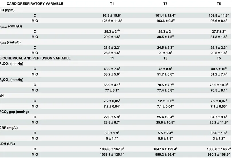
differences noted from T3 and T1when compared to the control group, respectively. There was a significant increase in PPV (Fig 4) from T1 in the MIO group in comparison with the control group. The SVRI (Fig 4) and CVP (Fig 5) were significantly higher in the MIO group from T3 and T5 onwards, respectively.
Respiratory parameters. Ppeakand Pplatincreased in the MIO group (Table 1), although
there were no significant differences compared to the control group.A decrease in Cdynand
increased Rawwere found (Fig 6) with significant differences between the MIO and control
groups.
Fig 5. Central venous pressure (CVP, mmHg) values up to 5 hours after IAH stabilization.(*) Indicates significant differences between the C and MIO groups at the same sampling interval (p<0.05).
doi:10.1371/journal.pone.0148058.g005
Fig 6. Airway resistance (Raw, cmH2O/L.sec) and pulmonary compliance (Cdyn, ml/cmH2O) values up to 2 hours after IAH stabilization.(*) Indicates significant differences between the C and MIO groups at the same sampling interval (p<0.05).
Biochemical and perfusion parameters. With the initiation of IAH, there were no signifi-cant increases or differences in PaCO2, PgCO2or pHiobserved between the MIO and control
groups (Table 1).The PCO2-gap progressively increased from T1whilethe CRP hardly changed
throughout the experiment (Table 1). LDH decreased in the MIO group but not significantly
Fig 8. Peak pressure (Ppeak, cmH2O) and plateau pressure (Pplat, cmH2O) values up to 5 hours after IAH stabilization.(*) Indicates significant differences between the C and MIO groups at the same sampling interval (p<0.05). (†) Indicates significant differences in MIO at either T7, T9 or T11 compared to T1 (p<0.05).
doi:10.1371/journal.pone.0148058.g008
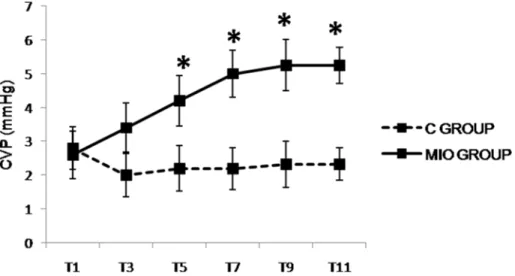
Fig 7. Abdominal perfusion pressure (APP, mmHg) and lactate (Lc, mmol/L) values up to 2 hours after IAH stabilization.(*) Indicates significant differences between the C and MIO groups at the same sampling interval (p<0.05). (†) Indicates significant differences in MIO between T5 compared to T1 (p<0.05).
when compared to the control group (Table 1). Lactate values increased and the APP decreased significantly from T1 (Fig 7) in the MIO group as compared to the control group.
Late study phase (T7 to T11)
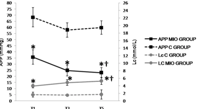
The results obtained from the third to fifth hour in hemodynamic, respiratory, laboratory and blood perfusion parameters showed the same trend as during the first two hours ( Experi-ment1), with the exception of the Ppeakand Pplat(Fig 8) that increased significantly and pHi
(Fig 9) that decreased significantly in the MIO group from T7 when compared to the control group.
Discussion
The porcine model in our study enabled the simulation of a commonly encountered IAH situa-tion, resulting from MIO due to increased colonic intraluminal pressure, despite anatomical differences in the colon between humans and pigs [30,31]. Hemodynamic, respiratory and lab-oratory parameters helped describe the IAH effects on the cardiovascular, pulmonary and renal systems, which are affected when IAP exceeds 18–20 mmHg [32–35].
Hemodynamic changes associated with IAH have previously been well described in humans [32,33,36–39]. According to these studies, increased IAP exerts direct pressure on the inferior cava vein and the heart which in turn decreases preload and venous return (while CVP increases due to increased intrathoracic pressures) resulting in a decline in CI [39,40]. The MAP may initially increase due to blood sequestration from the abdominal vessels (mesenteric capacitance veins), but if sustained, may stabilize and even decrease. Unlike the preload, the after load increases by activation of the renin-angiotensin-aldosterone system causing an increase in the SVRI. In summary IAH reduces cardiac contractility and preload and increases cardiac afterload [40]. In our study, all these effects were observed in the MIO group: CI and MAP decreased (51.4% and 31.7%, respectively), while an increase of 49.4% was obtained in the SVRI 2hours after stabilization of the IAP. These results were consistent with previous studies simulating IAH via fluid instillation into the abdominal cavity [41,42] or a
Fig 9. Gastric intramucosal pH (pHi) values up to 5 hours after IAH stabilization.(*) Indicates significant differences between the C and MIO groups at the same sampling interval (p<0.05).
pneumoperitoneum model [43–47], and showing a decrease in CI and increase in SVRI com-pared to the baseline. With regard to the MAP, although an initial increase has been described [38,41], a significant decrease is a common finding in different IAH models [42–49]. In con-trast to previous works [42–49] where such a decrease was only observed after several hours of increased IAP, we noticed a drop in MAP just after stabilization of IAH. This may be explained by a greater effect of the MIO model on MAP compared to previously studied models. On the other hand, as was already shown in clinical as well as in other experimental studies [41,42,
44–47], CVP increases are common during IAH. Our results revealed significant CVP increases from 2 hours after stabilization of the IAP, similar to that observed in others works with higher IAP [44,45,47]. This rise, as well as the increased observed in the MAP, was more noticeable in those with an IAP of 25 mmHg [41,42,46], indicating a greater effect of the MIO. Our data on functional hemodynamic parameters, such as PPV and SVV, are consistent with previous animal experiments that demonstrated IAH either abolishes or increases thresh-old values for PPV and SVV to predict fluid responsiveness [50–52]. Using porcine models of simulated IAH, these studies demonstrated that PPV, SVV, as well as caudal cava vein flow fluctuations, in relation to positive pressure ventilation, were dependent on IAP [50]. There-fore, higher thresholds may need to be used among those with IAH to indicate fluid respon-siveness [53]. Compared to the normovolemia conditions referred to these previous studies, our results showed increases of around 80% of the PPV at the beginning of IAH.
The respiratory changes seen in clinical studies on intestinal occlusion are thought to be caused by the upward displacement of the diaphragm caused by the increased IAP. The pulmo-nary Cdynas well as the respiratory rate are usually reduced while the Ppeak, Pplatand Raware
increased. Furthermore, atelectasis in the dorsobasal and caudal areas of the lungs has been described because of limited pulmonary inflation and recruitment [33,36,37,54]. In our study an early Cdyndecrease of 56% was observed from the onset of IAH, which is consistent with
previous work where IAP was increased up to 25 mmHg [41,42]. In addition, increases in Raw,
Ppeakand Pplatwere observed from the beginning of IAH in our model. Regardless of the
exper-imental model for IAH of 20–25 mmHg [41,42,46], significant increases in Ppeakand Pplat
have been observed in the period between 1 to 3 hours after commencing the experiment. In addition, those studies where IAP was as high as 30 mmHg after 1 hour of IAH, the Ppeakand
Pplatrose by 50% compared to controls [42,44]. However, in our model these effects only
became significant after 3 hours of IAH.
The anaerobic metabolism occurring in patients with IAH results from changes in the cardiorespiratory dynamics that are determined by a PaCO2increase and PaO2decrease. This
may stimulate lactate production, the endpoint of respiratory and metabolic acidosis [55,56]. In our results PaCO2increased by around 25% which is in keeping with the results found by
Diaz et al [41] and similar to other work using a porcine model of pneumoperitoneum up to 30 mmHg [45,47]. In previous IAH models using 20 mmHg and 30 mmHg [43,44,46,48,49], lactate increases were noted but only became apparent and significant after 3 to 6 hours. Lac-tate levels in our MIO model increased by up to 5 times compared to the control group before 3 hours.
significantly altered soon after IAH stabilization. Compared with the control group, significant decreases in APP of 40% and 61% were observed after the onset and 2 hours of IAH, respec-tively. This rapid and important deleterious effect on perfusion was consistent with other por-cine models at either 20 or 30 mmHg. In ascetic and pneumoperitoneum models of 20 mmHg [48,49], significant decreases in APP were also observed from the beginning of the study. Fur-thermore, in models subjected to 30 mmHg [45,46,48], significant decreases in APP of 30 to 60% at the beginning were also observed, with further decreases up to 80% at 6 h.
The stomach and intestines are among the most sensitive abdominal organs that express the deleterious effects of IAH. In particular, the use of gastric air tonometry has been established as a useful method to estimate PgCO2and pHi[28,29]. IAH related decreases in gastrointestinal
blood flow may cause ischemic events [44,61],and as a result, pHimay decrease [62] in the
early stages of IAH. In this study, pHionly decreased significantly after 3 hours of
experimenta-tion. This is not unusual as effects were even more delayed in a pneumoperitoneum model of 20 mmHg [48] where no significant differences were observed up to 5 hours after commencing. However, in two ascetic models with similar IAP, pHiwas seriously affected and significantly
decreased from the beginning of the study [49] or reduced by more than 4% after 1 hour [42]. Further pHichanges were observed when IAP was increased to 30 mmHg. As a 7% decrease,
and absolute values below 7, were recorded after 30 minutes of IAH [42,48].Thus, the effect of IAP on pHiis influenced by both the method used to achieve the hypertension and the value of
abdominal pressure itself. Some clinical studies found an inverse relation between IAP and pHi
[62,63].
The present study has some limitations. The number of animals studied was relatively small and the time course of events was short (up to a maximum of 5 hours). Future studies could try to determine the effects of improved perfusion, including the effects of abdominal decompres-sion, and how this influences endorgan function. Data collection for esophageal pressure, volu-metric preload or extravascular lung water, as well as measurements of other perfusion parameters, for example indocyanine green plasma disappearance rate, microdialysis and bio-markers such as citrullin, intestinal fatty acid binding protein, N-Gal or cystatin C, could help to clarify the changes.
In conclusion, we found that the present MIO model affecting the large bowel with a com-petent ileocaecal valve may be a useful simulation of human intestinal obstruction scenarios. Hemodynamic, respiratory, laboratory and perfusion alterations were similar to those described previously in IAH and ACS studies both in humans and animals, thus supporting the model studied. The most relevant parameters to evaluate the deleterious effects of IAH are monitoring of APP, Cdyn, pHiand lactate.
Acknowledgments
We acknowledge Minimally Invasive Surgery Centre Jesús Usón’s staff of Cáceres (Spain) and department of Veterinary Anatomy and Embryology at the University of Murcia (Spain) for their assistance in conducting this research.
Author Contributions
References
1. Markogiannakis H, Messaris E, Dardamanis D, Pararas N, Tzertzemelis D, Giannopoulos P, et al. Acute mechanical bowel obstruction: clinical presentation, etiology, management and outcome. World J Gastroenterol. 2007; 13(3):432–7. PMID:17230614
2. Laurell H, Hansson LE, Gunnarsson U. Why do surgeons miss malignancies in patients with acute abdominal pain? Anticancer Res. 2006; 26(5B):3675–8. PMID:17094384
3. Küçük HF, Sikar HE, Uzun H, Tutal F, Kaptanoğlu L, Kurt N. Acute mechanical intestinal obstructions. Ulus Travma Acil Cerrahi Derg. 2010; 16(4):349–52. PMID:20849053
4. Fauci B, Kasper, Hauser, Longo, Jameson, Loscalzo. Harrison. Medicina interna. 17 ed: McGraw Hill; 2008.
5. Lopez-Kostner F, Hool GR, Lavery IC. Management and causes of acute large-bowel obstruction. Surg Clin North Am. 1997; 77(6):1265–90. PMID:9431339
6. Madl C, Druml W. Gastrointestinal disorders of the critically ill. Systemic consequences of ileus. Best Pract Res Clin Gastroenterol. 2003; 17(3):445–56. PMID:12763506
7. Malbrain ML, Chiumello D, Cesana BM, Reintam Blaser A, Starkopf J, Sugrue M, et al. A systematic review and individual patient data meta-analysis on intra-abdominal hypertension in critically ill patients: the wake-up project. World initiative on Abdominal Hypertension Epidemiology, a Unifying Project (WAKE-Up!). Minerva Anestesiol. 2014; 80(3):293–306. PMID:24603146
8. Malbrain ML, Chiumello D, Pelosi P, Bihari D, Innes R, Ranieri VM, et al. Incidence and prognosis of intraabdominal hypertension in a mixed population of critically ill patients: a multiple-center epidemio-logical study. Crit Care Med. 2005; 33(2):315–22. PMID:15699833
9. Kirkpatrick AW, Roberts DJ, De Waele J, Jaeschke R, Malbrain ML, De Keulenaer B, et al. Intra-abdominal hypertension and the Intra-abdominal compartment syndrome: updated consensus definitions and clinical practice guidelines from the World Society of the Abdominal Compartment Syndrome. Intensive Care Med. 2013; 39(7):1190–206. doi:10.1007/s00134-013-2906-zPMID:23673399
10. Cheatham ML, Malbrain ML, Kirkpatrick A, Sugrue M, Parr M, De Waele J, et al. Results from the Inter-national Conference of Experts on Intra-abdominal Hypertension and Abdominal Compartment Syn-drome. II. Recommendations. Intensive Care Med. 2007; 33(6):951–62. PMID:17377769
11. Malbrain ML, Cheatham ML, Kirkpatrick A, Sugrue M, Parr M, De Waele J, et al. Results from the Inter-national Conference of Experts on Intra-abdominal Hypertension and Abdominal Compartment Syn-drome. I. Definitions. Intensive Care Med. 2006; 32(11):1722–32. PMID:16967294
12. Holodinsky JK, Roberts DJ, Ball CG, Blaser AR, Starkopf J, Zygun DA, et al. Risk factors for intra-abdominal hypertension and intra-abdominal compartment syndrome among adult intensive care unit patients: a systematic review and meta-analysis. Crit Care. 2013; 17(5):R249. doi:10.1186/cc13075
PMID:24144138
13. Wu CC, Lu YZ, Wu LL, Yu LC. Role of myosin light chain kinase in intestinal epithelial barrier defects in a rat model of bowel obstruction. BMC Gastroenterol. 2010; 10:39. doi:10.1186/1471-230X-10-39
PMID:20403206
14. Yuan ML, Yang Z, Li YC, Shi LL, Guo JL, Huang YQ, et al. Comparison of different methods of intestinal obstruction in a rat model. World J Gastroenterol. 2013; 19(5):692–705. doi:10.3748/wjg.v19.i5.692
PMID:23430052
15. Zanoni FL, Benabou S, Greco KV, Moreno AC, Cruz JW, Filgueira FP, et al. Mesenteric microcircula-tory dysfunctions and translocation of indigenous bacteria in a rat model of strangulated small bowel obstruction. Clinics (Sao Paulo). 2009; 64(9):911–9.
16. Leite R Junior, Mello NB, Pereira LeP, Takiya CM, Oliveira CA, Schanaider A. Enterocyte ultrastructural alterations following intestinal obstruction in rats. Acta Cir Bras. 2010; 25(1):2–8. PMID:20126879
17. El-Awady SI, El-Nagar M, El-Dakar M, Ragab M, Elnady G. Bacterial translocation in an experimental intestinal obstruction model. C-reactive protein reliability? Acta Cir Bras. 2009; 24(2):98–106. PMID:
19377777
18. Pittner A, Nalos M, Theisen M, Ploner F, Brückner UB, Georgieff M, et al. Inhaling nitrous oxide or xenon does not influence bowel wall energy balance during porcine bowel obstruction. Anesth Analg. 2002; 94(6):1510–6. PMID:12032017
19. Walters EM, Prather RS. Advancing swine models for human health and diseases. Mo Med. 2013; 110 (3):212–5. PMID:23829105
20. Giardino R, Faenza S, Spighi M, Fini M, Giavaresi G, Morrone G, et al. In vivo experimental models on the evaluation of haemoperfusion. Boll Soc Ital Biol Sper. 1993; 69(10):625–32. PMID:8198804
22. Schachtrupp A, Wauters J, Wilmer A. What is the best animal model for ACS? Acta Clin Belg Suppl. 2007(1: ):225–32. PMID:17469725
23. AVMA. Guidelines for the Euthanasia of Animals: 2013 Edition.
24. Correa-Martín L, Castellanos G, García M, Sánchez-Margallo FM. Renal consequences of intraabdom-inal hypertension in a porcine model. Search for the choice indirect technique for intraabdomintraabdom-inal pres-sure meapres-surement. Actas Urol Esp. 2013; 37(5):273–9. doi:10.1016/j.acuro.2012.06.001PMID:
23122948
25. Malbrain ML. Different techniques to measure intra-abdominal pressure (IAP): time for a critical re-appraisal. Intensive Care Med. 2004; 30(3):357–71. PMID:14730376
26. Martín Vivas A, Saboya Sánchez S, Patiño Rodríguez M, Silva Obregón JA, Gómez Rosado S, Blanco
García JJ.Hemodynamic monitoring: PiCCO system. Enferm Intensiva. 2008; 19(3):132–40. PMID:
18840328
27. Cheatham ML, White MW, Sagraves SG, Johnson JL, Block EF. Abdominal perfusion pressure: a superior parameter in the assessment of intra-abdominal hypertension. J Trauma. 2000; 49(4):621–6; discussion 6–7. PMID:11038078
28. de Tomás J TF, Bardina A, Perea J. Utilidad de la tonometría por aire en el diagnóstico de la isquemia intestinal experimental. Cirugía Española; 2001. p. 129–32.
29. Mäkinen M-J HP, Klemola UM, Yli-Hankala A. Gastric air tonometry during laparoscopic cholecystec-tomy: a comparison of two PaCO2 levels. Canadian Journal of Anesthesia; 2000. p. 121–8.
30. Swindle MM, Smith AC. Comparative anatomy and physiology of the pig. Sacand J Lab Anim Sci. 1998; 25.
31. Swindle MM, Makin A, Herron AJ, Clubb FJ, Frazier KS. Swine as models in biomedical research and toxicology testing. Vet Pathol. 2012; 49(2):344–56. doi:10.1177/0300985811402846PMID:21441112
32. Sánchez-Miralles A, Castellanos G, Badenes R, Conejero R. Abdominal compartment syndrome and acute intestinal distress syndrome. Med Intensiva. 2013; 37(2):99–109. doi:10.1016/j.medin.2011.11. 019PMID:22244213
33. Deenichin GP. Abdominal compartment syndrome. Surg Today. 2008; 38(1):5–19. PMID:18085356
34. de Laet IE, Malbrain M. Current insights in intra-abdominal hypertension and abdominal compartment syndrome. Med Intensiva. 2007; 31(2):88–99. PMID:17433187
35. Malbrain M. Abdominal compartment syndrome. F1000 Med Rep. 2009; 1.
36. Piacentini E, Ferrer Pereto C. Intraabdominal hypertension and abdominal compartment syndrome. Enferm Infecc Microbiol Clin. 2010; 28 Suppl 2:2–10. doi:10.1016/S0213-005X(10)70024-0PMID:
21130924
37. Lee RK. Intra-abdominal hypertension and abdominal compartment syndrome: a comprehensive over-view. Crit Care Nurse. 2012; 32(1):19–31. doi:10.4037/ccn2012662PMID:22298715
38. Malbrain ML, Vidts W, Ravyts M, De Laet I, De Waele J. Acute intestinal distress syndrome: the impor-tance of intra-abdominal pressure. Minerva Anestesiol. 2008; 74(11):657–73. PMID:18636062
39. Cheatham ML, Malbrain ML. Cardiovascular implications of abdominal compartment syndrome. Acta Clin Belg Suppl. 2007(1: ):98–112. PMID:17469707
40. Ameloot K, Gillebert C, Desie N, Malbrain ML. Hypoperfusion, shock states, and abdominal compart-ment syndrome (ACS). Surg Clin North Am. 2012; 92(2):207–20, vii. doi:10.1016/j.suc.2012.01.009
PMID:22414408
41. Díaz F DA, Carvajal C, Salomon T, Torres MF, Erranz B, Cruces P. Consecuencias hemodinámicas y respiratorias del síndrome compartimental abdominal en un modelo experimental.2012; 83(5):454–
61 pp.
42. Pattillo JC RC, Storaker M, Anastasiadis Z, Llanos O, Urenda J, López F et al. Desarrollo de un modelo experimental de hipertensión intra-abdominal. Revista Chilena de Medicina Intensiva. 2004:7–12. 43. Gudmundsson FF, Gislason HG, Dicko A, Horn A, Viste A, Grong K, et al. Effects of prolonged
increased intra-abdominal pressure on gastrointestinal blood flow in pigs. Surg Endosc. 2001; 15 (8):854–60. PMID:11443466
44. Toens C, Schachtrupp A, Hoer J, Junge K, Klosterhalfen B, Schumpelick V. A porcine model of the abdominal compartment syndrome. Shock. 2002; 18(4):316–21. PMID:12392274
45. Kaussen T, Srinivasan PK, Afify M, Herweg C, Tolba R, Conze J, et al. Influence of two different levels of intra-abdominal hypertension on bacterial translocation in a porcine model. Ann Intensive Care. 2012; 2 Suppl 1:S17. doi:10.1186/2110-5820-2-S1-S17PMID:22873417
47. Otto J, Afify M, Jautz U, Schumpelick V, Tolba R, Schachtrupp A. Histomorphologic and ultrastructural lesions of the pancreas in a porcine model of intra-abdominal hypertension. Shock. 2010; 33(6):639–
45. doi:10.1097/SHK.0b013e3181cb8be0PMID:19940813
48. Correa-Martín L, Castellanos G, García-Lindo M, Díaz-Güemes I, Sánchez-Margallo FM. Tonometry as a predictor of inadequate splanchnic perfusion in an intra-abdominal hypertension animal model. J Surg Res. 2013; 184(2):1028–34. doi:10.1016/j.jss.2013.04.041PMID:23688792
49. Correa-Martín L, Castellanos G, García-Lindo M, Díaz-Güemes I, Piñero A, Sánchez-Margallo FM.
Intra-abdominal hypertension: Effects on the splanchnic circulation. Preliminary study in a model of ascites. Gastroenterol Hepatol. 2014; 37(2):51–7. doi:10.1016/j.gastrohep.2013.08.002PMID:
24238726
50. Duperret S, Lhuillier F, Piriou V, Vivier E, Metton O, Branche P, et al. Increased intra-abdominal pres-sure affects respiratory variations in arterial prespres-sure in normovolaemic and hypovolaemic mechani-cally ventilated healthy pigs. Intensive Care Med. 2007; 33(1):163–71. PMID:17102964
51. Renner J, Gruenewald M, Quaden R, Hanss R, Meybohm P, Steinfath M, et al. Influence of increased intra-abdominal pressure on fluid responsiveness predicted by pulse pressure variation and stroke vol-ume variation in a porcine model. Crit Care Med. 2009; 37(2):650–8. doi:10.1097/CCM.
0b013e3181959864PMID:19114894
52. Jacques D, Bendjelid K, Duperret S, Colling J, Piriou V, Viale JP. Pulse pressure variation and stroke volume variation during increased intra-abdominal pressure: an experimental study. Crit Care. 2011; 15(1):R33. doi:10.1186/cc9980PMID:21247472
53. Malbrain ML, de Laet I. Functional hemodynamics and increased intra-abdominal pressure: same thresholds for different conditions. . .? Crit Care Med. 2009; 37(2):781–3. doi:10.1097/CCM.
0b013e318194c397PMID:19325388
54. Pelosi P, Quintel M, Malbrain ML. Effect of intra-abdominal pressure on respiratory mechanics. Acta Clin Belg Suppl. 2007(1: ):78–88. PMID:17469705
55. Baigorri-González F. LJ. Oxigenación tisular y Sepsis2005; 29(3), 178–84 pp.
56. van Noord D, Mensink PB, de Knegt RJ, Ouwendijk M, Francke J, van Vuuren AJ, et al. Serum markers and intestinal mucosal injury in chronic gastrointestinal ischemia. Dig Dis Sci. 2011; 56(2):506–12. doi:
10.1007/s10620-010-1303-5PMID:20628816
57. Malbrain ML, De laet IE. Intra-abdominal hypertension: evolving concepts. Clin Chest Med. 2009; 30 (1):45–70, viii. doi:10.1016/j.ccm.2008.09.003PMID:19186280
58. Diebel LN, Wilson RF, Dulchavsky SA, Saxe J. Effect of increased intra-abdominal pressure on hepatic arterial, portal venous, and hepatic microcirculatory blood flow. J Trauma. 1992; 33(2):279–82; discus-sion 82–3. PMID:1507294
59. Diebel L, Saxe J, Dulchavsky S. Effect of intra-abdominal pressure on abdominal wall blood flow. Am Surg. 1992; 58(9):573–5; discussion 5–6. PMID:1388005
60. Malbrain ML. Abdominal perfusion pressure as a prognostic marker in intra-abdominal hypertension. Berlin: Springer-Verlag; 2002.
61. Schachtrupp A, Toens C, Hoer J, Klosterhalfen B, Lawong AG, Schumpelick V. A 24-h pneumoperito-neum leads to multiple organ impairment in a porcine model. J Surg Res. 2002; 106(1):37–45. PMID:
12127806
62. Sugrue M, Jones F, Lee A, Buist MD, Deane S, Bauman A, et al. Intraabdominal pressure and gastric intramucosal pH: is there an association? World J Surg. 1996; 20(8):988–91. PMID:8798353
63. Balogh Z, McKinley BA, Cocanour CS, Kozar RA, Valdivia A, Sailors RM, et al. Supranormal trauma resuscitation causes more cases of abdominal compartment syndrome. Arch Surg. 2003; 138(6):637–