Liliana Mónica Santos Capinha
Licenciada em Biologia Humana
Interplay between Rac1b and Sodium
Iodide symporter expression in thyroid
and breast cancers
Dissertação para obtenção do Grau de Mestre em Genética
Molecular e Biomedicina
Orientador: Doutora Ana Luísa Silva, Instituto Português
de Oncologia de Lisboa Francisco Gentil
Júri:
Presidente: Prof. Doutora Paula Maria Theriaga Mendes Bernardo Gonçalves Arguente: Doutora Vânia Marina Cristovão Gonçalves
Setembro 2015
I
Interplay between RAC1b and Sodium Iodide symporter in thyroid and breast cancers.Copyright © reserved to Liliana Mónica Santos Capinha, FCT/UNL, UNL
The Faculty of Science and Technology and the New University of Lisbon have the perpetual right, and without geographical limits, to archive and publish this dissertation through press copies in paper or digital form, or by other known form or any other that will be invented, and to divulgate it through scientific repositories and to admit its copy and distribution with educational or research objectives, non-commercial, as long as it is given credit to the author and editor.
A Faculdade de Ciências e Tecnologia e a Universidade Nova de Lisboa têm o direito, perpétuo e sem limites geográficos, de arquivar e publicar esta dissertação através de exemplares impressos reproduzidos em papel ou de forma digital, ou por qualquer outro meio conhecido ou que venha a ser inventado, e de a divulgar através de repositórios científicos e de admitir a sua cópia e distribuição com objectivos educacionais ou de investigação, não comerciais, desde que seja dado crédito ao autor e editor.
III
An arrow can only be shot by pulling it backwards. So when life is dragging you back with difficulties, it means that it’s going to launch you into something great.V
Agradecimentos
Chegando ao fim desta etapa, quero agradecer às inúmeras pessoas que de uma forma ou de outra me ajudaram e ensinaram a crescer.
Em primeiro lugar, um especial agradecimento à Doutora Ana Luísa Silva, minha orientadora, por me ter recebido no grupo de Endocrinologia do UIPM-IPO. Conseguiu sempre transformar o que poderia parecer Rocket Science em algo simples e interessante. Obrigada por todos os conhecimentos transmitidos, por toda a paciência e boa disposição contagiante.
Um muito obrigado à Márcia, por todo o acompanhamento, ajuda, dicas e apoio! Sem ti, não teria sido possível.
À Doutora Branco Cavaco pelo voto de confiança. Obrigada pela oportunidade e simpatia. Obrigada às pessoas fantásticas que conheci no CIPM; Inês Marques, pela amizade e motivação nos dias menos bons, Sofia Nunes e Teresa Matias. Margarida Moura, obrigada por toda a ajuda e amabilidade! À Rita Domingues, às “LHOs” que tornaram a estadia no CIPM sempre tão agradável e divertida.
Obrigada a quem me tem acompanhado nestes últimos anos da minha vida académica: David Francisco, Francisca Pereira, Mariana Dias. Pelos momentos de boa disposição assim como os das palestras de estudo. À Filipa Ferreira, por ser incansável, em tudo. Pela amizade, compreensão, e imensa ajuda nesta fase.
À Gui, por ser o buffer da minha vida e ainda por ser a irmã que nunca tive. Obrigada pelo apoio e força nos momentos de break-down. Pelas repetidas conversas durante os mil Km que percorremos juntas este ano - Tarefas difíceis para pessoas capazes. Obrigada por isto e por tudo.
Ao meu irmão que sempre foi, para mim, um exemplo a seguir. Obrigada por tudo, devo a ti muito do que sou hoje.
E por último aos meus pais. Ao meu pai, que mesmo longe, esteve sempre perto. Por ter feito tudo para me proporcionar aquilo que não lhe pode ser dado. A minha mãe, por ser a melhor pessoa que conheço e me apoiar incondicionalmente. Obrigada por me terem deixado sempre escolher o meu rumo, pela confiança e palavras de coragem. Espero um dia poder compensar-vos em 1/10 do que fizeram por mim. Sei que não é meu hábito fazer demonstrações de afeto mas... Amo-vos muito .
VII
Abstract
Rac1b, an alternative isoform of the small GTPase RAC1, has recently be shown to be present in thyroid tissue and overexpressed in thyroid cancer cells, particularly in a subset of papillary thyroid carcinomas carrying the activating mutation BRAFV600E that are associated with an unfavorable
outcome. On the other hand, RAC1 seems to be involved in the upregulation of NIS, the glycoprotein responsible for iodide uptake that allows the use of 131 I as a diagnostic and therapeutic tool, in thyroid cancer. However, NIS expression levels and iodine uptake in thyroid cancer cells are reduced when compared to normal tissue. Also, B-Raf V600E mutation has been shown to correlate with a lower expression of NIS. RAC1b overexpression has also been documented in breast cancer. This hyperactivatable variant was shown to be able to compete with and inhibit RAC1 endogenous activity in several signaling pathways. Breast carcinomas also express NIS but at levels too low to warrant treatment with 131I. Thus, in order to understand the regulatory mechanisms of NIS expression we aimed to evaluate the balance of RAC1/1b effect in NIS mRNA expression in follicular cell derived thyroid tumor samples, as well as, in a cell line derived from normal thyroid and in breast cancer cell lines. Understanding the necessary switch to increase NIS expression in cancer cells, would open a new window of opportunity to fight thyroid tumor resistance to radioiodine therapy and develop and possible treatment by the radiodide uptake therapy in breast cancer in a selective way.
IX
Resumo
A proteína Rac1b é uma variante hiperativavel da pequena GTPase RAC1, tendo sido recentemente demonstrada a sua sobreexpressão em carcinomas da tiróide, em particular num subgrupo de carcinomas papilares da tiróide positivos para a mutação BRAFV600E que apresentam uma
progressão clínica da doença mais desfavorável. Por outro lado, RAC1 parece estar envolvida no estímulo da expressão de NIS, a glicoproteína responsável pela entrada de iodo nas células que permite a utilização de iodo 131 como diagnóstico e terapêutica em carcinomas da tiróide. No entanto, os níveis de expressão do NIS e entrada de iodo nas lesões malignas são reduzidos comparativamente ao tecido normal. Verificou-se também uma relação entre a mutação V600E em BRAF e menores níveis de expressão de NIS. A sobrexpressão de RAC1b foi igualmente documentada em cancro da mama. Esta variante hiperativavel mostrou ser capaz de competir e inibir a atividade endógena de RAC1 em diversas vias de sinalização. Carcinomas da mama também apresentam expressão de NIS, todavia em níveis demasiado baixos para viabilizar o tratamento com iodo 131. Assim de forma a compreender os processos regulatórios associados a expressão de NIS em tumores propusemo-nos a avaliar de que forma o balanço RAC1/1b poderá contribuir para a expressão de NIS em tumores da tiróide derivados de células foliculares, assim como em linhas celulares de tiróide normal e de carcinoma da mama. A compreensão de um mecanismo regulatório que permita o aumento dos níveis de NIS nas células cancerígenas poderá abrir uma nova janela de oportunidade para combater a resistência à terapêutica com iodo radioativo e desenvolver um possível tratamento através da incorporação de iodo radioativo em carcinoma de mama de uma forma seletiva.
XI
Table of Contents
Agradecimentos V Abstract VII Resumo IX Table of Contents XIList of Figures XIII
List of Tables XV
List of abbreviations XVII
I- Introduction
1. Cancer 1
2. Thyroid Gland 2
3. Thyroid Neoplasia 2
3.1 Papillary Thyroid Carcinoma 4
3.2 Follicular Thyroid Carcinoma 4
4. RAC1: a member of the RAS superfamily of small GTPases 5
4.1 RAC1b, an hyper-activatable splicing variant of RAC1 6
5. The Sodium/Iodide Symporter (NIS) 8
5.1 NIS biochemical characterization 8
5.2 NIS functional characterization 9
5.3 NIS regulation 9
5.3.1 Transcriptional regulation of NIS 10
5.3.2 Posttranscriptional regulation of NIS 10
5.3.3 Molecular Pathways modulating NIS expression 11
5.3.3.1 PI3K 11
5.3.3.2 BRAF 12
5.3.3.3 RAC1 12
5.3.4 RAC1/RAC1b and NIS 13
6. Iodide Therapies 13
6.1 Thyroid Cancer 13
6.2 Iodide related therapies in non-thyroid tissues 14
7. Aim of the project 15
II - Material and Methods 1. Patient samples 15
2. Cell culture 16
XII
4. RNA extraction and cDNA synthesis 16
5. RT-PCR 17
6. qRT-PCR 17
7. Protein extracts, SDS-PAGE and Western blotting 17
8. Statistical Analysis 18
III - Results 1. NIS expression levels in RAC1b-positive and RAC1b-negative follicular cell derived thyroid carcinomas 19
2. NIS expression in cell lines 20
2.1 Effect of RAC1/1b in NIS expression in Nthy-ori 3-1 20
2.2 Effect of RAC1/1b in NIS expression in MDA-MB-231 21
2.3 RAC1 effect on NIS expression levels in MCF-7 23
IV - Discussion 25
V – References 29
VI – Appendix 41
XIII
List of Figures
Figure I.1 - Thyroid gland 2
Figure I.2 -Thyroid Cancer Prevalence 3
Figure I.3 - Alternative splicing of the small GTPase Rac1 in colorectal cells 6
Figure I.4 - Iodide uptake function of NIS 8
Figure III.1 - NIS expression levels in RAC1b‐positive and RAC1b‐negative PTCs and FTCs. 19
Figure III.2 - Regulation of NIS expression by RAC1 and RAC1b in N-thyroid cells A and B, effects of RAC1 and RAC1b (both L61 and WT) on NIS expression 20
Figure III.3 - RAC1/1b protein expression in Nthy-ori 3-1 transfected cells 21
Figure III.4 - Regulation of NIS expression by RAC1 and RAC1b in MDA-MB-231 cells A and B, effects of RAC1 and RAC1b (both L61 and WT) on NIS expression. 22
Figure III.5 - RAC1/1b protein expression in MDA-MB-231 transfected cells. 22
Figure III.6 - Regulation of NIS expression by RAC1 and RAC1b in MCF-7 cells A and B, effects of RAC1 and RAC1b (both L61 and WT) on NIS expression. 23
XIV
XV
List of Tables
Supplementary Table 1 - Conditions of PCR amplification of NIS cDNA 43
Supplementary Table 2 – Final concentrations for RIPA buffer 43 Supplementary Table 3 - Final concentrations for Western Blotting 43
XVII
List of abbreviations
AA – Aminoacid Ab – Anti-body
AKT – Protein kinase B
ASF/SF2 – Serine/Arginine-Rich Splicing Factor 1 ATC – Anaplastic thyroid carcinoma
BRAF- v-raf murine sarcoma viral oncogene homolog B1 cAMP – Cyclic adenosine monophastase
DNA – Deoxyribonucleic acid
DMEM - Modification of Basal Medium Eagle EMT – Epithelial-mesenchymal transition ERK – Extracellular signal-regulated kinase ESEs – Exonic splicing silencers
ESSs – Intronic splicing silencers FA – Follicular Adenomas FBS – Fetal Bovine Serum FTC – Follicular thyroid carcinoma GAP – GTPase-activating protein GDP – Guanosine diphosphate
GEF – Guanine nucleotide exchange factor GTP – Guanosine triphosphate
IGF- I - Insulin-like growth factor-I ISEs – Intronic splicing silencers
MAPK – Mitogen-activated protein kinase MEK – MAPK/ERK kinase
MMP-3 – Matrix metalloproteinase-3 mRNA – messenger Ribonucleic Acid NF-kB – Nuclear Factor-Kappa Beta NIS – Sodium/iodide symporter
Nkx2.1 – NK2 homebox 1 transcription factor NUE – Nis upstream enchancer
XVIII
Pax-8 – Paired box 8PCR – Polymerase Chain Reaction
PDTC – Poorly differentiated thyroid carcinoma PI3K – Phosphoinositide 3-kinase
PTC – Papillary thyroid carcinoma
RAS – Rat sarcoma viral oncogene homolog RAR – Retinoic Acid Receptor
RET – Rearranged during transfection Gene RNA – Ribonucleic Acid
RIPA - Radio Immunoprecipitation Assay
ROS – Reactive Oxygen especies
RPMI - Roswell Park Memorial Institute medium RXR – Retinoid X Receptor SR – Serine-Rich T3 – L-triiodothyronine T4 – L-thyroxide TC – Thyroid Cancer Tg – Thyroglobulin TPO – Thyroperoxidase tRA – Trans Retinoic Acid
TSH – Thyroid stimulating hormone V - Volume
W – Weigh
WDTC – Well-differentiated thyroid carcinoma WHO – World Health Organization
1
I – Introduction
1. Cancer
Cancer remains one of the most complex diseases affecting humans in our century (Grizzi et
al., 2006). After decades of research, several biological and biochemical processes have been revealed,
providing a start point to better understand the genesis of this malignancy and also to improve its treatment.
According to WHO (World Health Organization, 2012) cancer is characterized by an abnormal growth of cells and it can develop from any cell in the body and spread to other organs. In 2012, there was a total of 14,1 million new cancer cases, 8.2 million cancer deaths and 32,6 million people living with cancer (within 5 years of diagnosis) worldwide.
A normal, healthy body comprises over 30 trillion cells in a complex, interdependent agglomerate, regulating one another’s proliferation. Indeed, normal cells reproduce only when instructed to do so by other surrounding cells which are being choreographed by regulatory genes. Such collaboration enables the maintenance of the proper size and architecture of the organism needs (Weinberg et al., 1996).
Somatic mutations found in cancer genomes may be the consequence of the intrinsic slight infidelity of the DNA replication machinery, exogenous or endogenous mutagen exposures, enzymatic modification of DNA, or defective DNA repair.
The complexity of tumor pathways have been studied over the years; a set of hallmarks have been proposed as a comprehensive and also simplified look at this complex disease, displaying an active role of the recruited normal cells in tumorigenesis rather than simple passive spectators.
The main alterations in cell physiology proposed included sustaining proliferative signaling, insensitivity to growth inhibition, resistance to cell death, replicative immortality, angiogenesis and tissue invasion (metastasis) (Hanahan & Weinberg, 2011; Seyfried & Shelton, 2010). Additionally, two other important emerging hallmarks in cancer recently raised were tumor reprogramming of energy metabolism and the ability to evade the immune system.
Two mechanisms have been proposed as the main inducers of the cancer hallmarks: genomic/chromosomal instability and mutation, which generates random mutations at high frequency; tumor-promoting inflammation, allowing the recruitment of important molecules for the tumoral microenvironment (Hanahan and Weinberg, 2011). Malignant transformation is always conditioned by tumor microenvironment, where the tumor and surrounding cells act as a functional network (Serpa & Dias, 2011).
2
2. Thyroid Gland
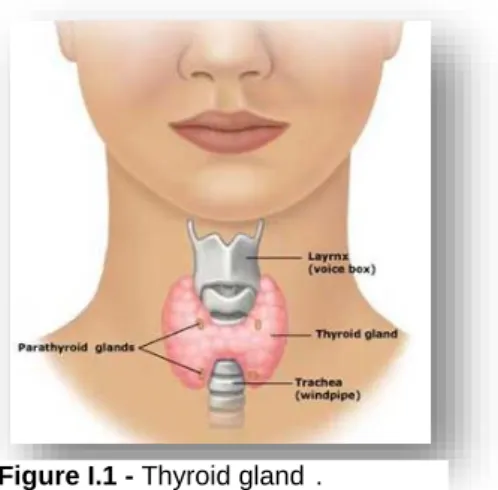
The thyroid gland is the main organ of the endocrine system. Embryologically, the thyroid anlage begins as a bilobed vesicular structure at the foramen cecum of the tongue and descends as a component of the thyroglossal duct to reach its definitive position in the anterior aspect of the trachea below the cricoid cartilage in the neck (Figure I.1). It is supplied by major arteries (Camacho et al., 2011), has also a rich lymphatic network and is composed of two lobes joined by an isthmus from which, in a significant number of normal individuals, the “pyramidal lobe” extends upward. The adult gland varies in size and appearance according to functional activity, gender, hormonal status, and iodine intake (Muro-Cacho & Ku, 2000).
This gland is composed of two distinct hormone-producing cell types: the follicular and the parafollicular cells. These last parafollicular, or C cells, are dedicated to the production of the calcium-regulating hormone calcitonin. Follicular cells comprise most of the epithelium and are responsible for iodine uptake and thyroid hormone synthesis;
The principal secretory products of the thyroid gland are L-triiodothyronine (T3) and L-thyroxide (T4), the only iodinecontaining hormones in vertebrates. These hormones are essential for growth, development and survival of vertebrates, since they regulate vital organism functions, such as body temperature, cardiac frequency, arterial pressure and intestinal function (Muro-Cacho & Ku, 2000). Thyroglobulin (Tg), thyroperoxidase (TPO), sodium/iodide symporter (NIS), and TSH receptor (TSHr) are genes necessary for the synthesis of such hormones which takes place in the fully differentiated thyroid cell, called the thyrocite function (Nitsch et al., 2010; Fagman & Nilsson, 2011, Damante & Lauro, 1994. Because I- is an essential constituent of T3 and T4, both thyroid function and its systemic ramifications depend on an adequate supply of I to the gland. In turn, this supply, depends on sufficient dietary intake of I and proper function of the membrane symporter, NIS, responsible for this ion uptake (Doan et al., 2003).
3. Thyroid Neoplasia
Similar to all other organs of the body, the thyroid gland also exhibits a variety of developmental and acquired diseases.
According to the WHO (2012), thyroid malignancies are classified as carcinomas, which are by far the most common thyroid malignancies, sarcomas, lymphomas and even less frequent tumors including metastases to the thyroid (Gimm, 2000).
Figure I.1 - Thyroid gland .
(http://www.medical1stop.com/overac tive-thyroid-gland-hyperthyroidism-symptoms causes-and-treatment/)
3
Thyroid cancer (TC) is the most common endocrine cancer, and its incidence has increased continuously over the last three decades worldwide. Although thyroid cancer is perceived to have a high survival rate, the sustained increase in thyroid carcinoma incidence worldwide has recently attracted considerable attention. Notwithstanding the good prognosis, a small portion of patients show recurrence and distant metastasis (Pak et al., 2015).Despite the significant increase in its incidence, mortality for TC has not increased in equal measure. It appears two times more in female subjects, with a rate of the annual mortality between 0.4– 2.8 and 0.2–1.2/100.000for women and men, respectively (Edwards, 2005).
Factors involved in the etiology of thyroid cancer include hormonal imbalance, radiation exposure, iodine deficient diets, or other environmental factors. Genetic alterations, at germinal or somatic level, also have an important role in thyroid carcinogenesis (DeLellis, 2006).
Thyroid cancer typically occurs in thyroid nodules, which are common and can be detected by palpation and imaging in a large proportion of adults, particularly in the elderly individuals (Nikiforov, 2004).
The thyroid gland contains two major types of epithelial cells - follicular and parafollicular or C cells. Therefore, Thyroid tumors may arise from these two main types of cells. Parafollicular-derived tumors represent only 3 to 5% of cases and are designated as medullary thyroid carcinomas (Nikiforov & Nikiforova, 2011).
Tumors arising from follicular thyroid cells may be benign forms, designated as follicular adenomas (FA). Based on morphological and clinical features, malignant tumors are subdivided into well-differentiated thyroid carcinomas (WDTC), poorly differentiated carcinoma (PDTC) and anaplastic carcinomas (ATC). As such, it has been suggested that follicular thyroid tumors represent a classical tumorigenesis model, where a unique epithelial cell may originate several types of tumors, each with distinctive clinico-pathological features (DeLellis, 2006).
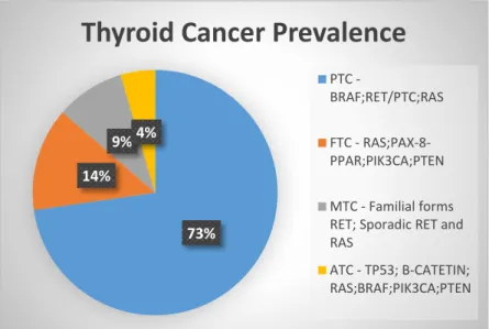
73% 14%
9%4%
Thyroid Cancer Prevalence
PTC
-BRAF;RET/PTC;RAS FTC - RAS;PAX-8-PPAR;PIK3CA;PTEN MTC - Familial forms RET; Sporadic RET and RAS
ATC - TP53; B-CATETIN; RAS;BRAF;PIK3CA;PTEN
Figure I.2 – Thyroid Cancer Prevalence worldwide and their respectively
4
The present study focused essentially in WDTC. This class of carcinoma is defined as deriving from the follicular epithelium and retaining basic biological characteristics of healthy thyroid tissue, including expression of the sodium iodide symporter (NIS), the key cellular feature for specific iodine uptake. In most cases WDTC share a relatively indolent natural history and good responsiveness to surgery and radioiodine (Muro-Cacho & Ku, 2000). This class is composed of two major tumor subtypes, the Papillary thyroid carcinoma (PTC) and the Follicular thyroid carcinoma (FTC).
3.1 Papillary thyroid carcinoma (PTC)
PTC is defined as a malignant epithelial tumor that shows evidence of follicular cell differentiation and that is characterized by a set of distinctive nuclear features. PTC is the most common thyroid carcinoma (about 90% of the cases), its prevalence is higher in patients between 20 and 50 year of age with a female to male ratio of 4–5:1. The incidence of thyroid cancer has increased from 3.6 to 8.7 per 100,000 population between 1973 and 2000, primarily due to the increased detection of small (subclinical) papillary carcinomas (DeLellis, 2006).
PTC is mainly characterized by the distinctive nuclei, which are overlapped, larger than normal, present inclusions or grooves and have an empty appearance. Metastases are commonly spread to lymph nodes and less frequently disseminated through the blood (Muro-Cacho and Ku, 2000).
A variety of different genetic alterations, including rearrangements and point mutations have been implicated in the development of PTC. Targets of these genetic events include RET, BRAF and
RAS (point mutations) (Moura et al., 2011) (Figure I.2). In general, rearrangements have been linked
with radiation exposure while the origin of point mutations has remained unknown.
Regarding to RET, BRAF, and RAS alterations it is in all commonly involved with signaling along with the MAPK pathway which is involved in signaling from a variety of growth factors and cell surface receptors. Mutations or rearrangements of these genes are present in approximately 70% of PTCs and they rarely overlap in the same tumor (DeLellis, 2006).
3.2 Follicular Thyroid Carcinoma (FTC)
In the respective chapter of the WHO Classification of Tumors of Endocrine Organs, follicular thyroid carcinoma (FTC) is defined as ‘A malignant epithelial tumor showing follicular cell differentiation and lacking the diagnostic nuclear features of papillary thyroid carcinoma (PTC) (WHO).
FTC accounts for 10% of all cases of thyroid malignancy in iodine-sufficient areas and 25%– 40% of thyroid malignancies in areas of iodine deficiency (D’Avanzo, 2004). Conventional follicular carcinomas hardly ever involve regional lymph nodes and have distant metastases, most commonly to the lungs and bones (10–20% of cases). Follicular carcinoma has microscopic features that are similar to a follicular adenoma. However, a follicular carcinoma tends to be more cellular with a thick irregular capsule, and often with areas of necrosis and more frequent mitoses (McHenry, 2011).Follicular
5
carcinoma typically presents as a solitary “cold” nodule without cervical adenopathy or signs of hyperthyroidism (Muro-Cacho & Ku, 2000).FTCs have been known to harbor activating point mutations in RAS genes (H-RAS, K-RAS, and
N-RAS). Somatic missense mutations in codons 12/13 and 61 of one of these three RAS genes have
been found in 18-52% of FTC, which lead to constitutive activation of downstream signaling pathways. This group encode highly related 21-kDa proteins located at the inner surface of the cell membrane and has a key role in the transduction of signals from tyrosine kinase and G protein-coupled receptors
(Nikiforova, 2003). A recent study highlights this hypothesis, given the use of Selumetinib, a MAPK pathway inhibitor, as co-adjuvant in iodide therapy, showing an increase in NIS expression and iodide uptake in a mouse model of thyroid cancer (Ho et al., 2013).
4. RAC1: a member of the Ras superfamily of small GTPases
The RAS (for rat sarcoma virus) superfamily of small guanosine triphosphatases (GTPases) comprises over 150 members (Ras super family) and was first discovered as the transforming genes of rat-derived Harvey and Kirsten murine sarcoma retrovirus in the early 1980s (Hankins, 1981). Mutations in the cellular RAS genes, which may alter the structure of RAS protein or even increase this gene expression levels, were thereafter demonstrated in human tumor cell lines, suggesting a role in growth and development, since their alterations lead to uncontrolled growth (Madaule, 1985).
This superfamily is divided into five major classes, accordingly with their sequence and function similarities: Ras, Rho, Rab, Ran and Arf.Regarding to the different classes, the Ras are generally related to cell proliferation, differentiation, morphology and apoptosis. TheRho family is involved in signaling networks that regulate actin, cell cycle progression and gene expression, influence cytoskeletal organization (Heasman & Ridley, 2008) and cell polarity (Park & Bi, 2007) as well as hematopoiesis. The Rab family, being the largest of the Ras superfamily, is involved in vesicular cargo trafficking between different organelles via endocytic and secretory pathways, facilitating budding from the donor compartment, transport to acceptors, vesicle fusion and cargo release. The Ran family, on the other hand, has only one member present in all eukaryotes, except for plants, being the most abundant in the cell and influence nuclear transport. Finally, the Arf protein family is also related to vesicle trafficking (Wennerberg et al., 2005).
All of these members function as signaling nodes that are activated by different extracellular stimuli and regulate intracellular signaling. These signaling control gene transcription which ultimately determine crucial processes such as cell growth and differentiation (Rojas et al., 2012).
The Ras superfamily are small GTP-binding proteins, with a common enzymatic activity. Members of this superfamily perform a general switch function that is based on active GTP-bound state, and an inactive GDP-bound state. The transition between the active and inactive states occurs in a cyclic process. In both inactive and active states, the proteins of the GTPase superfamily have a specific
6
affinity to other signaling proteins that are upstream or downstream parts of the reaction chain (Krauss & Haucke, 2014).
In this study we focused on RAC1, a member of the Rho GTPases subfamily. RAC1 is one of the members of the Rho family of small GTPases. Rho stands for Ras homologous and like Ras proteins, functions as molecular switches; they are 'ON' in the GTPbound state and 'OFF' in the GDP-bound state. The switching between the active and inactive forms is controlled by several accessory proteins: the guanine nucleotide exchange factors (GEFs), GTPase-activating proteins (GAPs) and GDP-dissociation inhibitory factors. (Symons, 1996). RAC1 is a key intervenient in a wide variety of cellular processes, comprising actin remodeling, cell migration and cell cycle progression. (Ridley et al., 2003, Vega and Ridley, 2008). It is also related with cancer, including anchorage-independent growth, cell transformation, survival and invasion (Cerezo et al. 2009, Bid et al., 2013, Moshfegh et al., 2014). Under normal conditions RAC1 activity is controlled by the opposing activities of GEFs (which exchange GDP for GTP and activate RAC1), and GAP (which stimulate the conversion of bound GTP to GDP and inactivate RAC1) (Ridley, 2001).
4.1
RAC1b, a hyper-activatable splicing variant of RAC1
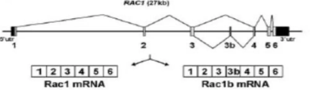
In 1999, the Rac1 spliced isoform, Rac1b, was described by Jordan and colleagues. This intriguing isoform, results from an in-frame insertion of 19 amino acid residues immediately C-terminal to the “switch II” domain (residues 60-76) within the reading frame of the GTPase, due to its 57 additional nucleotides (Matos et al., 2000).These extra nucleotides result from an alternative splicing mechanism, carried out by the spliceosome, a massive structure and a wide number of auxiliary proteins cooperating to accurately recognize the splice sites and catalyze de splicing reaction (Zhou et al., 2013).
Figure I.3 - Alternative splicing of the small GTPase Rac1 in colorectal cells. Schematic representation of the human RAC1 gene with its seven coding exons, including the alternative exon 3b, and of the two alternative transcripts Rac1 and Rac1b (from Gonçalves et al., 2009).
7
The decision of which exon should be removed and which should be included, involves RNA sequence elements and protein regulators. Depending on different characteristics, such as position and function of the cis-regulatory elements, this mechanism can be divided into four classes: Exonic splicing enhancers (ESEs), exonic splicing silencers (ESSs), intronic splicing enhanchers (ISEs) and intronic splicing silencers (ISSs) (Chen et al., 2012). In the current case, Gonçalves et al. (2009), demonstrated that RAC1b splicing occurs through the regulation of two antagonistic splicing factors (SR proteins), ASF/SF2 and SRp20, in colorectal tumor cells, which elucidate us for the need of a concert of different signaling pathways to provide the correct combination to regulate alternative splicing of small GTPase Rac1.Comparably to Rac1, Rac1b presents its self predominantly in a GTP-bound active form, due to an accelerated GDP/GTP exchange activity, a lower rate of GDP/GTP hydrolysis and its impairment in interaction with RhoGDI (Matos et al., 2003).
These features render this protein predominantly in its active state, leading to several alterations in cells. In fact, it has been shown that this spliced isoform is a potent inducer of cyclin D1 transcription and transformation of cells through NF-Kb pathway, responsible for apoptotic response and cell cycle progression regulation (Matos et al., 2003). In fibroblasts, the expression of RAC1b, seems to stimulate cell-cycle progression and survival in serum starvation conditions. (Matos et al., 2005). A role of RAC1b has also been discovered in MMP-3 epithelial to mesenchymal transition (EMT) in cultured cells, when inducing Reactive Oxygen Species (ROS) (Radisky, 2005). High levels of cellular ROS causes oxidative damage and induce genomic instability, stimulating carcinogenesis. In the same year, Esufali et al. reported that Rac1b promotes an effect on the Wnt signaling, a pathway often deregulated in colon cancer. Recent papers have corroborated results from about two decades ago, suggesting an oncogenic role for Rac1b, when upregulated in breast (Schnelzer et al., 2000), colon (Jordan, 1999) and more recently, lung cancer (Zhou et al., 2013) and thyroid (Silva et al., 2013).
Another important feature of RAC1b in what concerns its tumorigenic properties, is its synergistic interplay with BRAFV600E. The association of BRAF V600E mutation with several human
cancers have been reported along the years. Melanoma, sarcomas and colorectal carcinoma are among the group of cancers reported with this mutation. Interestingly, in 2008, a subgroup of colorectal tumors showed a cell survival dependency on a functional cooperation between mutant BRAF V600E and the
overexpression of RAC1b (Matos et al., 2008). Later, Silva and co-workers, (2013,) showed that thyroid tissues also express RAC1b, and that RAC1b overexpression was also significantly associated with BRAFV600E in PTC samples. This association suggests a possible synergistic relation between these two
8
5. The sodium/iodide Symporter (NIS)
Sodium iodide symporter gene or Solute carrier family 5 (SLC5A5) gene encodes for a glycoprotein responsible for the iodide uptake in follicular cells of the thyroid gland. Several DNA mutations in this protein are related to hypothyroidism and thyroid dyshormonogenesis. Lack of NIS expression and defective iodide transport to follicular cells are associated with goiters and cancer treatment resistance.
Besides the thyroid being the main organ in which NIS is expressed at high levels, several other tissues have been found to express NIS protein or to contain NIS mRNAs. Those include lactating breast, salivary gland, gastric mucosa and kidney (Petrich et al., 2002).
5.1
NIS biochemical characterization
NIS is an intrinsic plasma membrane glycoprotein with 13 transmembrane domains, with the COOH-terminal in the intracellular compartment and the NH2-terminal facing the extracellular space. NIS is highly expressed in thyroid and lactating breast (Carrasco, 2000). This protein is responsible for the iodide uptake into the thyroid follicular cells as a fundamental first step for thyroid hormone biosynthesis. The human NIS (hNIS) gene is localized on chromosome 19p12-13.2 and encodes a glycoprotein of 643 amino acids (aa) with a molecular mass of approximately 70-90 kDa. Though a NIS anti-body generated immunoreacts with mature (approximately 87-kD) polypeptide and a partially glycosylated (56kDa) polypeptide (Levi et
al., 1997). This anti-COOH-terminal NIS Ab was the first available tool to experimentally probe the NIS
secondary structure model, and it was used to confirm the model-predicted cytosolic-side location of the COOH terminus by immunofluorescence experiments in permeabilized thyroid rat cells (FRTL-5). Six years later, Dohan et al., (2003) reported the identification of three potential Asn-glycosylation sites in the deduced aa sequence at position 225, 485 and 479.
The role of NIS protein in thyroid hormone biosynthesis has been unequivocally confirmed after the cloning of NIS gene and the description of mutations in patients with dyshormonogenetic goiters, together with the functional studies of the mutated proteins (Carvalho & Ferreira, 2007).
Besides the thyroid being the main organ in which NIS is expressed at high levels, several other tissues have been found to express NIS protein or to contain NIS mRNAs. These include lactating breast, salivary gland, gastric mucosa and kidney (Petrich et al., 2002).
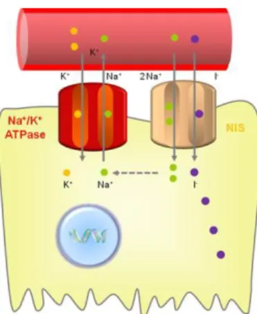
Figure I.4 - Iodide uptake
9
5.2 NIS functional characterization
NIS transports negatively-charged solutes into the cytoplasm using an electrochemical Na+ gradient. This mechanism acts as the driving force for iodide uptake providing energy transfer generated by ouabain-sensitive Na+/K+ adenosine triphosphatase (Na+/K+-ATPase) pump (Spitzweg et al.,, 2001). This route begins with the entrance of Iodide into lumen of the thyroid follicle, where it is organified in the course of thyroid hormone biosynthesis by subsequent oxidation and transfer onto tyrosol residues of thyroglobulin (TG) catalyzed by the thyroid peroxidase (TPO) (Carrasco, 2000; Vieja et al., 2000).
Iodide organification is a particular and unique characteristic of the thyroid gland, and long-term
retention of iodide does not occur in extrathyroidal tissues expressing NIS (Ahn, 2012). In thyroid, NIS can be found at the basolateral membrane of the follicular cells, whereas for some tissues an apical localization has been described, suggesting that the NIS membrane targeting in polarized cells is cell-type dependent (Darrouzet et al., 2014).
5.3 NIS regulation
The NIS gene regulation occurs through different pathways according to tissue. Thyroid and breast are the two main tissues where NIS can be found. Several studies showed the differences in NIS expression and regulation in each of these tissues. In thyroid the regulation ensues mainly by TSH, followed by cAMP accumulation. In breast tissues, however, the cAMP levels do not influence NIS expression. On the other hand, while Retinoic Acid is the principal NIS inducer in breast cancer cells, it was shown to decrease NIS expression in rat thyroid cells (FRTL-5). (Kogai et al., 2000; Schmutzler et
al., 2007)
In thyroid cells, in the absence of TSH stimulation, NIS is not retained at the membrane, leading to a decrease in iodide uptake. However, there must exist alternative regulation mechanisms for the NIS trafficking, since several studies reported a NIS withdrawn at the membrane of non-thyroid tissues without TSH stimulation. Phosphorylation of NIS at serine residues in the carboxy terminus, and protein-protein interactions are some alternative mechanism that were suggested to regulate NIS trafficking (Carrasco, 2000). Down-regulation or mis-targeted NIS expression is commonly found in thyroid carcinomas and frequently correlated with tumor dedifferentiation (Liang et al., 2005). Without a proper regulation, both transcriptional and post-transcriptional, this membrane protein become incapable to perform its key role on up taking iodide, a feature that make NIS a precious cancer therapy and diagnosis tool.
10
5.3.1 Transcriptional regulation of NIS
The transcriptional regulation of NIS expression is complex and involves different regulatory regions. The activity of both thyroid-enriched and ubiquitous transcription factors is required. The upstream enhancer (NUE), located in human cells between 9470 and 9046 relative to ATG (initiation site) and its sequence, is conserved among several species (Kogai et al., 2006). The human (h)NUE can be highly stimulated by TSH and contains putative cis-elements for the Paired box gene 8, (Pax-8) and NK2 homebox 1 (Nkx2.1) transcription factors. These two proteins are required for thyroid development and differentiation (Kogai et al., 2012; Kogai et al., 2006).
Pax-8 sites co-operate with an adjacent cAMP-responsive element to ensure complete hNUE activity and also leads to the repression of hNIS expression performed by Pituitary tumor-transforming gene-binding factor (PTTG) (a proto-oncogene implicated in thyroid cancer). Previously to this process, an activation of adenylyl cyclase through the Gs-protein, is needed so the cAMP levels increase and accumulate in thyroid cells, which leads to an indirect hNUE stimulation (Ohno et al., 1999; Taki et al, 2002).
Retinoic acid (RA) is a metabolite from vitamin A, it plays a pivotal role in development, differentiation, and cell growth. RA role is mediated through two families of nuclear receptors, retinoic acid receptors (RARs) and retinoid X receptors (RXRs). All-trans RA (tRA) inhibits cell cycle progression and induces apoptosis in many tumor cell lines (Evans & Kaye, 1998).In clinical studies, tRA and its analogues showed to stimulate NIS expression in breast cancer and has an element located at position 1375 relative to NIS ATG that mediates the activation of NIS expression in human follicular thyroid carcinoma cells. However, in breast cancer cells the effect is through a downstream intronic enhancer, which binds RAR and RXR (Tanosaki et al., 2003) Moreover, the cardiac homeobox transcription factor Nkx2.5, which is induced upon tRA stimulation, binds two cis-acting elements in the hNIS promoter located at 446 and 154 relative to ATG (Kogai, et al., 2006).
5.3.2 Posttranscriptional regulation of NIS
Besides being finely regulated in a transcriptional level, NIS is also regulated in a pos-translational level, especially regarding to its subcellular localization. NIS needs to be localized in the plasma membrane to be fully functional. This is important not only for iodide transport into the thyroid gland, but also for radioiodide therapy in thyroid cancer. The decrease in iodide uptake observed in most thyroid cancers is due to impaired NIS targeting of, or retention at, the plasma membrane.
A common cellular mechanism for modulating activity, subcellular activation and/or degradation of proteins is Phosphorylation. More recently has been reported as a posttranscriptional regulatory mechanism for the activity of transporters as well. Therefore, one can speculate that TSH might be also be involved in the NIS subcellular distribution (Ramamoorthy, et al., 1999; Glavy et al., 2000). Doahn et
al., (2003) showed that NIS is phosphorylated in vivo and that serines are the principal residues of aa
11
(Riedel et al., 2001; Riedel et al., 1999) have identified five in vitro phosphorylation sites in rat NIS: S227, which is functionally silent; S43 and S581 which modulate transport velocity; T577 and T49 that modulate NIS stability. It was also proposed that hsp90 may function as a chaperone for NIS folding (Marsee et al., 2004).More recently, a studied performed with a two hybrid yeast system revealed the interaction of NIS with LARG, a Rho-guanine nucleotide-exchange factor and its role in the regulation of cancer cell motility and invasiveness through the PDZ binding site at the C-terminal extremity of NIS and the PDZ domain of LARG (Huc-Brandt et al., 2011).
In 2007, Vadysirisack et al., revealed that the MEK pathway has an effect on NIS regulation in thyroid cells, and a more recent study showed that MEK inhibition leads to lysomal degradation in human breast cancer cells (Zhang et al., 2014.)
Under physiological conditions, besides TSH, iodide control also controls the protein expression level and its localization to the thyroid plasma membrane (stimulated by TSH and inhibited by an excess of iodide). Interestingly, it was also showed that the increased of NIS mRNA,does not necessarily increase iodide uptake activity, as previously exemplified by the PI3K-selective inhibitor, PI-103 (Kogai
et al., 2008; Riedel et al., 2009)
5.3.3 Molecular Pathways modulating NIS expression
Besides the mechanisms mentioned above which are responsible for transcriptional and translational modifications, there are specific molecules responsible for regulating NIS expression.
5.3.3.1 PI3Kinase
Insulin and Insulin-like growth factor-I (IGF-I), reduces TSH-induced iodide uptake in FRTLL-rat thyroid cells (Saji & Kohn, 1991). However, insulin, a major regulator of Akt, was not required for the exogenous NIS induction in Ret/PTC1-expressing BHP2-7 cells, although the induction was mimicked by pharmacological inhibition of Akt. Expression of Ret/PTC stimulates the PI3K/Akt pathway in thyroid cells even in the absence of insulin or serum (Miyagi et al., 2004) PI3K is one of the major mediators of insulin/IGf signaling. Selective inhibitors of PI3K, significantly increase NIS mRNA expression in rat thyroid cells by stimulating its transcription. The PI3K inhibitors have a potential to increase the radioiodide accumulation, (Liu et al., 2007), in some differentiated thyroid cancer tissue. Kogai et al., (2008) also demonstrated that PI3K inhibitition significantly increases the iodide uptake in TSH-stimulated and non-TSH TSH-stimulated FRTL-5 cells. However, the mechanisms of inhibition of iodide uptake by PI3K are distinct from rat thyroid cells to thyroid cancer cells. These authors also verified that the effect of PI3K in NIS-expressing BHP human papillary thyroid cancer cells was mimicked by an inhibitor of AKT, a major effector of PI3K, hence indicating a contribution of the canonical PI3K-AKT pathway (Kogai et al., 2008)
12
Additional studies with human thyroid cells are therefore necessary to evaluate the impact of inhibitors of the PI3K pathway on NIS expression in thyroid cancer.
An in vitro study with BHP 2–7 papillary thyroid cancer cells demonstrated stimulatory effects of sunitinib on NIS mRNA expression in the presence of an adenylyl cyclase activator, forskolin (Fenton et al., 2010). Since sunitinib down-regulates the PI3K-AKT pathway (Keefe et al., 2010), it likely mimics the effects of PI3K inhibition on NIS expression, at least partially through PAX8 induction (Fenton et al., 2010).
5.3.3.2 BRAF
The BRAF gene encodes the b-raf protein belonging to the raf family of serine/threonine kinases, responsible for regulating the MAP kinases/ERKs signaling pathway known for regulation of cell differentiation, proliferation, senescence and survival (Choi et al., 2014).
The predominant mutation in the BRAF gene involves a thymidine to adenosine transversion at nucleotide 1,799, being the most observed mutations in BRAF, about 90% of cases (Davies et al., 2002). This results into an activating mutation due to substitution of valine with glutamic acid at aa 600 (Cantwell-Doris et al., 2011). Significant progress has been made in understanding the carcinogenic role of BRAFV600E mutation.
Numerous studies have reported an association of BRAFV600E with decreased or lost expression
of thyroid iodide-handling genes in PTC, particularly NIS (e.g. Zhang et al., 2013), while others have shown that introduced expression of BRAFV600E in thyroid cells could induce the silencing of various
thyroid iodidehandling genes, most prominently the NIS gene (e.g. Liu et al., 2007). BRAF V600E
expression could also cause misallocation of NIS in the cytoplasm in addition to its decreased expression in thyroid cells (Riesco-Eizaguirre et al., 2006). In in vitro cell line assays, inhibition of the BRAFV600E/MEK pathway or silencing of BRAFV600E expression could restore the expression of thyroid
genes, particularly NIS in thyroid cells (Liu et al., 2007), which provided important therapeutic implications for targeting the BRAFV600E/MAPK pathway to restore thyroid gene expression and
radioiodine avidity of radioiodine- refractory thyroid cancer. A recent clinical trial highlights this hypothesis, since the use of Selumetinib, a MAPK pathway inhibitor, as co-adjuvant in iodide therapy, showed an increased in NIS expression and iodide uptake in thyroid cancer cells (Ho et al., 2013).
5.3.3.3 RAC1
Besides its role in tumor associated processes, new roles of Rac1 are still emerging. In addition, the differential regulation of Rac1 is still not fully understood (Navarro-Lérida et al., 2015).
In a recent study Kogai et al. (2012), using MCF-7 cells, showed a role for RAC1 in NIS modulation. Since the activity of the p38 MAPK cascade is regulated by tRA through Rac1, and tRA is the main stimulator of NIS in breast cancer cells, they used a pharmacological Rac1 inhibitor,
13
NSC23766. The p38 kinase is a MAPK, regulating cell proliferation, differentiation and migration. Four p38 isoforms, α, β, γ, and δ, are found in mammalian cells with variable tissue distribution and substrate specificity, producing differential activation of downstream effector pathways (Pramanik et al., 2003). tRA-induced iodide uptake was significantly decreased by the inhibitor. Induction of NIS mRNA by tRA was also significantly decreased by the Rac1 inhibition at every time point tested. These data indicate a critical role of Rac1 in the tRA-induced NIS expression in MCF-7 cells. This regulation occurs through a tRA up-regulation of p38 β phosphorylation through Rac1. A previous studie demonstrated that the p38 isoform which influences NIS expression is the p38 α in thyroid, while p38 β modulates NIS in breast cancer cells (Kogai et al., 2012). Therefore, although the rac1-p38 pathway stimulates NIS expression in both thyroid cells and breast cancer cells, (Pomerance et al., 2000), the Rac1 signal diverges into two pathways through the different isoforms of p38. Over-expression of p38β, as well as of Rac1, significantly enhances the tRA-induced NIS expression and iodide uptake requirement of p38β for the NIS expression in breast cancer cells, therefore, it may provide a strategy for relatively specific induction of NIS in some breast cancer cells (Kogai et al., 2012).5.4 RAC1/RAC1b and NIS
RAC1b protein expression has been described in normal thyroid cells, as well as its overexpression in a number of papillary thyroid carcinomas, carrying the activating mutation BRAFV600E,
with unfavorable outcome (Silva et al. 2013). Also, BRAF V600E has been associated with decreased or
lost expression of NIS in PTC.
On the other hand the GTPase Rac1 was shown to have the ability to stimulate NIS expression through induction of the p38 mitogenic kinase activity. More interestingly, it was shown that while hyperactive, the Rac1b variant has a very selective downstream signaling (Matos et al., 2003) and may even compete with and inhibit Rac1 endogenous activity in signaling pathways where Rac1b does not participate (Matos et al., 2003, Matos et al., 2006).
One such pathway leads precisely to the activation of p38 kinase (Orlichenko et al., 2010). This led us to hypothesize that Rac1b overexpression in thyroid and breast tumors might be involved in the inhibition of NIS expression, through an RAC1 antagonistic effect.
6. Iodide Therapies
6.1 - Thyroid Cancer
Although thyroid nodules are common, differentiated thyroid carcinomas are relatively rare. Managing differentiated thyroid carcinomas can be a challenge, because no prospective randomized trials of treatment have been performed and results from ongoing trials will not be available for many
14
years given the rareness of these tumors. Nonetheless, most thyroid cancer patients have a great outcome. The preferred treatment starts with the need of total thyroidectomy whenever possible. Therefore, the thyroid gland and the lymph nodes affect are resected thyroid hormone suppressive therapy, and in more advanced stages of the disease, radioactive iodine (I-131) for either remnant ablation or therapeutic treatment (Schulumberger, 1998; Legakis and Syrigos, 2011).
By the end of the 19th century Baumann (1896) discovered, for the first time, the ability of thyroid gland to uptake and concentrate iodide; two times more than in plasma compartment. This knowledge was applied in thyroid diagnose and therapy only 50 years later (1942). The transport of the iodide is a crucial step in thyroid hormone biosynthesis and occurs through NIS. This features allows the use of Radioactive Iodine (I-131) as a tool to diagnosis and treatment of thyroid carcinoma. Radioiodine ablation of remnant thyroid following thyroidectomy is performed as an adjuvant therapy for potential residual tumors. Radioiodine is used to ablate any residual remnant thyroid tissues after surgical resection, which increases the sensitivity of subsequent tests based on serum TG measurement and radioiodine whole body scanning. The delivery of high radiation doses to the thyroid tissue due to the latter’s ability to concentrate iodine after TSH stimulation and the ability to destroy residual thyroid tissue cells, allows the early detection of recurrences based on serum TG (Cooper et al., 2009; Pacini et al., 2006; Chung et al., 2010).
Recurrence in the thyroid bed or cervical lymph nodes develops in 5% to 20% of patients with WDTC. Some patients develop distant metastatic disease, which decreases the 10-year survival rate of patients by 50% (Van Nostran, 2009; Haugen, 2010). Postoperative 131 I thyroid remnant ablation is performed when a tumor has the potential to recur. Several studies showed the decreased recurrence and disease-specific mortality when post-operative 131 I therapy is performed as a part of the initial treatment. However, significant controversy persists regarding which patients may benefit from radioactive iodine treatment and the method of radioiodine application (Chung et al., 2014). Nonetheless, in a study with 1004 patients with differentiated thyroid carcinoma, tumor recurrence was approximately 3-fold higher in patients either treated with thyroid hormone alone or given no postoperative medical therapy when compared with those who underwent postoperative thyroid remnant 131 I ablation (Mazzaferri et al., 1997)
6.2 Iodide related therapies in non-thyroid tissues
The cloning and sequencing of the human NIS gene (hNIS) has allowed investigations into the effect of induced NIS expression by non-thyroidal cells (Smanik et al., 1996). Several studies have shown that NIS expression confers the ability to concentrate iodine in a variety of cell types, such as lactating mammary gland, gastric mucosa, salivary and lacrimal glands, choroid plexus, skin, placenta, and thymus, among other tissues (Dohan et al., 2003). Focusing on breast tissue, it is known that during late pregnancy and lactation, active transport of iodide takes place in the mammary gland in order to provide an adequate supply of iodide to the newborn, through NIS. This delivery of iodide is essential for thyroid hormone production and proper development of the newborn’s nervous system, skeletal
15
muscle, and lungs. More interestingly, recent finding on NIS demonstrated that more than 80% of the human breast cancer samples studied expressed NIS, whereas none of the normal samples did (non-lactating tissue) (Kogai et al., 2012). NIS expression within other cell types could be used for diagnostic procedures and treatment of malignant diseases in other tissues such as breast cancer. However, NIS expression and NIS mediated radioiodine uptake (RAIU) activity are often low in breast cancer since the radioiodine concentration is not sufficient in such tumor cells, making the treatment unfeasible. Therefore, it becomes demanding to find and understand the specific regulatory mechanisms beyond NIS gene transcription to post-translational processing and cell trafficking. These investigations will eventually lead to strategies that can be used to increase functional NIS expression in breast cancer.7. Aim of the project
Given the context described previously, the main objective of this work is to test for a relation between Rac1b and NIS in thyroid and breast cancers. More specifically, given that Rac1 is involved in NIS regulation our aim is to understand if the overexpression of RAC1b could interfere with this process.
The testing will focus in the mRNA and protein expression of NIS through qRT-PCR in PTCs and FTC, as well as thyroid cell lines and breast cancer cell lines in order to understand the possible differences in RAC1/1b–NIS modulation in diverse biological systems.
17
II – Material and Methods
1. Patient samples
A total of 64 samples of primary tumors from PTC and FTC patients who underwent partial/total thyroidectomy in IPOLFG were analyzed. These samples were collected at surgery and immediately frozen and stored in liquid nitrogen. Tissue sample collection was carried out in accordance with protocols approved by the institutional review board and informed consent was obtained for the study, together with the consent for surgery.
2. Cell Culture
In the present study, three human cell lines were used: Nthy-ori 3-1, MCF-7 (ATCC®-HTB-22™and MDA-MD-231 (ATCC® CRM-HTB-26™), commercially available cell lines. These three cell lines are adherent and where maintained at 37ºC in a humidified 5% CO2 environment in an appropriate
chamber (NUAIRE TM US AUTOFLOW CO2 water-jacketed incubator).
At least twice a week, when cultures reached confluence of about 80-95%, cells were subcultured. Different culture mediums were used accordingly to cell line nature. N-thy-ori 3-1 were cultured in a RPMI 1640 (GIBCOTM) medium supplemented with 10% (v/v) Fetal Bovine Serum
(FBS).The mammary gland/breast cell lines (MCF-7 and MDA-MD-231) were cultured in DMEM (GIBCOTM) also supplemented with 10% (v/v) FBS and 1% (v/v) glutamine. Cells were detached with 1x
0.05% trypsin-EDTA (InvitrogenTM).
3. Cell Transfection
Transfection assay is the process of introducing nucleic acids into eukaryotic cells by nonviral methods. In transient transfections, foreign DNA does not integrate into the cell genome and transfected gene are expressed for a limited time. In stable transfections, however, cell have integrated foreign DNA into their genome, and consequently, descendants of these transfected cells will also express the transfected gene (Kim & Eberwine, 2010). In the context of this thesis, transient transfections were used throughout the work.
Transfections were performed using the cationic lipid-based transfection reagent LipofectamineTM 2000 (InvitrogenTM) in 35-mm dishes (NunclonTM). Just before transfection, cells were
fed with new fresh complete DMEM medium. Plasmid DNA (2 μg) was diluted into 125 μl OPTI-MEM (GIBCOTM); in an additional tube, 4 μl of transfection reagent was also diluted in 125 μl of OPTI-MEM
and incubated 5 minutes at room temperature (RT). Diluted DNA and lipofectamine were then mixed gently and incubated for 20 minutes at RT to allow the formation of DNA-lipofectamine complexes. After
18
incubation, 250 µl of transfection mixture was added to each culture dish.
The expression vectors used in this study were kindly supplied by Doctor Peter Jordan, from Instituto Nacional de Saúde Dr. Ricardo Jorge: RAC1WT, RAC1L61, pEGFP-RAC1bWT, and pEGFP-RAC1bL61. The pcDNA3.1 (+)-empty vector was used as MOCK control.
4. RNA extraction and cDNA synthesis
Total RNA was obtained from frozen tissues or from transfected cell lysates using RNeasy Mini Extraction Kit 74106, Qiagen), according to manufacturer’s instructions, and 2 μg were reverse transcribed using random primers and SuperScript II (InvitrogenTM).
5. Reverse Transcription (RT) Polymerase Chain Reaction (PCR)
After the cDNA synthesis, PCR for NIS were performed. Primers for specific amplification of NIS were NISF (5’-CCATGTATGGCGTGAACC) and NISR (5’CTTCGAAGATGTCCAGCACC), generating PCR product of 234 bp (Appendix supplementary Table 1).
PCR products were resolved by electrophoresis on a 2% (w/v) agarose gel in 1X Tris-borate-EDTA (TBE) buffer (diluted from 10X TBE, EC-860, National diagnostics) and stained with 0.05% (v/v) ethidium bromide. The separated DNA was visualized under UV light and image acquired in a ChemiDoc XRS System (BIO-RAD).
6.
Quantitative (q) RT-PCR
The Rac1b and total Rac1 expression levels were quantified by qRT-PCR on an ABI Prism 7900HT Sequence Detection System using specific primers and TaqMan probes from the Assay_on_Demand products [Hs00251654_ml (selectively amplifies Rac1b), Hs01902432_sl (amplifies both variants); Applied Biosystems], according to manufacturer's instructions. cDNA samples were diluted 20 times and 4 μl were used in each real-time PCR reaction. Amplification reactions were performed in triplicate for each sample. Rac1b levels were normalized to total Rac1 (Rac1b + Rac1) expression level. For each sample, Rac1b normalized values were then expressed relative to Rac1b expression level present in RNA obtained from a pool of normal thyroid tissues, used as reference sample.
19
The NIS and β-actin expression levels were also quantified using specific primers and TaqmanTM probes from Assay_on_Demand products (Hs.584804; Applied Biosystems). cDNA samples(both obtained from fresh tissues or transfected cell pools) were diluted 4 times and 4μL were used in each real-time PCR. Amplification reactions were performed in triplicate for each sample. NIS levels were normalized to β-actin expression level (endogenous control. For each sample, NIS normalized values were then expressed relative to NIS expression levels of reference sample. For NIS assessment in transfected cells, pCDNA3-empty transfected samples (MOCK samples) were used as reference; for NIS quantification in tumors, we used as reference RNA obtained from a pool of normal thyroid tissues. Expression values correspond to arbitrary units representing fold differences relative to the reference sample.
7. Protein extracts, SDS-PAGE and Western blotting
Protein extracts were obtained from cell lysates performed with RIPA (radio immunoprecipitation assay) Lysis buffer (appendix supplementary Table 2) and stored at -80ºC.
The proteins were separated in a 12% (RAC1/b) sodium dodecyl sulfate-polyacrylamide gel electrophoresis (SDS-PAGE) and the protein bands were then transferred electrophoretically to a PVDF membrane (Polyvinylidene fluoride; BIO-RAD). Membranes were blocked in blocking buffer (20 mM Tris–HCl, pH 7.6, 140 mM NaCl, 0.1% Tween-20, 5% skim milk powder) and probed with primary RAC1 (Upstate, EMD Milipore Bioscience, Billerica, Massachusetts, United States) at 1:2000 dilution. Detection was carried out using secondary peroxidase-conjugated anti-mouse IgG (BioRad) at 1:5000 dilution. The Immunoreactive bands were detected by chemiluminescence using ECL Western blotting substrate followed by exposure and image acquisition in a ChemiDoc XRS System (BIO-RAD).
8. Statistical Analysis
Statistical analyses weres performed with GraphPad Prism statistical software (San Diego, CA, USA). Values are expressed as mean ± SD. Comparisons of rates and proportions were made using the unpaired two-tailed Student’s t-test. Statistical significance was accepted at P < 0.05.
21
III - Results
1. NIS expression levels in RAC1b-positive and RAC1b-negative follicular
cell derived thyroid carcinomas.
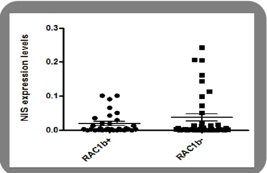
As a primary approach to test whether the RAC1 isoform, RAC1b, has a role in NIS expression modulation, we evaluated the NIS expression levels among samples from FTC and PTC (n=64) comparing tumors presenting RAC1b overexpression (n=32) to those that do not (n=32). In order to make a distinction between RAC1b -overexpressing and non-overexpressing tumors, we defined a threshold of expression above which we considered RAC1b to be overexpressed. We set the threshold at the value corresponding to the mean plus two standard deviations of the Rac1b expression level in a set of normal thyroid tissues. Based on this threshold, 32 out of 64 PTCs (50%) were found to overexpress Rac1b.
NIS expression assessment by quantitative RT-PCR indicated higher levels of NIS in samples negative for RAC1b expression: 0.04402 ±0.01271 comparatively to 0.01916±0.005423 in RAC1b positive samples ( P = 0.0384; two-tailed Student’s t-test) (Figure III.1), allowing to suggest that tumors overexpressing RAC1b are associated with NIS downregulation.
Figure III.1 - NIS expression levels in RAC1b‐positive and RAC1b‐negative PTCs and
FTCs. NIS expression levels quantified by qRT‐ PCR, correspond to arbitrary units representing fold differences relative to a reference sample Values are the mean ± S.D.(error bars) (n= 64).
22
2. NIS expression in cell lines
2.1 Effect of RAC1/1b in NIS expression in Nthy-ori 3-1
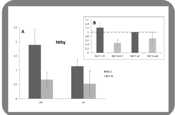
In Nthy-ori 3-1 cells, a follicular epithelial normal tissue cell line where it is supposed to exist substantial levels of NIS, we observed a considerably difference among NIS expression. This was found for cells transfected with RAC1 and RAC1b. The present data suggests that RAC1 is able to stimulate
NIS expression, while RAC1b seems to decrease NIS levels. These results are more evident when
comparing RAC1-L61 and RAC1b-L61 (the constitutively active variants of the GTPases RAC1/1b) effect on NIS. These data suggest a role of RAC1 in NIS stimulation while RAC1b seems to inhibit it (Figure III-2.A). In fact, when we compare the effect on NIS expression levels among the different variants with the effect of RAC1 WT, we found a minor expression of NIS in RAC1b L61 transfected cells, dropping to half. RAC1b WT also seem to decrease NIS levels, but in a weaker way, about 30% (Figure III-2.B).
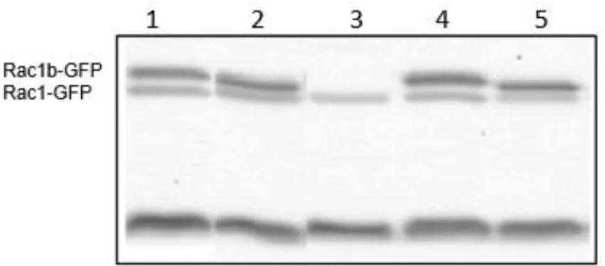
Figure III.3 - RAC1/1b protein expression in Nthy-ori 3-1 transfected cells.
Representative western blot of RAC1 and RAC1b expression in Nthy-ori 3-1 transfected cells: 1 – RAC1b-L61-GFP; 2 – RAC1-L61-GFP; 3 – MOCK; 4 – RAC1b-WT-GFP; 5 – RAC1-WT-GFP; transfection monitorization.
Figure III.2 - Regulation of NIS expression by RAC1 and RAC1b in N-thyroid cells A and B, effects of
RAC1 and RAC1b (both L61 and WT) on NIS expression. A, NIS expression levels were accessed through quantitative RT-PCR in cells transfected with RAC1/RAC1b-WT or with the constitutively dominant mutants RAC1/1b-L61 and normalized to MOCK transfections. B, Comparison of the effect of the different RAC1/1b variants to that of RAC1-WT.Cells were transfected with: pEGFP-GFP-RAC1WT, pEGFP-GFP-RAC1L61, pEGFP-GFP-RAC1bWT, pEGFP-RAC1bL61 and pcDNA3.1 (+)-empty vector used as MOCK control. A Western blot was performed to monitor (RAC1/b) transfected levels.
23
2.2 Effect of RAC1/1b in NIS expression in MDA-MB-231
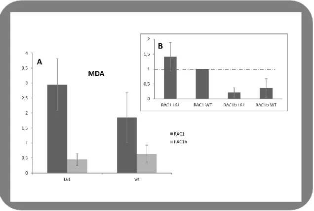
Our quantitative RT-PCR showed that in MDA-MB-231 cells NIS expression is 2.5 fold higher in cells transfected with RAC1 L61 comparatively with RAC1b L61 transfection. Regarding to WT transfections, cells still showed ~1.5 times higher levels (of NIS, for RAC1 comparatively with RAC1b (Figure III.4.A). When comparing the effect on NIS expression to that of RAC1 -WT, both L61 and WT variants of RAC1b induced a notorious decrease on NIS expression: NIS levels drop to more than a half, in this breast cancer cell line when transfected with RAC1b comparatively with RAC1 transfections (Figure III.4.B).
Figure III.3 - RAC1/1b protein expression in Nthy-ori 3-1 transfected cells.
Representative western blot of RAC1 and RAC1b expression in Nthy-ori 3-1 transfected cells: 1 – RAC1b-L61-GFP; 2 – RAC1-L61-GFP; 3 – MOCK; 4 – RAC1b-WT-GFP; 5 – RAC1-WT-GFP; transfection monitorization.
24
Figure III.4 - Regulation of NIS expression by RAC1 and RAC1b in MDA-MB-231 cells A and B, effects of RAC1 and RAC1b (both L61 and WT) on NIS expression. A, NIS expression levels were accessed
through quantitative RT-PCR in cells transfected with RAC1/RAC1b-WT or with the constitutively dominant mutants RAC1/1b-L61 and normalized to MOCK transfections. B, Comparison of the effect of the different RAC1/1b variants to that of RAC1-WT. Cells were transfected with: RAC1WT, pEGFP-GFP-RAC1L61, pEGFP-GFP-RAC1bWT, pEGFP-RAC1bL61 and pcDNA3.1 (+)-empty vector used as MOCK control. A Western blot was performed to monitor (RAC1/b) transfected levels.
Figure III.5 - RAC1/1b protein expression in MDA-MB-231 transfected cells. Representative western blot of RAC1 and RAC1b
expression in MDA-MB-231 transfected cells: 1 – RAC1b-L61-GFP; 2 – RAC1-L61-GFP; 3 – MOCK; 4 – RAC1b-WT-GFP; 5 – RAC1-WT-GFP; transfection monitorization.