1
FUNCTIONAL ACTIVITY OF SEAWEED EXTRACTS FROM THE NORTH PORTUGUESE
COAST
by
Marta Sofia de Almeida Mendes
i
FUNCTIONAL ACTIVITY OF SEAWEED EXTRACTS FROM THE NORTH PORTUGUESE
COAST
Thesis presented to
Escola Superior de Biotecnologia
of the
Universidade Católica
Portuguesa
to achieve the Master of Science degree in
Microbiology
by
Marta Sofia de Almeida Mendes
Place: CBQF/Escola Superior de Biotecnologia da Universidade Católica Portuguesa
Supervision: Prof. Ana Maria Pereira Gomes, Doutora Ana Paula Santos de Carvalho
iii
Resumo
A utilização de algas marinhas como fontes potenciais de compostos nutracêuticos e farmacêuticos tem aumentado recentemente devido à constatação de que estas contêm compostos bioactivos, com actividades antioxidante e antimicrobiana (entre outras actividades), que podem inibir o crescimento de algumas bactérias contaminantes e/ou patogénicas e de leveduras prevenindo a deterioração de alimentos ou a infecção ou contribuindo mesmo para o seu melhor controlo. O litoral português alberga uma grande biodiversidade no que concerne a algas marinhas, porém muitas encontram-se por caracterizar em termos de propriedades funcionais. Neste contexto, o objectivo deste trabalho foi estabelecer um procedimento melhorado para a obtenção de extractos de algas marinhas e testar a sua actividade antimicrobiana contra espécies selecionadas de leveduras, bactérias Gram positivas e Gram negativas, bem como a sua actividade antioxidante. Para tentativamente atestar o seu comportamento, os perfis lipídico e fenólico foram testados.
As algas utilizadas neste estudo, incluindo as de aquacultura integrada e as de habitat natural, foram obtidas no Norte de Portugal. A alga Gracilaria vermiculophylla foi usada para os ensaios de optimização do processo de extracção, enquanto as Gracilaria vermiculophylla, Porphyra dioica e Chondrus crispus foram utilizadas para os ensaios de actividade antimicrobiana e antioxidante. Os estudos de optimização centraram-se na definição dos pré-tratamentos (secagem) e da temperatura a utilizar durante o processo de extracção. Os resultados revelaram que os organismos testados foram mais sensíveis aos extractos obtidos com algas secas, continuamente processados a temperaturas mais elevadas. Posteriormente, extratos obtidos com três diferentes solventes (acetato de etilo, éter dietílico e metanol:água) foram testados.
No que diz respeito à avaliação da actividade antimicrobiana, as espécies testadas incluíram (i) bactérias Gram negativas - Escherichia coli, Salmonella enteritidis e Pseudomonas aeruginosa; (ii) bactérias Gram positivas – Listeria innocua, Bacillus cereus, Enterococcus faecalis, Lactobacillus brevis, Staphylococcus aureus, todas de origem alimentar, e uma estirpe de Staphylococcus aureus de origem clínica, e (iii) a levedura Candida spp. também de origem clinica. Os testes para avaliar a actividade antimicrobiana dos extractos foram realizados utilizando o método de difusão em agar e os resultados indicaram uma forte actividade antimicrobiana dos extractos de acetato de etilo, quando comparado com os extractos de metanol e éter dietílico e mostraram uma tendência fraca para a inibição de microrganismos Gram positivos.
O perfil de ácidos gordos de extractos de acetato de etilo revelou uma predominância de ácidos gordos saturados (SFA), especialmente o acido palmítico (16:0), seguido por ácidos gordos polinsaturados (PUFA) e ácidos gordos monoinsaturados (MUFA) e mostrou um teor mais elevado de ácidos gordos em G. vermiculophylla e P. dioica de aquacultura.
Tendo em conta os resultados obtidos para a actividade antioxidante, foi demonstrado que os extractos metanólicos apresentaram actividade mais elevada quando comparada com os outros solventes testados.
O perfil fenólico revelou que os extractos metanólicos mostraram quantidades mais elevadas de compostos fenólicos, tais como catequinas e ácido protocatecuico, o que pode indiciar o seu papel na actividade antioxidante.
v
Abstract
The use of marine algae as potential sources of pharmaceutical and nutraceutical compounds has been increasing recently, due to the realization that they contain bioactive compounds, with antioxidant and antimicrobial activities (among others), which could inhibit the growth of some contaminant and/or pathogenic bacteria and yeasts, preventing food spoilage or infection and even contributing to its better control. The Portuguese coastline is home to a great diversity in terms of seaweed however, many of them are not yet characterized in terms of functional properties. In this context, the aim of this work was to establish an improved procedure for obtaining extracts from marine algae and to test its antimicrobial activity against selected species of yeasts, Gram positive and Gram negative bacteria. Furthermore, the antioxidant activity of the extracts was also assayed. Finally, an in order to correlate between the composition of the extracts and its bioactivity, their characterization was tentatively established through the determination of lipidic and phenolic profiles. Seaweeds used in this study including those from integrated aquaculture and from their natural habitat, were obtained in the North of Portugal. Gracilaria vermiculophylla was used for the assays of optimization of the extraction procedure, whereas Gracilaria vermiculophylla, Porphyra dioica and Chondrus crispus were used for antimicrobial and antioxidant assays.
Optimization studies were focused on the definition of the pre-treatments of the algae (drying) and the temperature used during the extraction process. Results revealed that test organisms were more sensitive to extracts obtained with dried algae, continuously processed at higher temperatures. Subsequently, extracts obtained with three different solvents (ethyl acetate, diethyl ether and methanol:water) were tested.
Concerning antimicrobial capacity evaluation, species tested included (i) Gram negative bacteria – Escherichia coli, Salmonella enteritidis and Pseudomonas aeruginosa; (ii) Gram positive bacteria – Listeria innocua, Bacillus cereus, Enterococcus faecalis, Lactobacillus brevis, Staphylococcus aureus, all from food origin and a strain of Staphylococcus aureus from clinical origin and (iii) the yeast Candida spp. from clinical origin as well. Tests to assess the antimicrobial activity of the extracts were performed using the agar diffusion method, and results indicated a stronger antimicrobial activity of the ethyl acetate extracts when comparing with the diethyl ether and methanolic ones, and a weak tendency for inhibition of Gram positive microorganisms.
The fatty acid profile of ethyl acetate extracts revealed a predominance of saturated fatty acids (SFA), especially palmitic acid (16:0), followed by polyunsaturated fatty acids (PUFA) and monounsaturated fatty acids (MUFA), and showed a higher content of fatty acids on aquaculture extracts in Gracilaria vermiculophylla and Porphyra dioica.
Taking into account the results for antioxidant activity tested with the ABTS•+ method, it was shown that methanolic extracts had highest activity when compared to the other solvents tested. The phenolic profile revealed that these extracts had highest amounts of phenolic compounds such as catechin and protocatechuic acid, which could take a role in the antioxidant activity.
vii
Acknowledgments
Firstly, I would like to acknowledge Escola Superior de Biotecnologia for having provided the facilities and all necessary conditions for this work, and CIIMAR for supplying the seaweeds I worked with.
To Professora Doutora Ana Gomes and Doutora Ana Carvalho, a special acknowledgement for all support, monitoring and words of encouragement, even in more difficult times and for providing me all the necessary conditions for the execution of this work, always with a loving smile.
To Professora Doutora Manuela Pintado that always give me a supporting smile although I didn’t work directly with her.
To all people that works or worked in the 6th floor laboratory, which were always there for me.
To all my friends, especially Sara Silva, Guilherme Ribeiro, Alexandra Correia and Nádia Silva, Eduardo Costa and Ana Oliveira, who always supported me, helped me and made me laugh!
Finally, to all my family members, especially my parents who always supported me and loved me unconditionally in all stages of my life and my grandparents, especially my grandfather for the support in all the decisions I made.
Despite being a bit silly, I want also to thank my pets: Izzy and especially Yuri (2002-2012) for having always patience with me (although sometimes I didn’t have with them!), for always being by my side and always willing to play with me!
ix
Table of Contents
Resumo ... iii
Abstract
... v
Acknowledgments
... vii
Table of Contents ... ix
Figure Index
... xi
Table Index
... xiii
List of Abbreviations ... xv
1
Introduction ... 1
1.1
Historical perspective and importance of seaweeds
... 1
1.2
Cultivation environment ... 4
1.3
Marine Algae ... 5
1.3.1
Gracilaria vermiculophylla
... 5
1.3.2
Porphyra dioica
... 6
1.3.3
Chondrus crispus
... 7
1.4
Methods of extraction of added-value compounds
... 8
1.5
Antimicrobial activity ... 9
1.6
Antioxidant activity ... 11
1.7
Work objectives
... 12
2
Materials and Methods ... 14
2.1
Algae
... 14
2.2
Preparation of seaweed extracts
... 14
2.2.1
Optimization of extraction procedure ... 14
2.2.2
Preparation of extracts to analyze
... 14
2.3
Determination of antimicrobial activity
... 15
2.4
Determination of lipid profile ... 16
2.5
Determination of antioxidant activity
... 16
2.6
Determination of phenolic profile
... 17
2.7
Statistical analysis ... 17
3
Results and Discussion
... 18
3.1
Optimization of extraction procedure
... 18
3.2
Antimicrobial activity ... 19
3.3
Determination of the lipid profile - Fatty acids
... 28
x
3.5
Determination of the phenolic profile ... 34
4
Conclusions ... 38
5
Future work
... 39
6
Appendixes ... 40
6.1
Lipid standards profile ... 40
6.2
Calibration curve of the ABTS
•+method
... 41
6.3
Phenolic standards profile ... 41
xi
Figure Index
1. Introduction
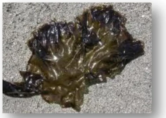
Figure 1.1 - Gracilaria vermiculophylla (adapted from: Jenneborg, HydroGIS).
Figure 1.2 - Porphyra dioica (adapted from: Holmes, 2007).
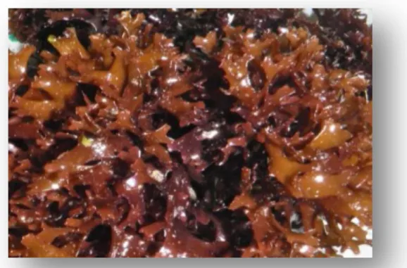
Figure 1.3 - Chondrus crispus (adapted from: Guiry, 2005).
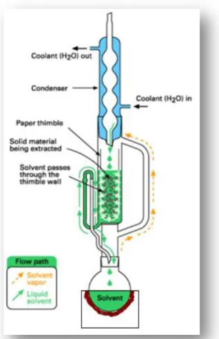
Figure 1.4 - Soxhlet extraction apparatus (adapted from: Kou, 2003).
3. Results and Discussion
Figure 3.1 - Effect of type of processing and physical state of G. vermiculophylla extracted with ethyl acetate on test microorganisms (CI – Clinical Isolate; FI – Food Isolate). Inhibition zones marked with the same letter are not significantly different (p < 0.05).
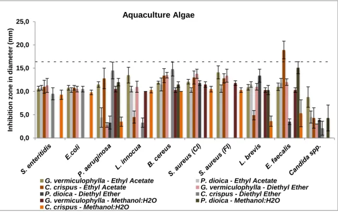
Figure 3.2 – Overview of the results for the antimicrobial activity of aquaculture algae extracts against tested microorganisms. Dotted line represents the inhibition halos average for positive control (lactic acid at 30% (v/v)) – (17 mm).
Figure 3.3 – Overview of the results for the antimicrobial activity of wild algae extracts against tested microorganisms. The dotted line represents the average of inhibition halos of the positive control (lactic acid at 30 % (v/v)) - (17 mm).
Figure 3.4 - Effect of ethyl acetate extracts of different seaweed from aquaculture and wild in the inhibition of tested microorganisms.
Figure 3.5 - Effect of diethyl ether of different seaweed from aquaculture and wild in the inhibition of tested microorganisms.
Figure 3.6 - Effect of methanol:H2O extracts of different seaweed from aquaculture and wild in the inhibition of tested microorganisms.
Figure 3.7 - Antioxidant activity of the wild and aquaculture (Aq.) extracts of G. vermiculophylla, P. dioica and C. crispus. Samples marked with the same letter are not significantly different (p < 0.05).
Figure 3.8 - Chromatograms representing the richness of compounds in aquaculture P. dioica extracted with ethyl acetate (A), diethyl ether (B) and methanol:H2O (C).
Figure 3.9 - Chromatograms representing methanolic extracts of aquaculture (A) and wild (B) Gracilaria vermiculophylla.1 -Protocatechuic acid; 2- Catechin.
xii
Figure 3.10 - Chromatograms representing methanolic extracts of aquaculture (A) and wild (B) Porphyra dioica. 1- Protocatechuic acid; 2- Catechin.
Figure 3.11 - Chromatograms representing methanolic extracts of aquaculture (A) and wild (B) Chondrus crispus. 1 - Protocatechuic acid; 2- Catechin.
6. Appendix
Figure 6.1 - Chromatogram representing the standards of fatty acids. 1- 12:0; 2 – 14:0; 3 – 14:1; 4 – 16:0; 5 – 16:1 (n-7); 6 – 18:0; 7 – 18:1 (n-9); 8 – 18:2 (n-6); 9 – 18:3 (n-6); 10 - 18:3 (n-3); 11 – 20:0;
12 – 20:1 (n-9); 13 – 20:2 (n-6); 14 – 20:3 (n-6); 15 - 20:3 (n-3); 16 - 20:4 (n-6); 17 - 20:5 (n-3); 18 –
21:0; 19 - 22:0; 20 – 22:1 (n-9); 21 – 22:2 (n-6); 22 – 22:6 (n-3).
Figure 6.2 – Calibration curve obtained using standard ascorbic acid and solutions to calculate the result obtained in the ABTS radical cation assay.
Figure 6.3 - Chromatograms representing the standards of phenolic compounds. 1 - Protocatechuic acid; 2 - Catechin; 3 - Chlorogenic acid; 4 - p - Coumaric acid; 5 - Quercetin; 6 - Ascorbic acid; 7 - Gallic acid; 8 - Dihydroxybenzoic acid; 9 - Caffeic acid; 10 - Hydroxycoumarin; 11 - Ascorbic acid; 12 - Ferrulic acid; 13 - Rutin; 14 - Cinnamic acid.
xiii
Table Index
1. Introduction
Table 1.1 - Polarity Index and boiling points of some solvents (adapted from Sidel, 2006).
2. Materials and Methods
Table 2.1 - List of the microorganisms tested.
3. Results and Discussion
Table 3.1 – Different algae extracts tested.
Table 3.2 - Inhibition halo (mm) for each combination of extract-microorganism tested.
Table 3.3 - Fatty acid content (µg/g sample) of wild and aquaculture (Aq.) extracts of G. vermiculophylla, P.dioica and C. crispus.
xv
List of Abbreviations
Abs - AbsorbanceABTS – 2,2’-azino-bis (3-ethylbenzothiazoline-6-sulphonic acid) diammonium salt ABTS•+ - 2,2’-azino-bis (3-ethylbenzothiazoline-6-sulphonic acid) radical cation AIDS – Acquired Immunodeficiency Syndrome
BC – Before Christ BHI – Brain Heart Infusion BHT – Butylated HydroxyToluene CFU – Colony Forming Units CI – Clinical Isolate
DMSO – Dimethyl Sulfoxide FI – Food Isolate
GC – Gas Chromatography
HIV – Human Immunodeficiency Virus
HPLC – DAD – High Performance Liquid Chromatography with Diode-Array Detection MRSA - Methylcilyn Resistant Staphylococcus aureus
MUFA – Monounsaturated Fatty Acids NA – Nutrient Agar
NB – Nutrient Broth OD – Optical Density PI – Percentage of Inhibition
PUFA – Polyunsaturated Fatty Acids ROS – Reactive Oxygen Species SFA – Saturated Fatty Acids
1
1
Introduction
1.1
Historical perspective and importance of seaweeds
Since the beginning of times, mankind tried to control diseases in order to maintain their survival. The first information concerning the practice of medicine dates from the 5th millennium BC with Egyptian priests; they joined medicine with superstition and used medicinal plants, herbs and other products such as milk, honey, salt or beer. However, none of these treatments were considered completely effective for the infectious diseases, one of the biggest problems that affected humanity and responsible for killing millions of people (Ferreira et al., 2008). It was by accident that, in 1928, Alexander Fleming discovered a substance that revolutionized medicine and promised an end to infectious diseases of bacterial origin. It happened when Fleming accidentally left a culture of Staphylococcus aureus to be contaminated with spores of a fungus. He noticed that around the fungus, an inhibition halo was formed, making impossible the growth of the bacterial strain. In addition, in that year, the environmental conditions were atypical, with heat and humidity above normal, which allowed that specific fungus to develop. After numerous researches, Fleming showed that the fungus, later identified as Penicillium notatum, was responsible for the production of a substance with bactericidal effect: penicillin. Years later, and after being purified, this substance was administered to human beings, having saved the lives of thousands of soldiers wounded in World War II. This gave rise to the development of industrial production processes of penicillin and the beginning of the era of antibiotics (Sykes, 2001).
From the moment that the use of antibiotics became widespread, the number of deaths from infectious diseases decreased dramatically. However, nowadays there is a downside: the overuse of these drugs has led to a serious public health problem, due to the acquired antibiotic resistance by microorganisms. Furthermore, there has also been an increase in the number of infections by fungi, especially with regard to patients with compromised immune systems, such as HIV patients and patients who have received chemotherapy treatments, among others (Stirk et al., 2007).
To overcome these emerging problems of microorganisms’ resistance to antibiotics, new natural sources of drugs have been investigated. Marine organisms have caught the attention of researchers because they are rich sources of structurally new and biologically active metabolites (Ely et al., 2004). Algae or seaweeds have been extensively studied and have proven its great potential as a source of primary and secondary metabolites (Tuney et al., 2006). In fact, there are several compounds obtained from macroalgae that have been used in traditional medicine for a long time; in particular, some red macroalgae belonging to the Phylum Rhodophyta are able to synthesize halogenated metabolites such as ketones, low molecular weight hydrocarbons and phenols (Taskin et al., 2007). In addition, several recent studies have described that many substances derived from seaweeds have been associated with a broad range of biological activities such as antibacterial, antiviral, antifungal, antifouling and anti-inflammatory effects as well as cytotoxic and antimitotic activities (del Val et al., 2001). Such activities are probably related with their composition: seaweeds are known to contain reactive antioxidant molecules such as ascorbate and glutathione when fresh, as well as secondary
2
metabolites, including carotenoids (e.g., α- and β-carotene, fucoxanthin, astaxanthin), mycosporine-like aminoacids (e.g., mycosporine palythine), catechins (e.g., catechin, epigallocatechin, epigallocatechin gallate) phlorotannins (e.g., phloroglucinol, eckol) and tocopherols (e.g., α-, χ- δ- tocopherols) (Demirel et al., 2009). Antibacterial halogenated compounds such as bromophenols have also been isolated from many types of seaweeds as well as some fatty acids and sterols such as fucosterol. Some derivatives of diterpenoids obtained from seaweeds are also known to possess antimicrobial activity against some bacteria. Besides, some seaweeds have proven to have also significant antitumoral, antileukemic, antiprotozoan and hypolipidemic activities (Demirel et al., 2009).
In places like China, Korea and Japan, there is evidence that the incidence of some cancers is lower due to regular consumption of seaweeds (Kumar et al., 2011; Vijayavel and Martinez, 2010). In traditional Chinese medicine, seaweeds have been widely used for centuries for healing a large variety of diseases such as tuberculosis, arthritis, colds and flu, worm infestations, among others. For example, it is described that a specific red algae (Digenea spp.) produces an effective vermifugal agent (i.e. kainic acid), the aqueous extracts from two other red alga of the Dumontiaceae family may inhibit the herpes simplex virus and carrageenan has been described as a potent anti-viral agent. Porphyra spp. is used in the treatment of urinary infections, sore throat, beriberi and edema (Guiry, 2011), whereas Sargassum spp. and Saccharina spp. have been used for the treatment of cancer, since the inhibition of cancerous tumors seems to be caused by long-chain polysaccharides. Besides this use, Sargassum spp. can be applied for treating edema, scrofula (a swelling associated with tuberculosis), goiter (a thyroid swelling), testicular pain, other swellings and tumors.
Nowadays, seaweeds have been incorporated in medicinal formulas in pharmaceutical industry for treating other disorders like ovarian cysts, breast lumps, lymph node swellings and obesity, among others. The intake of minerals such as calcium, sodium and potassium are associated with lower systolic pressure and consequently, lower risk of hypertension. Seaweeds’ sterols and related compounds have the ability to lower blood plasma cholesterol level. Moreover, seaweeds are the best source of bimolecular iodine, providing di-iodotyrosin which is a precursor to forming essential thyroid hormones that regulate body metabolism. This could control and prevent many endocrine deficiency conditions such as breast and uterine fibroids, tumors, prostrate inflammation and toxic liver and kidney states. Moreover, there is great potential for developing drugs to treat cancer, AIDS and other diseases that affect millions of people (Dhargalkar and Pereira, 2005).
Algae also have other applications, such as in cosmetics: as an ingredient in creams and toothpaste and in seaweeds baths and thalassotherapy that are very popular in some cultures ever since ancient times. Currently, more importance has been given to this kind of treatments, as they can improve life quality and well-being. They consist essentially in the medicinal use of seawater, mud and algae as a form of therapy. Some of them include wrapping some algae such as Fucus serratus around the body. Seaweeds are generally steam-treated prior to use so that they release minerals, trace elements and polysaccharides. These treatments can relieve pain and are often effective in cases of rheumatism and arthritis; besides, they relieve fatigue, tiredness and tension, and are also very good for skin, since they eliminate impurities and balance pH. Furthermore, seaweeds have become an
3
important ingredient for cosmetic products such as soaps, shampoos, creams and sprays since they are naturally revitalizing, moisturizing and full of aminoacids, minerals and vitamins that nourish the skin. Based on all these properties it’s no wonder their popularity these days (Dhargalkar and Pereira, 2005).
Due to their richness in protein, fiber, vitamins (A, B1, B12, C, D, E) and minerals such as calcium, sodium, potassium and magnesium and especially iodine, many algae are widely used in cuisine in many parts of the world, especially in Asia where they have been cultivated for centuries and have become a major industry representing one of the most profitable. Seaweeds are often used in soups, salads and others recipes like jelly and puddings (Sachindra et al., 2009). In some parts of Asia like Japan, China and Korea, sheets of the dried red alga Porphyra spp. are used in soups or to wrap sushi. In Ireland, the main species used for food are dulse (Palmaria palmata), carrageen moss (Chondrus crispus) and various kelps. These algae are generally sold in their dried form and eaten in various food and drink recipes after soaking in water. Due to their nutritional qualities, the trend today is to refer to marine macroalgae as “sea-vegetables” as they are as good as any land-vegetable and, in some cases, superior in their vitamin, trace element and protein contents (Dhargalkar and Pereira, 2005).
Food industry also uses alginates (essentially from brown algae), carrageenan and agar (from red algae) which are phycocolloids, or marine hydrocolloids, that are extracted from seaweed and are used as thickeners, gelling agents, emulsifiers and stabilizers. These compounds could replace synthetic additives, considered as more harmful to health (Patra et al., 2008). As an example, alginate can be used as an ingredient for ice creams, drinks and cosmetics, as well as in pharmaceutical preparations, dental care and prosthetic materials among other uses; carrageenan can be used in salad dressings and sauces, dietetic foods and as a preservative in meat, fish, dairy and baked products; agar can be used in confectionery, meat and poultry products, molded foods, desserts and beverages (Guiry, 2011).
Nowadays, industrial uses of macroalgae are largely confined to extraction of phycocolloids and, at a lesser extent, to the extraction of fine chemicals. Many of these products are used in several other industries such as components of paints, dyes, adhesives, gels, explosives as well as in paper, leather and textile industries (Guiry, 2011).
Besides, seaweeds are widely used in agriculture as an organic fertilizer, in agrochemicals, compost for landscaping or a means of combating beach dunes erosion, since they are biodegradable, non-toxic, non-polluting and non-hazardous for the environment. Seaweeds can also be used as a food supplement for daily meals for farm animals such as cattle and poultry, since it has been established that they increase fertility and birth rate, and also improve eggs quality (Dhargalkar and Pereira, 2005).
In future, seaweed fermentation may lead to the production of some fuels like bioethanol and other alcohols as well as some acids and esters which may be an important source for industry. Seaweed biomass could be used for biogas production, through anaerobic digestion to methane and, through
4
pyrolysis, seaweeds could generate some sorts of gas and chemicals and coal-like materials used as fuel. Moreover, some algae are being studied as an alternative source of biofuel such as biodiesel. This alternative fuel is produced through a transesterification process and can be used for vehicles in its pure form or as a diesel additive to reduce levels of particulates, carbon monoxide and hydrocarbons from diesel-powered vehicles. This type of biofuel has already been used in some countries and is one of the most common in Europe (Guiry, 2011).
Nevertheless, algae nutrient content shows a large variation related to the environmental conditions such as water temperature, salinity, light, and nutrients availability. These environmental parameters change according to the location and the season, which can lead to an increased or decreased synthesis of compounds of interest (Sasidharan et al., 2009).
1.2
Cultivation environment
Seaweeds are found throughout the world’s oceans and seas and none is known to be harmful. Many of them are even considered a delicacy, especially in Asia. As already mentioned, there are several studies on marine algae collected from their natural habitat: the sea. However, nowadays, due to the increase on fish consumption by human population, as well as the decrease in the number of species in their natural habitat due to overfishing, pollution and other causes, fish industries have been focusing on growing fish under aquaculture regime. In this system, fishes or other aquatic organisms like crustaceans and mollusks are cultivated in water tanks or in sea cages and are fed in an intensive mode. This system proved to be unsustainable since it generates large amounts of by-products that are harmful to the environment, particularly phosphorus and nitrogen derived from the fish metabolism that could be discharged into coastal waters and lead to eutrophication or other environmental issues. To overcome this problem, the integrated aquaculture regime emerged. This system includes, besides the cultivated species of interest (i.e. fish), other organisms (i.e. algae, crustaceans or others) that will take advantage of the by-products from the first organism to their own benefit, as they are going to feed themselves from this waste. Thus, it will be possible to remove the harmful products from the environment, by metabolizing them in a natural way. In this mode, it is possible to obtain a sustainable system with practically no waste, an important issue nowadays (Chopin et al., 2001).
Seaweeds are of extreme importance to the environment where fish is grown, because they act as ecological purifying agents for water tanks, removing essentially phosphorus, nitrogen and carbon dioxide and providing oxygen. When they are inserted into the contaminated water to metabolize these residues, this will cause them stress that will probably change their metabolic behavior, which could lead to the production of other interesting compounds. Thus, the antimicrobial and antioxidant potential of algae can also be changed (Bansemir et al., 2006). This issue was studied along the experimental work, by using species both from natural habitats and from integrated aquaculture.
5
1.3
Marine Algae
The term algae includes a large and diverse group of aquatic organisms that comprehend simple, tiny, unicellular organisms such as microalgae and more complex, large, multicellular organisms with differentiated tissues, commonly known as seaweeds. They are typically autotrophic organisms since they possess chloroplasts and perform photosynthesis to obtain energy. Algae are primitive nonflowering plants without true stems and leaves. They are abundant in intertidal, shallow, coastal estuaries and backwaters and flourish wherever the substratum is available. Furthermore, they grow on rocks, dead corals, stones, pebbles, solid substances and other plants (Seenivasan et al., 2010). About 9500 species of seaweeds have been identified and are distributed into three distinct groups in which the greatest difference between them lies in the photosynthetic pigments as well as in nutrient and chemical composition that each one possesses:
Phaeophyta or brown algae (about 1800 species) – chlorophylls a and c; Chlorophyta or green algae (about 1500 species) – chlorophylls a and b; Rhodophyta or red algae (about 6200 species) - chlorophylls a and d.
Marine macroalgae used in this study belong to the red algae Phylum, Rhodophyta. Rhodophyta is one of the oldest groups of eukaryotic algae and is characterized by the accessory photosynthetic pigments phycoerythrin, phycocyanin and allophycocyanins arranged in phycobilisomes. This Phylum contains a large group of species that predominate in the coastal and continental shelf areas of tropical, temperate and cold-water regions. Red algae are ecologically significant as primary producers, providers of structural habitat for other marine organisms and they play an important role in the primary establishment and maintenance of coral reefs. Some of them are economically important as providers of food and gels. For this reason, extensive farming and natural harvest of red algae occur in numerous areas of the globe (Guiry and Guiry, 2012; Kumar et al., 2008).
On the following pages there’s a short summary of the characteristics of different algae studied in this work:
1.3.1 Gracilaria vermiculophylla
Gracilaria vermiculophylla, also known as Gracilaria asiatica, is a red seaweed belonging to the Phylum Rhodophyta. It is a cartilaginous, cylindrical alga that can grow up to 50 cm long. This macroalgae is distributed along the east and west coasts of the Atlantic Ocean, being found from the coast of Denmark to the coast of Morocco and also appearing off coasts of United States and Mexico as well as in Baltic Sea, Japan and East Asia. This seaweed can
be found
in
shallow-bottom bays, lagoons, estuaries and harbors, in the intertidal zone and upper Figure 1.1 - Gracilaria vermiculophylla (adapted from: Jenneborg, HydroGIS).6
sublitoral zones, stuck in the fine sand, rocks or shells, up to 4 - 5 meters deep. Its color varies from brown to gray to dark red, depending on the availability of sunlight. Sometimes it can be confused with some brown algae (Guiry and Guiry, 2012).
This seaweed is well-adapted to stress conditions since it is able to grow in a wide range of temperatures (5 - 35 °C), light intensities and salinities. Its optimum growing conditions are between 15 - 25 °C and it is also tolerant to other stress conditions including sedimentation, desiccation and low nutrient availability. It was also found that this alga was able to survive in complete darkness for more than five months in the laboratory.
Gracilaria vermiculophylla reproduces by non-motile spores, with male and female gametophytes and tetrasporophytes with similar morphology. This species is perennial with alternating generations (isomorphic life cycle). Dioecious haploid gametophytes produce either male or female gametes that fuse to create a diploid zygote which grows into a diploid tetrasporophyte (Pagad, 2011).
Mainly in Asia, this alga is economically very important, since it is cultivated for production of agar, a thickener and gelling agent. This product is widely used in Filipino, Hawaiian and Japanese cuisine, being called ogonori or ogo in Japan and gulaman or guramam in Philippines. In addition to its culinary uses, it is also used in industry due to its thickener characteristics and in the laboratory in the preparation of culture media for bacteria.
Nevertheless, this alga can be a problem since it can form dense mats shading other seaweeds and other marine organisms inhibiting their growth as well as affect the fishery industries by entangling round the propellers of small boats, clogging some pipes and fouling of nets(Jenneborg, 2006).
1.3.2 Porphyra dioica
Porphyra dioica is a red alga belonging to the Phylum Rhodophyta. This is a membranous, monostromic, blade-like fronds alga that can reach up to 70 cm long and 29 cm broad, although in Portugal they are typically smaller, and 48 - 80 µm thick. Its appearance is shiny, smooth glossy, flat and usually with ruffled margins which correspond to the gametophyte phase and its color varies from green to purple passing through brown.
This alga has a heteromorphic alternation of generations since its haploid phase is different from its diploid phase. It can reproduce asexually by forming spores which grow to replicate the original thallus (haploid generation) and sexually (mainly in spring) by forming male and female gametes on the thallus (each one produces only one type of gametes). The female gametes are fertilized by the non-motile male gametes. The structure formed after fertilization (diploid) is called carposporangia that after mitosis produce spores (shell-shaped carpospores) that when settled in a substrate, germinate
Figure 1.2 - Porphyra dioica (adapted from: Holmes, 2007).
7
and give origin to a filamentous structure (sporophyte phase also known as “chonchocelis”). By the end of summer, this structure begins to form a different structure that through meiosis origin the thallus phase, restarting the cycle (Chen, 1999).
This alga grows in the cold waters of temperate oceans around the world and it can be found on both sides of the Atlantic Ocean. In the east side it is found from Northern Europe down to Portugal. Although it is easy to find throughout the year, it is more abundant from late winter until early summer. This seaweed is often found attached in the rocks of areas partly covered by sand in the intertidal and on semi-exposed shores (Guiry and Guiry, 2012).
Economically, the culture of the genus Porphyra is one of the most important worldwide, under an aquaculture, due to its importance in feeding especially in Asian countries. In these countries, it is known as "nori" (Japan) or “gim” (Korea) which is mainly used in "sushi" or “gimbap” to wrap the rice and fish, but also in soups and salads because of its nutritional constitution. Besides that it is also used as a drug in Traditional Chinese Medicine. This alga is rich in proteins (50% (w/w)), vitamin C and several other important amino acids (Zhang et al., 2003).
1.3.3 Chondrus crispus
Chondrus crispus, also known as Irish moss is a red alga belonging to the phylum Rhodophyta. This seaweed is relatively small (20 cm in length) and has a highly variable morphology, especially the extent of the thalli that can reach 2 – 15 mm wide. Its constitution is soft and cartilaginous but firm in texture, varying in color from greenish-yellow when exposed to sunlight, through reddish brown to dark purple. The fronds grow dichotomously from a narrow, unbranched stripe and are flat and wide with
rounded tips. This seaweed is highly variable in appearance depending on the level of wave exposure of the shore. Underwater, the tips of the frond can be iridescent.
This alga grows abundantly along the Atlantic coast of Europe and North America and can also be found in Iceland, Baltic Sea and Japan, with preference for middle intertidal into the sublitoral of rocky areas (Guiry, 2011).
As is common with other seaweeds, Chondrus crispus has an alternation of generation in its life cycle with two distinct stages: the sexual haploid gametophyte stage and the asexual diploid sporophyte stage. Furthermore, there is a third stage (called carposporophyte) which is formed on the female gametophyte after fertilization. The male and female gametes formed by the gametophytes fuse and form a diploid carposporophyte, forming carpospores which originate sporophytes. This structure suffers meiosis producing haploid tetraspores that lead to the development of gametophytes (Rayment and Pizzola, 2008).
Figure 1.3 - Chondrus crispus (adapted from: Guiry, 2005).
8
This seaweed is composed mainly of the polysaccharide carrageenan (55% (w/w)), 10% (w/w) protein and about 15% (w/w) of minerals, including iodine and sulfur. Due to its high carrageenan constitution it is widely used, together with Mastocarpus stellatus, in the industry as the primary source of this product, including the food industry where carrageenan is often used as a stabilizer, thickener and gelling agent, in alternative to animal gelatin, in products such as ice cream, gelatin-like desserts and other processed foods and as a clarifying agent in homebrewing of beer (Guiry, 2011).
1.4
Methods of extraction of added-value compounds
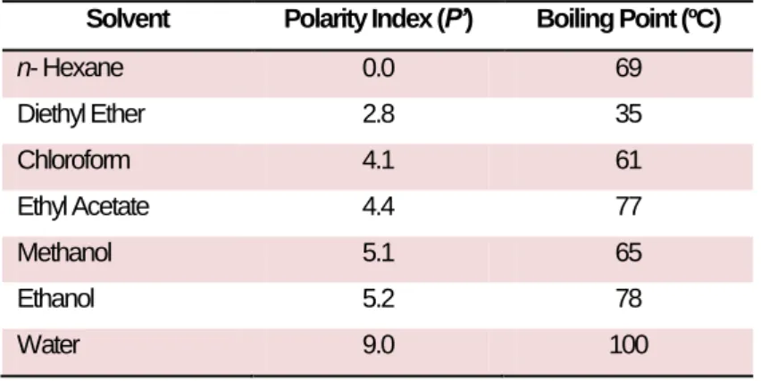
Several methodologies have been used for extracting compounds from herbs, plants and algae, some of which are more effective than others. It is of extreme importance to choose properly the solvent to be used in the extraction process, in order to increase its affinities to the compounds that are going to be extracted from the algae, as it is known that “equal dissolves equal” or, in other words, a solvent with a certain polarity dissolves compounds with the same polarity. When there is no available information on the polarity of the desired bioactive compounds to be extracted, a safe methodology consists in choosing several solvents, with different polarity indices, in order to cover a wide range of polarities (Table 1.1).
Table 2.1 - Polarity Index and boiling points of some solvents (adapted from Seidel, 2006).
Solvent Polarity Index (P’) Boiling Point (ºC)
n- Hexane 0.0 69 Diethyl Ether 2.8 35 Chloroform 4.1 61 Ethyl Acetate 4.4 77 Methanol 5.1 65 Ethanol 5.2 78 Water 9.0 100
Another important parameter is the temperature during extraction. Extractions can be conducted at room temperature (cold extractions) or at high temperature (hot extractions), and different values of temperature can promote the extraction of different compounds. The type of extraction chosen depends on the compound (currently secondary metabolites) that we wanted to extract as well as the type of raw material (very often plant material) that was going to be used for that purpose. Therefore, cold extractions such as maceration or soaking the tissue in water or other organic solvents are often recommended when the compounds of interest are sensible/unstable to high temperatures, e.g. volatile compounds. Hot extractions are used mainly because the increase in temperature leads to an increased of the substance solubility, which is why the hot extractions methods are generally faster than those performed at room temperature. Infusions and steam distillations are commonly hot extractions methods used to extract, e.g. essential oils from plants (Seidel, 2006).
9
Cold extractions can be prepared with a flask containing the sample and an appropriate solvent in a shaking device for several hours or days, whereas hot extractions may need more complex equipment. Some hot extractions are also simple to prepare, as they can be done by simply pouring a hot solvent (e.g. water) over the sample (such as happens in an infusion); other types of hot extractions require a more complex apparatus such as a heating mantle, a Soxhlet extractor and a condenser (as shown in Figure 1.4). In this type of extraction process (continuous solid/liquid extraction), the solid sample is placed in a sort of thimble, made from thick filter paper, inside the chamber of the Soxhlet extractor, while the extraction solvent, placed in a balloon, is heated by the heating mantle. The solvent evaporates and the vapor travels through the exterior canal until it reaches the condenser. There, the vapor condenses as it reaches a cold site due to the cold water that isconstantly passing through the condenser, and drops above the paper thimble that contains the sample. The chamber slowly fills with solvent and it is in this site that the extraction takes place since the desired compounds pass from the sample to the solvent. When the Soxhlet’s chamber is almost full of solvent, the chamber is automatically discharged by a siphon side arm, through communicating vessels principle, with the solvent running back down to the flask and the process starts over. This cycle can be repeated many times, over several hours or days. After the extraction process, it is necessary to evaporate the solvent in order to recover the extracted compounds. This is accomplished using a rotatory evaporator (Kou et al., 2003).
1.5
Antimicrobial activity
Seaweed extracts are being investigated for antimicrobial activity, among others, due to their composition in various compounds. In these research studies several substances were identified as antimicrobial agents: chlorellin derivatives, acrylic acid, terpenes, phenolic inhibitors, halogenated aliphatic compounds and sulfur-containing heterocyclic compounds (Espeche et al., 1984). These last two are toxic to microorganisms and therefore responsible for the antimicrobial activity of some seaweeds (Salem et al., 2011). Beyond these compounds, antimicrobial activity can also be derived from the presence of some amino acids, phlorotannins, steroids, halogenated ketones and alkanes,
Figure 1.4 - Soxhlet extraction apparatus (adapted from Kou and Mitra, 2003).
10
cyclic polysulphides and fatty acids (Watson and Cruz-Rivera, 2003). However, many questions still need to be answered and therefore new studies are continuously appearing in the literature:
Gerasimenko et al. (2010) have been dedicated to the study of diverse activities derived from algae such as antimicrobial and hemolytic activities. This author studied the effect of ethanolic extract of brown seaweed (Laminaria cichorioides) on two yeasts (Candida albicans and Safale), two fungus (Aspergillus niger and Fusarium oxysporum) and two bacteria (S. aureus and E. coli). Results demonstrated that the extracts had antimicrobial activity on the yeasts and on the Gram negative bacteria under assay. Moreover, hemolytic activity of the seaweed extract was evaluated via the addition of the extract to an erythrocyte suspension. The results obtained showed its effectiveness since it caused hemolysis of the cells.
Vijayavel and Martinez (2010) stated that ethanolic extracts of two Hawaiian marine algae (Ulva fasciata and Gracilaria salicornia) exhibited antimicrobial and antioxidant activity and could be used as bioactive compounds in nutraceutical agents.
Mtolera and Semesi (1996) tested the effect of diethyl ether extracts of six green algae collected in the coast of Tanzania, on Gram positive and Gram negative bacteria and yeast, as well as their cytotoxic activity, using Artemia salina larvae, concluding that both antimicrobial and cytotoxic activities were effective and dependent on the alga species.
Ely et al. (2004) studied the effect of methanolic extracts of some marine organisms (sponges and seaweeds – Stoechospermum marginatum and Cladophora prolifera) from the south east coast of India, against clinical isolates of bacteria and fungi, and found that some of them showed good antimicrobial activity.
Another study conducted by Patra et al. (2008) revealed that Sargassum spp., a brown alga collected in Goa, could be used against several diseases and as preservative in food industry, since it proved to have antimicrobial and antioxidant activity.
Seenivasam et al. (2010) tested acetone, methanol and ethanol extracts from some green algae from the coast of India against several microorganisms from clinical and food origin and concluded that they could inhibit both Gram positive and negative bacteria depending on the alga species.
Demirel et al. (2009) conducted a study to evaluate methanol, dichloromethane and hexane extracts from several brown algae collected along the Izmir coast (Turkey) for their antimicrobial and antioxidant activity. Results showed that dichloromethane was more effective than the two other solvents tested, both in the antimicrobial and antioxidant activities.
Plaza et al. (2010) tested hexane, ethanol and water extracts from brown algae for antimicrobial activities, and discovered that ethanol was the most appropriate solvent to extract fatty acids and volatile compounds with antimicrobial activity. These compounds were then identified as phytol, fucosterol, neophytadiene, palmitic, palmitoleic and oleic acids.
11
Finally, Salem et al. (2011) tested methanolic and ethyl acetate extracts of eight different seaweeds from the red sea (Egypt), against both Gram positive and Gram negative bacteria, and concluded that the ethyl acetate extracts had a higher effect on microorganisms tested, mainly on Gram positive bacteria, when compared with the methanolic ones.
1.6
Antioxidant activity
Antioxidant activity has been show to play an important role on several pharmacological issues such as anti-aging, anti-inflammatory, anti-atherosclerosis and anti-cancer activities. The inhibition of the free radical induced damage by supplementation of antioxidants has become an attractive therapeutic strategy for reducing the risk of diseases (Amornlerdpison et al., 2007). It is known that free radicals can be generated in the biological systems by the form of reactive oxygen species (ROS). The reactive forms can be superoxide anion radicals (O2
-), hydroxyl radicals (HO-), hydrogen peroxide (H2O2) and singlet oxygen (
1
O2). These molecules belong to a class of highly reactive molecules that derive from the normal metabolism of oxygen or from exogenous factors and agents (Heo et al., 2006). Many pathological conditions such as atherosclerosis, arthritis, diabetes, muscular dystrophy, pulmonary dysfunction, ischemia reperfusion tissue damage and neurological disorders such as Alzheimer’s disease are associated with the oxidative damage of DNA, protein, lipid and other molecules caused by the reactive oxygen species (Amornlerdpison et al., 2007). In these pathological conditions, the antioxidant mechanisms are often inadequate since excessive quantities of ROS can be generated. The ROS formed may cause cellular damage by peroxidation of membrane lipids by denaturating cellular proteins and by breaking DNA strands, disrupting cellular functions (Patra et al., 2008).
It is also known that antioxidants are used as a preservative of food quality, mainly by preventing the oxidative deterioration of lipid constituents in food, increasing associated shelf life. Nowadays there is an increasing interest in antioxidants from natural sources, because of the safety and toxicity problems of synthetic antioxidants such as butylated hydroxylanisol (BHA) and butylated hydroxitoluene (BHT), which are commonly used in lipid-containing food. Thus, natural antioxidants can protect the human organism from reactive oxygen species and free radicals and retard the progress of many chronic pathologies as well as lipid oxidative rancidity in foodstuff (Amornlerdpison et al., 2007; Heo et al., 2006).
Seaweeds are considered to be a rich source of antioxidants and many species of these marine organisms have been investigated to identify new and effective antioxidant compounds, as well as to explain the mechanisms of cell proliferation and apoptosis. In recent times, active antioxidant compounds from some algae were identified as fucoxanthin, phlorotannins and other polyphenolic compounds (Heo et al., 2006).
Many studies have been performed with algae all over the world to search for antioxidant and other activities that these marine organisms could offer to mankind. Authors like López et al. (2011)
12
examined water, water/methanol (1/1), methanol and ethanol crude extracts from brown algae for antioxidant activity and total phenolic contents and found a significant association between these two. The aqueous extract showed the highest antioxidant activity and the highest phenolic content. Moreover, phenolic antioxidants were examined by RP-HPLC and it was found that gallic acid was the predominant polyphenol present in the algae extracts.
Onofrejová et al. (2010) studied the methanolic extracts of two macroalgae (Porphyra tenera and Undaria pinnatifida) and found that their antioxidant activity were due to their high content in phenolic compounds such as p-hydroxybenzoic acid, salicylic acid, protocatechuic acid, 2,3-dihydroxybenzoic acid, p-coumaric acid, cinnamic acid and some other derivatives.
In addition, Rodríguez-Bernaldo de Quirós et al. (2010) improved a new and simple extraction procedure using methanol-water-acetic acid (30:69:1, (v/v/v)) to prepare extracts from both red (Porphyra spp.) and brown algae. Their studies demonstrated that the antioxidant effect founded could be due to the content of polyphenols. They concluded that the concentration of polyphenols in algae depends on many variables such as habitat, harvesting season, environmental conditions such as light, temperature and salinity, as well as with the algae species. Results showed that epigallocatechin was the predominant catechin in the algae studied and epigallocatechin gallate, epicatechin gallate and catechin gallate were found in smaller quantity. Epicatechin gallate was only detected in brown algae and catechin was only found in Porphyra spp.
Furthermore, Amornlerdpsion et al. (2007) tested the aqueous extracts of fresh brown marine algae collected in Thailand for antioxidant activity by DPPH, ABTS•+ and lipid peroxidation of rat liver. These assays showed the antioxidant activity of the algae extracts reflected in their capacity to reduce both free radicals and oxidative damage.
Devi et al. (2011) studied the antioxidant activity of both methanolic and diethyl ether extracts of brown, green and red seaweeds, and found that they all contained phenolic constituents in various proportions and showed antioxidant activity in various degrees, whereas methanolic extracts exhibited higher antioxidant activities when compared to diethyl ether extracts.
For all these reasons, the supplementation of antioxidants in human diet has become an attractive therapeutic strategy for reducing the risks of diseases caused by the free radical induced damage (Amornlerdpison et al., 2007), and the active search for new and natural sources of antioxidant compounds is on the way.
1.7
Work objectives
The aim of this research work was to characterize the antioxidant and antimicrobial properties of red algae from the Portuguese coast, collected from natural habitats and from integrated aquaculture systems, in order to evaluate their potential as ingredients for food a/or nutraceutical or pharmaceutical industries. In order to accomplish this general goal three specific objectives were
13
established: (i) to implement an effective methodology for obtaining extracts from marine macroalgae, collected from both culture systems; (ii) to test the antimicrobial and antioxidant activities of the prepared extracts against selected species of Gram negative and Gram positive bacteria (from clinical and food origins), as well as against a yeast species; and (iii) to analyse the composition of the extracts, both by GC and HPLC, in order to identify possible compounds responsible for the activities found.
14
2
Materials and Methods
2.1
Algae
Seaweeds used in this study were collected in the North of Portugal. Some of them were from integrated aquaculture and others were collected directly in coastal areas, being all provided by CIIMAR (Centre of Marine and Environmental Research of the University of Porto).
The species studied were: Gracilaria vermiculophylla, Porphyra dioica and Chondrus crispus (Rhodophyta). Those from aquaculture were collected from December 2010 to May 2011, whereas algae collected along the coast followed the calendar: Gracilaria vermiculophylla and Porphyra dioica (May 2011), Chondrus crispus (March 2012).
All the samples were collected in plastic bags and brought to the laboratory to be washed with fresh water to remove all the epiphytes, necrotic parts and suspended materials. Then, part of the samples was dried at 37 ºC, shredded with a food processor (A327R1
,
Moulinex, Spain) and kept in the dark until use; the remaining fresh sample was immediately extracted.2.2
Preparation of seaweed extracts
2.2.1 Optimization of extraction procedureIn order to optimize the extraction procedure, initial conditions of the algae (fresh or dried samples) and extraction temperature (room temperature or boiling temperature of solvent) were simultaneously tested in G. vermiculophylla samples, using ethyl acetate as extraction solvent. This solvent was selected due to its behavior in previous extraction studies with other algal samples.
Extracts at room temperature were prepared by weighting 20 g of fresh or dried algae into a closed bottle wrapped with aluminium foil (to protect from light deterioration); 50 mL of ethyl acetate (Merck, Germany) were added, and the mixture was kept with agitation for 72 hours at room temperature.
Ethyl acetate extracts of G. vermiculophylla at higher temperature were obtained by weighting the same amounts of algae as for cold extracts (fresh and dried), and refluxing in Soxhlet apparatus for about 10 hours.
Final extracts, at room temperature and boiling temperature of solvent were evaporated under reduced pressure with a rotatory evaporator and re-dissolved in DMSO (Sigma-Aldrich, Missouri, USA).
2.2.2 Preparation of extracts to analyze
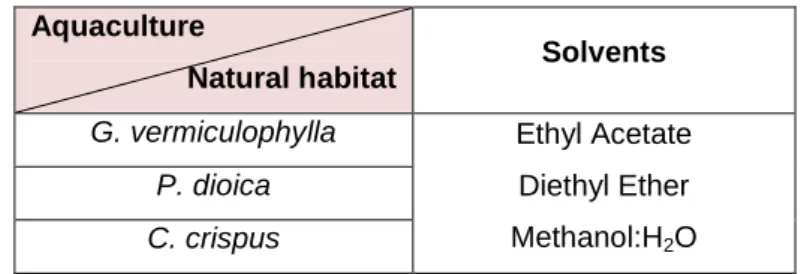
After choosing the most efficient extraction method, hot dried extracts from G. vermiculophylla, P. dioica and C. crispus (from aquaculture and wild) were prepared using solvents with different polarity:
15
diethyl ether (Sigma-Aldrich, Missouri, USA), ethyl acetate and methanol:H2O (1:1) (Sigma-Aldrich, Missouri, USA).
Thus, 15 g of each sample were extracted in 200 mL (in duplicate) of the three different solvents, using Soxhlet apparatus during approximately 18 hours. In the end of each extraction the solvents were evaporated, and the dry extract was re-dissolved in DMSO and stored at -30 ºC until use, as described by Lekameera et al. (2008). The concentrations of all the extracts were ca. 500 g/L.
2.3
Determination of antimicrobial activity
In this study the following microorganisms were used to evaluate the antimicrobial activity of the seaweed extracts:
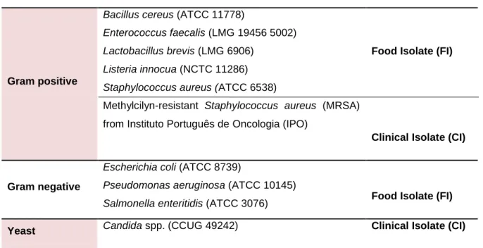
Table 2.1 - List of the microorganisms tested.
Gram positive
Bacillus cereus (ATCC 11778)
Enterococcus faecalis (LMG 19456 5002) Lactobacillus brevis (LMG 6906)
Listeria innocua (NCTC 11286) Staphylococcus aureus (ATCC 6538)
Methylcilyn-resistant Staphylococcus aureus (MRSA) from Instituto Português de Oncologia (IPO)
Food Isolate (FI)
Clinical Isolate (CI)
Gram negative
Escherichia coli (ATCC 8739)
Pseudomonas aeruginosa (ATCC 10145)
Salmonella enteritidis (ATCC 3076) Food Isolate (FI)
Yeast Candida spp. (CCUG 49242) Clinical Isolate (CI)
All bacteria were cultivated and stored in Brain Heart Infusion (BHI) or Agar (BHI agar) (BD, Maryland, USA) except Enterococcus faecalis and Lactobacillus brevis that used Nutrient Broth (NB) or Nutrient Agar (NA) (Oxoid, England), and Candida spp. that used Sabouraud Broth or Agar (Difco, Michigan, USA), as necessary.
In the present study, agar diffusion method was used to assess the antimicrobial activity of the extracts. So, overnight cultures of the microorganisms were adjusted at 0.5 of McFarland standard (1.5 x 10⁸ CFU/mL) before spreading 100 µL of the culture broth on the respective culture medium. Afterwards, algae extracts were applied directly on seeded agar plates using the drop method (20 µL). Negative control was performed with DMSO whereas positive controls were checked with ampicillin at 1000 µg/mL Aldrich, Missouri, USA) for L. innocua, cycloheximide at 1000 µg/mL
(Sigma-16
Aldrich, Missouri, USA) for Candida spp. and chloramphenicol at 1000 µg/mL (Sigma-Aldrich, Missouri, USA) for the remaining microorganisms. Lactic acid 30% (v/v) (Fluka, Missouri, USA) was also used as a complementary negative control for all the microorganisms.
All the tests were performed under sterile conditions and prepared in quadruplicate. Plates were incubated at 37 ºC for 16 hours and examined for inhibition zones around the drop points. Inhibition of microbial growth was determined as the diameter of these inhibition zones.
2.4
Determination of lipid profile
To determine the lipid profile of the samples the Lepage and Roy method (1994), modified by Carvalho et al. (2006) was used for sample derivatization for gas chromatography (GC). Thus, 1 mg of internal standard (5 mg/mL) and about 200 mg of algae extract were added to a Teflon-capped Pyrex tube. Then, 2 mL of a freshly prepared mixture of acetyl chloride and dried methanol (5:100, (v/v)) were also added. Nitrogen was blown up in each tube to remove all the oxygen, tubes were rapidly closed and protected from the light with aluminum foil and heated in a heating block at 90-100 ºC for 1 hour. After cooling to 30 - 40 ºC in the dark, 1 mL of hexane (with 0. 01 % (v/v) BHT) was added and mixed in vortex for a few seconds. Afterwards, 1 mL of pure water was added, the tubes were mixed gently to allow the phases to separate, and the upper phases were removed with a Pasteur pipette, filtered into another Pasteur pipette filled with cotton and anhydrous Na2SO4, and collect in a GC vial. Analysis of fatty acid methyl esters were performed in a gas chomatograph AutoSystem XL from HP (California, USA) and was equipped with a flame ionization detector. The separation was performed in a Supelcowax-10 (60 m, 0.32 mm and 0.25 µm) column from Supelco (Pennsylvania, USA). The temperature was programmed to increase from 170 to 220 ºC at a rate of 1 ºC min-1; the injector and detector temperatures were 250 and 270 ºC, respectively. Injections were performed under splitless mode, using helium as a carrier gas. Calculations were performed according to the AOCS official method Ce 1b-89. Pure standards (Sigma) were used for fatty acid identification, which was based on a comparison of peak retention times between samples and standards.
2.5
Determination of antioxidant activity
The total antioxidant capacity of the different algae extracts was determined with the ABTS•+ method, according with Gião et al. (2007). ABTS•+ (2,2-azino-bis(3-ethylbenzothiazoline-6-sulfonic acid) radical cation was generated by a reaction of 7 mM of ABTS (Sigma-Aldrich, Missouri, USA) with 2.45 mM potassium persulfate (Merck, Germany). The reaction mixture was allowed to stand in the dark for 16 hours at room temperature. This radical cation has blue color and absorbs light at 734 nm. Working solution was prepared by diluting ABTS•+ stock solution in ultra-pure water until the initial optical density (OD) was 0.700±0.020 nm using an UV mini 1240 UV-Vis spectrophotometer (Shimadzu, Japan). Then, 20 µL of the diluted (1:50) algae extracts were added to 1 mL of the ABTS•+ diluted
17
solution and the final optical density (OD) was measured after 6 minutes of reaction. Samples were measured in triplicate. The total antioxidant capacity was expressed as percentage of inhibition (PI) according to the equation:
PI
x 100
(2.1)
where AbsABTS•+ was the initial absorbance of diluted ABTS•+ and Abssample denotes the absorbance of the sample. Through the use of a calibration curve prepared with ascorbic acid as a standard, the final result was expressed as equivalent concentration of ascorbic acid (mg ascorbic acid equivalent/ g extract) (see Appendix 6.2).
2.6
Determination of phenolic profile
The phenolic profile of the seaweed extracts were determined by HPLC-DAD (Waters Series 600, Massachusetts, EUA). Detection was achieved by a diode array detector (Waters, Massachusetts, EUA) at wavelength intervals of 200 – 600 nm in intervals of 2 nm. Separation was performed in a reverse phase Symmetry® C18 column (250 x 4.6 mm i.d., 5 µm particle size and 125 Å pore size) with a guard column containing the same stationary phase (Symmetry® C18). Chromatographic separation of phenolic compounds was carried out with solvent A (water, methanol and formic acid at 92.5:5:2.5), and solvent B (methanol and water at 94:6), under the following conditions: linear gradient starting at 0 to 10% solvent B in 10 min at 0.5 ml/min, 10 to 30% in 40 min at 0.65 ml/min, 30 to 50% in 20 min at 0.75 ml/min and from 50 to 0% in 10 min at 0.75 ml/min. Injection volume was 50 µl and temperature was 30 ºC. Absorbance was measured at 260 - 280 nm for phenols. Retention times and spectra of compounds were analysed by comparison with pure standards of protocatechuic acid, (+)-catechin, (-)-epi(+)-catechin, chlorogenic acid, dihydroxibenzoic acid, p-coumaric acid, hydroxycoumarin, quercetin, ascorbic acid, gallic acid, ferrulic acid, caffeic acid, rutin and cinnamic acid.
2.7
Statistical analysis
To evaluate the normality of the distributions, the Kolmogorov-Smirnov test was used. The One Way ANOVA test in association with Scheffe’s test was used when a normal distribution was observed between the differences in sample groups. The differences were considered statistically significant at a 5% confidence degree level. All statistical analysis were performed using IBM SPSS Statistics v.19.0.0 (New York, USA) software.
18
3
Results and Discussion
3.1
Optimization of extraction procedure
Bioactive properties of marine algae have been empirically known for centuries, but only recently have been studied in more detail. These properties have been attributed to a wide range of different algal species, extracted under various conditions (Demirel et al., 2009; Patra et al., 2008; Vijayavel and Martinez, 2010); such miscellaneous data complicates the statement of correlations between algal compounds and bioactive effects, and therefore detailed studies are necessary. In order to test antimicrobial and antioxidant activities of algae it was necessary to previously obtain extracts rich in compounds with those properties. As extracts’ properties depend on the conditions in which they were prepared, the optimization of the extraction procedure was the first logical step to be performed. There are many methodologies to produce extracts, but the most common are infusions with hot water or extractions with organic solvents. Considering the fact that most of the consulted literature referenced extractions with organic solvents, this was the line of research followed in this study (Demirel et al., 2009; Patra et al., 2008).
Thus, initially, preliminary tests were performed with G. vermiculophylla from an integrated aquaculture system which was extracted with an organic solvent (ethyl acetate) at room temperature, to assess the most appropriate physical state of the alga (fresh or dried) for achieving the best extracts, i.e., those with higher antimicrobial activity. When algae were tested dried, the drying temperature employed was 37 ºC, as it was described that at a higher temperature (60 ºC) little or no activity against several microorganisms (S. aureus, B. subtilis, E. coli and C. albicans) was shown (Mtolera and Semesi, 1996). It was found that the most effective extracts were those prepared with dried algae, since they presented a larger growth inhibition zone. These results were not in accordance with Tuney et al. (2006), who observed less or no effects of dried algae extracts on bacteria, when compared to the fresh ones. However, this result could probably be related to the loss of volatile antimicrobial compounds present in fresh algae (hydrogen peroxide, terpenoid and bromo-ether compounds and volatile fatty-acids) during the drying process at higher temperatures.
Thereafter, the extraction temperature was tested with cold and hot extractions with ethyl acetate of fresh and dried G. vermiculophylla (also from aquaculture regime). Results of these tests are presented in Figure 3.1.