Contents lists available atScienceDirect
Industrial Crops & Products
journal homepage:www.elsevier.com/locate/indcrop
Croton argyrophyllus
Kunth and
Croton heliotropiifolius
Kunth: Phytochemical
characterization and bioactive properties
Sara Samanta da Silva Brito
a, Franceli Silva
a,⁎⁎, Ricardo Malheiro
b, Paula Baptista
b,
José Alberto Pereira
b,⁎aCenter for Agricultural, Environmental and Biological Sciences, Federal University of Recôncavo da Bahia (UFRB), University Campus, 44380-000, Cruz das Almas,
Bahia, Brazil
bCentro de Investigação de Montanha (CIMO), School of Agriculture, Polytechnic Institute of Bragança, Santa Apolónia Campus, 5300-253, Bragança, Portugal
A R T I C L E I N F O
Keywords:
Medicinal plants
Crotonspp Essential oil Chemical composition Bioactive properties
A B S T R A C T
Croton heliotropiifoliusKunth andCroton argyrophyllusKunth are endemic plant species from northeastern Brazil widely used in folk medicine and scarcely studied. In this context, the essential oils (EO’s) and methanolic extracts (leaves and stalk) of both species were chemically characterized, and their antioxidant and antimicrobial activities were assessed. The chemical characterization of the EO’s identified sixty components,being the major ones inC. argyrophyllusbicyclogermacrene (14.0%),β-pinene (8.9%) and spathulenol (8.7%), and inC. helio-tropiifoliuslimonene (16.9%),α-pinene (13.3%) and caryophyllene (12.1%). Essential oils and methanolic ex-tracts from leaves ofC. argyrophylluspossess greater antioxidant potential, which could be related to the high levels of total phenols andflavonols. The antimicrobial activity ofC. argyrophyllusessential oil proven to be more efficient than chloramphenicol (30μg mL−1), with a minimum inhibitory concentration (MIC) of 25μL mL−1 againstBacillus subtilis,Staphylococcus aureus, Escherichia coliand Pseudomonas aeruginosa and 10μL mL−1 againstB. cereus. Leaf extracts presented high activity against yeasts (MIC = 50 mg mL−1) beingC.
helio-tropiifoliuseffective againstCandida albicansandC. parapsilosis, whileC. argyrophylluswas effective againstC. glabrata. Overall results showed that these plant species are potential sources of phytochemicals with interest in thefields of both pharmacology (e.g., antimicrobial) and human health (e.g., antioxidant). Furthermore, in the socio-economic aspect, these results can improve and disseminate the cultivation of these species, inducing improvements in the rural populations.
1. Introduction
The genusCroton, the most diverse genus of the Euphorbiaceae fa-mily, contains about 1300 species distributed across the world’s tropical regions. In Brazil, around 350 of these species can be found, being 252 of them considered endemic (Secco et al., 2012).Crotongreat diversity in species is also reflected in their chemical constituents, most of them still unknown (Dória et al., 2010).Croton heliotropiifolius Kunth and Croton argyrophyllusKunth are popularly known in Brazil as“velame” or“velame-branco”and“cassutinga”. They are endemic species from northeastern Brazil and are frequently found in the vegetation of the Caatinga biome. These species are greatly used in folk medicine in Africa, Asia and South America, generally as stimulants, tranquilizers, insecticides, vermifuges and analgesics, among other uses (Compagnone et al., 2010).
Different chemical classes of compounds were identified inCroton
species, such as terpenoids and phenylpropanoids, which could be re-lated to the biological activity demonstrated by their species (Aguiar et al., 2016). Concerning toC. heliotropiifoliusandC. argyrophyllussome important terpenic compounds, monoterpenes and sesquiterpenes, were identified namely limonene, β-caryophyllene, spathulenol, bicyclo-germacrene and bicyclo-germacrene D (Fontenelle et al., 2008;Angélico et al., 2014;Souza et al., 2017).
Plant extracts and essential oils are good sources of molecules such as terpenoids,flavonoids, and tannins (Sun et al., 2015), that possess important biological activities. When present in food, due to their an-timicrobial and antioxidant activities prevent the deterioration and increase the nutritional value. The importance of this type of matrices increases nowadays with the knowledge of the rise of resistance to human pathogenic bacteria and fungi to the available synthetic pro-ducts (Grundmann et al., 2011). In this context, the use of plant extracts and essential oils in food and pharmaceutical industries has been
https://doi.org/10.1016/j.indcrop.2018.01.044
Received 25 August 2017; Received in revised form 23 December 2017; Accepted 19 January 2018
⁎Corresponding author at: Centro de Investigação de Montanha (CIMO), ESA, Instituto Politécnico de Bragança, Campus de Santa Apolónia, 5300-253, Bragança, Portugal.
⁎⁎Corresponding author. Centre for Agricultural, Environmental and Biological Sciences, Federal University of Recôncavo da Bahia, 44380-000, Cruz das Almas, Bahia, Brazil. E-mail addresses:[email protected](F. Silva),[email protected](J.A. Pereira).
0926-6690/ © 2018 Elsevier B.V. All rights reserved.
growing significantly and is an openfield with great potential. Recent works have demonstrated the antimicrobial, insecticidal and anti-oxidant activities ofCrotonspecies (Queiroz et al., 2014;Wijesundara et al., 2016) which are correlated with the chemical composition of their essential oils. Compounds present in the essential oils of some Crotonspecies, such as spathulenol and caryophyllene oxide, have the capacity to inhibit the growth of some filamentous fungi species (Wenqiang et al., 2006); whereas bicyclogermacrene, α-pinene and caryophyllene have been described to possess antimicrobial activity (Cavin et al., 2006;Morais et al., 2006;Ramos et al., 2013).
In this context, the main objective of this work is to contribute for the phytochemical characterization of two key plant species of estab-lished socio-economic importance in the Caatinga biome, namely C. heliotropiifoliusandC. argyrophyllus. Exploitation of their essential oil and aerial part for bioactive compounds (i.e. antioxidant and anti-microbial) is imperative to better know the real potential of these plants as a source for drug development and use as a health supplement.
2. Material and methods
2.1. Standards and reagents
DPPH (2,2-diphenyl-1-picrylhydrazyl), ABTS [2,2-azino-bis(3-ethylbenzothiazoline-6-sulfonic acid)], Trolox (6-hydroxy-2,5,7,8-tet-ramethylchroman-2-carboxylic acid), iron (III) chloride, potassium persulfate, trichloroacetic acid, gallic acid, quercetin, Tween 80, re-zasurin, dimethyl sulfoxide, sodium chloride, agar-agar and saturated alkanes series were obtained from Sigma-Aldrich (St. Louis, USA). Methanol (HPLC grade), ethanol absolute, sodium dihydrogen phos-phate dihydrate, glucose, and potassium hexacyanoferrate (III) were purchased from Merck (Darmstadt, Germany). Hydrochloric acid, di-sodium hydrogen phosphate 2-hydrate, and caffeic acid were obtained from Panreac (Barcelona, Spain). Chloramphenicol and fluconazole were obtained from Oxoid Ltd (Basingstoke, UK). Yeast extract, peptone and tryptone were obtained from Himedia (Mumbai, India). The water was treated in a Milli-Q water purification system (Millipore, Bedford, MA, USA).
2.2. Plant material
Both species were collected in the Caatinga biome (Bahia, Brazil) between February and April 2016.Croton argyrophylluswas recovered in São Domingos (11°27'56“S 39°31'34“W), and C. heliotropiifolius in Conceição do Coité (11°33′50′’S 39°16′58′’W). Voucher species were deposited in the Herbarium of the Federal University of Recôncavo da Bahia, Cruz das Almas, with the catalog numbers HURB 15400, forC. heliotropiifolius, and HURB 15401 forC. argyrophyllus. For each species, six independent samples were taken and prepared separately.
2.3. Distillation of essential oil and preparation of extracts
The plant material was dried in a heated chamber with forced cir-culation at 40 °C until a constant weight was reached. This took around three to four days. After drying, the leaves were ground up manually and 50 g of them were placed in a balloonflask and distilled water was added up to a total volume of three liters. This mixture was then sub-jected to a hydrodistillation process in steam using Clevenger apparatus (model TE 2762 from Tecnal) for two hours, to obtain the essential oil. Methanolic extracts were prepared from the leaves and stalk. Briefly, 500 mL of methanol (HPLC grade) was added to 50 g of plant material and the extraction was carried out during 72 h, being the solvent renewed every 24 h (500 mL of methanol) and recovered throughfiltration. Oncefinished the 72 h, the solvents were combined and the methanolic extract was obtained under vacuum in a rotary evaporator (RE300/MS, Bibby Scientific Limited, Staffordshire, UK) at a temperature of 40 °C. All the extractions, both for essential oils and
methanolic extracts were carried out in triplicate.
2.4. Characterization of essential oils
The essential oils of C. argyrophyllus andC. heliotropiifolius were determined through gas chromatography analysis using mass spectro-metry detection (GC/MS). Before injection, 10μL of the essential oil were diluted in 0.5 mL of methanol, and 1μL of the volume was in-jected.
2.4.1. Gas chromatography with mass spectrometry detection (GC/MS) The gas chromatograph used was a Shimadzu model GC-2010 coupled to a GC/MS-QP 2010 Shimadzu mass spectrometer (Kyoto, Japan). A TRB-5MS capillary column (30 m × 0.25 mm × 0.25μm; Teknokroma, Spain) was used. The injector port (model AOC20i + s) was heated to 220 °C, and the injections were performed in split mode (1:20). The oven temperature was programmed to start at 60 °C and then increase by 3 °C/min until reaching 240 °C, and this was then maintained for 20 min. The carrier gas was helium (Praxair, Portugal), at a linear velocity of 30 cm/s and a totalflow rate of 44.3 mL/min. The temperature of the ionization source was maintained at 240 °C, the ionization energy at 70 eV, and the ionization current at 0.1 kV. Compounds were identified by comparing their MS spectra with those obtained from a database (NIST 11; the minimum identification simi-larity is 80%), and with those of pure compounds analyzed under the same conditions, and by comparing the retention indices (in the form of Kovats indices, with the series of alkanes injected with the same chromatographic conditions used for the EO’s) with data in the litera-ture (Adams, 2007). Retention indices were calculated according tovan Den Dool and Kratz (1963). The results were expressed as the relative percentages of each compound, calculated via normalization of the chromatographic peak areas.
2.5. Antioxidant activity
Three methodologies were used to evaluate the antioxidant poten-tial of the species studied. For the essenpoten-tial oil, the 2,2-diphenyl-1-pi-crylhydrazyl (DPPH) and 2,2-azino-bis(3-ethylbenzothiazoline-6-sul-fonic acid) (ABTS) methods were used, at concentrations ranging from 10 to 50 mg mL−1. For the methanolic extracts from the leaves and
stalks, the DPPH, ABTS and reducing power methods were tested, at concentrations ranging from 0.1 to 2.0 mg mL−1. Trolox was used as a
reference for the three methods at concentrations ranging from 0.0025 to 0.25 mg mL−1
.
2.5.1. Scavenging effect on DPPH radicals
The capacity to scavenge the DPPH free radical was monitored in accordance with the method ofHatano et al. (1988)with minor mod-ifications. The extract solution/EO (0.3 mL) was mixed with 2.7 mL of a methanol solution containing DPPH radicals (6 × 10⁻⁵mol L−1). The
mixture was shaken vigorously and left to stand for 60 min and three hours, respectively for the methanolic extracts and EO’s, at room tem-perature in the dark (until stable absorbance values were obtained). The reduction of the DPPH radical was measured by continuous mon-itoring of the absorption decrease at 517 nm (Genesys 10UV, Thermo Electron Corporation). The DPPH scavenging effect was calculated as the percentage of DPPH discoloration, using the following formula: [(ADPPH−AS)/ADPPH] × 100, where ASwas the absorbance of the so-lution when the sample extract has been added at a particular level, and ADPPHwas the absorbance of the DPPH solution. The EO/methanolic extract concentration providing 50% inhibition (EC50) was calculated from the graph of percentage scavenging effect against extract con-centration in the solution.
2.5.2. Scavenging effect on ABTS radicals
(2007), based on the capacity of a sample to inhibit the ABTS radical. The ABTS scavenging effect was calculated as the percentage of ABTS discoloration, using the same formula as used for the DPPH method. The ABTS radical was generated by chemical reaction with potassium persulfate (K2S2O8). To 25 mL of ABTS (7 mmol L−1
) were added 440μL of K2S2O8(140 mmol L−1), being the solution kept in darkness
during 12–16 h at room temperature in order to form the radical. An accurate volume of the previous solution was diluted in absolute ethanol until an absorbance of 0.70 ± 0.02 atλ= 734 nm (Genesys 10UV, Thermo Electron Corporation). Once the radical was formed 2 mL of the ABTS radical solution were mixed with 100μL of the me-thanolic extracts/EO and the absorbance measured atλ= 734 nm. The ABTS scavenging effect and EC50values were calculated according to the previously mentioned for the DPPH method.
2.5.3. Reducing power
The reducing power was determined in accordance with the pro-cedure described by Berker et al. (2007). The extract solution (1 mL from 0.1 to 2 mg mL−1) was mixed with 2.5 mL of 200 mmol L−1
so-dium phosphate buffer (pH 6.6) and 2.5 mL of 1% potassium ferricya-nide. The mixture was incubated at 50 °C for 20 min. After cooling, 2.5 mL of 10% trichloroacetic acid (w/v) were added and the mixture was centrifuged at 1000 rpm for 8 min (Centorion K24OR-2003 re-frigerated centrifuge). The upper layer (2.5 mL) was mixed with 2.5 mL of deionised water and 0.5 mL of 0.1% ferric chloride, and the bance was measured spectrophotometrically at 700 nm (higher absor-bance readings indicate higher reducing power). Extract concentration providing 0.5 of absorbance (EC50) was calculated from the graph of absorbance at 700 nm (Genesys 10UV, Thermo Electron Corporation) against extract concentration in the solution.
2.6. Determination of different groups of phenolic compounds
The total phenols, hydroxycinnamic acid derivatives andflavonols content were determined in triplicate in accordance with the metho-dology described byBoulanouar et al. (2013). One mL of the metha-nolic extract (1 mg mL−1) was diluted with 1 mL of aqueous ethanol
(95% v/v) containing 0.1% hydrochloric acid and 8 mL of 2% hydro-chloric acid. The absorbance was measured at 280 nm to determine total phenols, 320 nm for hydroxycinnamic acid derivatives, and 360 nm for flavonols content (Genesys 10UV, Thermo Electron Cor-poration). The results were expressed as gallic acid equivalents (GAE) g−1of extract for total phenols, caffeic acid equivalents (CAE)
g−1
of extract for hydroxycinnamic acid derivatives and quercetin (QE) g−1of extract forflavonols. The calibration curves followed the
same methodology applied to the methanolic extracts, with con-centrations varying from 0.001 and 1 mM for gallic and cafeic acids, and concentrations from 0.001 to 0.5 mM for quercetin.
2.7. Antimicrobial activity
The antimicrobial activity of both the extracts and the essential oils obtained fromC. argyrophyllusandC. heliotropiifolius were evaluated against Gram-positive bacteria (Bacillus cereus ATCC 7064, Bacillus subtilis 48886 andStaphylococcus aureusATCC 6538), Gram-negative bacteria (Escherichia coliCECT 423 andPseudomonas aeruginosaATCC 10145), and yeasts (Candida albicansIGC 3436T,Candida glabrataIGC 2418T and Candida parapsilosis28B), which were obtained from the collection from the University of Minho, Portugal. The yeast strains were maintained at 4 °C in YEPDA medium [(Yeast Extract-Peptone-Dextrose-Agar) 1% (w/v) yeast extract, 2% (w/v) peptone, 2% (w/v) glucose and 2% (w/v) agar], and were subcultured periodically. Growth was promoted aerobically at 28 °C. Bacterial stock cultures were maintained at 4 °C in LBA medium [(Luria-Bertani Agar) tryptone 1% (w/v), yeast extract 0.5% (w/v), NaCl 1% (w/v) and agar 2% (w/ v)], and were subcultured periodically at 37 °C.
The minimum inhibitory concentration (MIC) values for both the extracts and the essential oils were determined using a resazurin mi-crotiter assay plate (Sarker et al., 2007) in YEPDA medium (for yeast) or LBA medium (for bacteria). Briefly, stock solutions of essential oils (100μL mL−1
in 5% Tween 80) and extracts (100 mg mL−1
in 10% dimethyl sulfoxide–DMSO) were diluted in broth medium, in 96-well microtiter plates, to yield final concentrations ranging from 10 to 50μL mL−1for essential oils and from 5 to 50 mg mL−1for plant
ex-tracts. Each well was further inoculated with bacterium/yeast suspen-sion to achieve the final concentration of 106 colony-forming units (CFU) mL−1, in a final volume of 150μL. Chloramphenicol
(30μg mL−1) and fluconazole (25μg mL−1) were used as positive
controls for antibacterial and antifungal activity, respectively. Negative controls were carried out with resazurin and culture medium, and with the presence of the microorganisms, and no color changes were re-corded, indicative that resazurin has no influence in the results ob-tained. Bacterium/yeast inoculum with 10% (v/v) DMSO or 5% (v/v) Tween 80 and without essential oils/extracts were also included as controls. All experiments were performed in triplicate. Antimicrobial activity was detected after 24 h of incubation at 25 °C (for yeast) or 37 °C (for bacteria), by adding 25μL (for bacteria) or 30μL (for yeast) of resazurin-staining aqueous solution (0.15 mg mL−1) to each well. Any
color changes from purple to pink or colorless were recorded as positive for bacterial/yeast growth. MICs were recorded as the lowest con-centration of extract/essential oil that inhibited the growth of bacteria/ yeast. The results were expressed as mg mL−1orμL mL−1for
metha-nolic extracts and essential oils, respectively.
2.8. Statistical analysis
Analysis of variance (ANOVA) was performed using the Sisvar sta-tistical software, version 5.6. Means were compared using Tukey’s test, with a 5% probability level.
Correlations between the antioxidant methods and the phenolic compounds in the plant extracts from the two species were performed by means of regression analysis using the Excel software (Microsoft Corporation).
Principal component analysis (PCA) was applied to reduce the number of variables corresponding to the chemical components of the essential oils ofC. heliotropiifoliusandC. argyrophyllus. This produced a smaller number of new derived variables (principal components or factors) that adequately summarized the original information. Overall, 60 variables corresponding to the essential oil components of the two Crotonspecies were used in PCA. This analysis was performed using the SPSS software, version 22.0 (IBM Corporation, New York, USA).
3. Results and discussion
3.1. Chemical composition of the essential oils
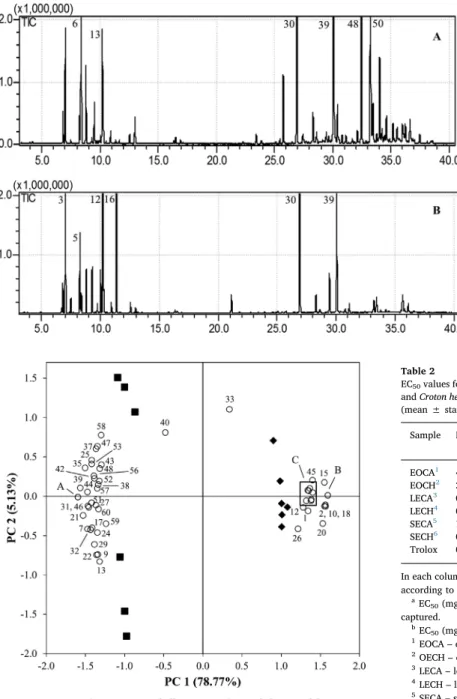
The essential oils composition of C. argyrophyllus and C. helio-tropiifoliusare described inTable 1. From the essential oil of the ana-lyzed species, a total of 60 compounds were identified, 51 and 37 inC. argyrophyllusandC. heliotropiifoliusrespectively (Fig. 1andTable 1). From those, 23 were exclusively identified inC. argyrophyllusand nine inC. heliotropiifolius, while 28 other compounds were present in both species. The composition of essential oils of the analyzed species dif-fered both quantitatively and qualitatively. In a general way, the re-lative abundance of each identified compound differed statistically (P < 0.001) among species, with the exception of humulene (P= 0.713) andα–muurolene (P= 0.501) (Table 1).
that represents 27.8% of the total identified components, followed by δ-elemene (8.7%),β-elemene (8.5%), and prenopsan-8-ol (8.5%). How-ever, in the work conducted by Fontenelle et al. (2008) the main compound was spathulenol (20.3%), whereas bicyclogermacrene (11.7%) appears in the second position. Also, spathulenol, with 14.3%, was a main component in the work conducted byMorais et al. (2006), with higher values comparatively to those reported in the present study (8.7%).
ConcerningC. heliotropiifoliusessential oil, limonene was the main compound, with 16.9%, followed byα-pinene (13.3%), caryophyllene (12.1%), bicyclogermacrene (10.3%) andγ-terpinene (9.6%) (Table 1). Once again, in general, other authors identified similar compounds although in different quantities. For instance, in the study conducted by Araújo et al. (2017)(E)-caryophyllene was the largest component, ac-counting for 23.8%, followed byγ-muurolene (10.5%) and viridiflorene (8.1%). Whereas, in other works the main compounds were 1,8-cineole (Angélico et al., 2014) andβ-caryophyllene (Neves and Camara, 2012). Still,Filho et al. (2017)characterized the essential oil of this species, in different seasons of the year, observed that theβ-caryophyllene, bicy-clogermacrene, germacrene-D, limonene, and 1,8-cineole were the major components, varying their relative abundance according to the season.
In general, our results are in agreement with literature forCroton species. Nevertheless, for each species, the composition of the essential oils varies. The observed differences could be attributed to a range of factors: geographical origin of plants, the characteristics of the soil and climatic conditions; season of sample collection (Gupta et al., 2011; Verma and Shukla, 2015); genetic factors and development of adaptive structures to the region; the time of collection along the day (Souza et al., 2017); and extraction conditions, among others. Other important aspect could be related to different chemotypes inside of the same species as noticed in other medicinal plants produced in the same biome. Such existence has already been proven inC. lichleriessential oils (Milanowski et al., 2002). Therefore, the results obtained may be indicative of new chemotypes, mainly in the case ofC. heliotropiifolius, rich inα-pinene (Table 1). This hypothesis need to be deeper studied in the future to verify the possible level of polymorphism withinC. he-liotropiifoliusandC. argyrophyllus.
The essential oil composition was used to verify whether the two species could be grouped according to their chemical components. For this, PCA was applied to the 60 components ofC. argyrophyllusandC. heliotropiifolius. The results obtained is depict inFig. 2. The PCA showed that the two species were completely separated by thefirst principal component (PC1).Croton heliotropiifoliuswas represented in the positive region of PC1, whileC. argyrophylluswas represented in the negative region. Three main characteristic groups of compounds (A, B, and C) could be perceived through the PCA results. Group A was composed of five compounds: β-pinene, linalool, elemene, alloaromadendrene and spathulenol (respectively numbers 6, 19, 28, 34 and, 50; Table 1). Thesefive components were represented in the negative region of PC1, near to the samples ofC. argyrophyllusessential oils, which means that those compounds were reported in both species, but at higher con-centrations inC. argyrophyllus, thus characterizing this species. Group B was located in the opposite region and was composed by a group of seven compounds: α-pinene, camphene, sabinene, p-cymene, γ-terpi-nene, caryophyllene and germacrene D (respectively numbers 3, 4, 5, 11, 16, 30, and 36;Table 1). These seven compounds were present in both species, but with significantly higher abundance in C. helio-tropiifolius, thus characterizing this species.
However, there was a third group of compounds that also char-acterized C. heliotropiifoliusessential oils, which was represented by letter C inFig. 2. Group C was composed of six compounds: α-phel-landrene, 1,8-cineole, bornyl acetate, germacrene A,δ-cadinol, and β-cadinol (respectively numbers 8, 14, 23, 41, 54, and 55;Table 1). These compounds characterized C. heliotropiifolius, since they were only identified in the essential oil of this species, and thus were absent from Table 1
Volatile composition of the essential oils ofCroton argyrophyllusKunth andCroton helio-tropiifoliusKunth (mean ± SD; n = 6) (only the components with a relative area greater than 0.1% were reported).
Oil composition LRI Lit EOCA (%)1 EOCH (%)2 P −value
1 Tricyclene 926 n.d. 0.29 ± 0.01 –
2 α-thujene 930 1.13 ± 0.05 1.71 ± 0.04 < 0.001 3 α-pinene 939 4.06 ± 0.13 13.30 ± 0.17 < 0.001 4 Camphene 954 0.14 ± 0.01 0.91 ± 0.02 < 0.001 5 Sabinene 975 1.05 ± 0.04 4.75 ± 0.04 < 0.001 6 β-pinene 979 8.92 ± 0.27 1.28 ± 0.03 < 0.001 7 β-myrcene 990 2.78 ± 0.09 2.52 ± 0.04 < 0.001 8 α-phellandrene 1002 n.d. 2.81 ± 0.03 –
9 3-carene 1011 1.61 ± 0.08 n.d. –
10 α-terpinene 1017 0.12 ± 0.03 0.63 ± 0.04 < 0.001 11 p-cymene 1024 0.33 ± 0.02 2.75 ± 0.03 < 0.001
12 Limonene 1029 n.d. 16.91 ± 0.13 –
13 β-phellandrene 1029 5.72 ± 0.13 n.d. –
14 1,8-cineole 1033 n.d. 1.14 ± 0.09 –
15 (Z)-β-ocimene 1037 0.51 ± 0.01 0.70 ± 0.01 < 0.001 16 γ-terpinene 1059 0.15 ± 0.03 9.55 ± 0.06 < 0.001
17 (E)-sabinene 1070 0.17 ± 0.03 n.d. –
18 α-terpinolene 1088 0.40 ± 0.03 0.75 ± 0.03 < 0.001 19 Linalool 1096 1.07 ± 0.03 0.36 ± 0.03 < 0.001 20 Terpinen-4-ol 1177 0.17 ± 0.01 0.25 ± 0.01 < 0.001 21 Naphthalene 1181 0.35 ± 0.01 0.22 ± 0.02 < 0.001
22 α-terpineol 1188 0.19 ± 0.02 n.d. –
23 Bornyl acetate 1285 n.d. 1.40 ± 0.03 –
24 δ-elemene 1338 0.54 ± 0.04 n.d. –
25 β-terpinyl acetate 1349 0.17 ± 0.02 n.d. –
26 α-copaene 1376 0.13 ± 0.01 0.16 ± 0.02 0.005 27 β-bourbonene 1388 0.25 ± 0.01 0.07 ± 0.05 < 0.001 28 Elemene 1390 3.00 ± 0.07 0.14 ± 0.01 < 0.001
29 Gurjunene 1409 0.12 ± 0.02 n.d. –
30 Caryophyllene 1419 7.65 ± 0.11 12.10 ± 0.17 < 0.001
31 β-gurjunene 1433 0.60 ± 0.06 n.d. –
32 Aromadendrene 1441 0.15 ± 0.05 n.d. –
33 Humulene 1454 1.53 ± 0.04 1.54 ± 0.02 0.713 34 Alloaromadendrene 1460 0.81 ± 0.02 0.36 ± 0.02 < 0.001 35 γ-muurolene 1479 0.29 ± 0.03 0.14 ± 0.02 < 0.001 36 Germacrene D 1485 0.76 ± 0.04 3.11 ± 0.06 < 0.001
37 Selinene 1490 0.48 ± 0.04 n.d. –
38 Viridiflorene 1496 0.23 ± 0.02 n.d. –
39 Bicyclogermacrene 1500 14.01 ± 0.26 10.30 ± 0.19 < 0.001 40 α-muurolene 1500 0.19 ± 0.02 0.18 ± 0.02 0.501
41 Germacrene A 1503 n.d. 0.12 ± 0.06 –
42 α-bulnesene 1505 1.92 ± 0.02 n.d. –
43 β-bisabolene 1509 0.24 ± 0.02 n.d. –
44 γ-cadinene 1513 0.52 ± 0.03 0.39 ± 0.02 < 0.001 45 δ-cadinene 1523 0.50 ± 0.03 0.70 ± 0.09 < 0.001
46 α-calacorene 1545 0.17 ± 0.01 n.d. –
47 Elemol 1549 0.73 ± 0.03 n.d. –
48 Sesquiterpene-like compound 1
– 6.23 ± 0.14 n.d. –
49 Germacrene B 1561 n.d. 0.18 ± 0.04 –
50 Spathulenol 1578 8.72 ± 0.10 1.26 ± 0.05 < 0.001 51 Caryophyllene oxide 1583 2.17 ± 0.17 1.52 ± 0.13 < 0.001
52 Guaiol 1600 4.14 ± 0.06 n.d. –
53 γ-eudesmol 1632 1.51 ± 0.03 n.d. –
54 δ-cadinol 1636 n.d. 2.11 ± 0.04 –
55 β-cadinol 1653 n.d. 0.89 ± 0.09 –
56 Sesquiterpene-like compound 2
– 1.08 ± 0.02 n.d. –
57 Sesquiterpene-like compound 3
– 1.21 ± 0.02 n.d. –
58 Sesquiterpene-like compound 4
– 1.14 ± 0.03 n.d. –
59 Bunesol 1666 1.13 ± 0.04 n.d. –
60 Sesquiterpene-like compound 5
– 0.53 ± 0.07 n.d. –
Total identified (%)* 91.71 97.91
LRI Lit.–linear retention index reported in literature (Adams, 2007); n. d.–not detected. 1EOCA
–essential oil ofC. argyrophyllus. 2EOCH
the chromatographic profile ofC. argyrophyllus(Table 1).
In the light of the results obtained and the interpretation derived from the PCA results, these two species may have different properties and bioactive potentials. This may explain the results that are presented in the next section.
3.2. Antioxidant activity
The antioxidant activity of the essential oils was evaluated using the DPPH and ABTS methods. These methods assess the capacity of es-sential oils to scavenge free radicals and thus provide a measurement of the potential for antioxidant activity. For DPPH, the essential oils from C. heliotropiifolius possess higher antioxidant activity, with an EC50 value of 34.8 mg mL−1. Nevertheless, when the ABTS method was
tested, no significant differences were found between both species (P= 0.185),C. argyrophylluspresented an EC50of 16.5 mg mL−1
andC. heliotropiifolius22.7 mg mL−1(Table 2). Other works demonstrated the
antioxidant activity ofCrotonessential oils (Ramos et al., 2013;Morais et al., 2006) with similar or better results than the obtained in this work.
For the methanolic extracts, and using the same methods, lower EC50values (mg mL−1) were observed than those of the essential oils.
So, a lower extract concentration was needed to neutralize 50% of the free radicals, thus generating a higher antioxidant potential. This fact could be related with the high temperatures and time used for the ex-traction of essential oils that could have negative impact on antioxidant molecules (Teixeira et al., 2007).
In a general way, the part of the plant used for extract preparation had a significant effect on the obtained results for the three methods used, being the leaf extracts more antioxidant than the stalk extracts (P < 0.001). For leaf extracts, in general,C. argyrophyllusis more ef-fective and present higher antioxidant potential thanC. heliotropiifolius with lower EC50 values (P <0.001), with the exception for ABTS Fig. 1.Chromatographic profile of the essential oils ofCroton argyr-ophyllus(A) andCroton heliotropiifolius(B) (numbers correspond to the compounds reported inTable 1).
Fig. 2.Principal component analysis on the volatile composition of the essential oils of
Croton argyrophyllusKunth andCroton heliotropiifoliusKunth. The two principal compo-nents (PC) explained 83.9% of the total variance. A–compounds 6, 19, 28, 34 and 50 of
Table 1; B–compounds 3, 4, 5, 11, 16, 30 and 36 ofTable 1; C–compounds 8, 14, 23, 41, 54 and 55.
Table 2
EC50values for the essential oils and methanolic extracts fromCroton argyrophyllusKunth andCroton heliotropiifoliusKunth through the DPPH, ABTS and reduction power methods (mean ± standard deviation).
Sample DPPHa(EC
50mg mL−1) ABTSa(EC50mg mL−1) Reduction powerb (EC50mg mL−1)
EOCA1 46.3 ± 2.75 b 16.6 ± 2.47 a
–
EOCH2 34.8 ± 3.75 a 22.7 ± 6.12 a
–
LECA3 0.222 ± 0.008 a 0.245 ± 0.037 a 0.658 ± 0.021 a
LECH4 0.357 ± 0.004 b 0.314 ± 0.071 a 0.791 ± 0.015 b
SECA5 1.036 ± 0.069 b 0.311 ± 0.112 a 1.781 ± 0.044 b
SECH6 0.782 ± 0.040 a 0.398 ± 0.011 a 1.281 ± 0.065 a
Trolox 0.037 ± 0.000 0.068 ± 0.001 0.159 ± 0.001
In each column, and for each type of extract, different letters mean statistical differences according to Tukey’s test.
aEC
50(mg mL−1)–concentration at which 50% of the DPPH and ABTS radicals are captured.
bEC
50(mg mL−1)–concentration at which the absorbance is 0.5. 1EOCA
–essential oil ofC. argyrophyllus. 2OECH
–essential oil ofC. heliotropiifolius. 3LECA
–leaf extract ofC. argyrophyllus. 4LECH
–leaf extract ofC. heliotropiifolius. 5SECA
–stalk extract ofC. argyrophyllus. 6SECH
method (P= 0.218) (Table 2). Using the DPPH method, the EC50values for leaf methanolic extracts were 0.222 mg mL−1and 0.357 mg mL−1
for C. argyrophyllus and C. heliotropiifolius, respectively. The values obtained with the other methodologies were higher but follow the same tendency, attesting the higher antioxidant potential forC. argyrophyllus essential oils (Table 2). For stalk methanolic extracts an opposite ten-dency was observed, with lower EC50values for the extracts obtained byC. heliotropiifoliusshowing greater antioxidant potential thanC. ar-gyrophyllus (in DPPH and reducing power). Nevertheless, for ABTS method no significant differences were observed between the two plant species (Table 2). Previous studies have already demonstrated the an-tioxidant potential of stalk fromCrotonspecies (Ndhlala et al., 2013) and for the same type of extracts and using the same methodologies, the extracts of the present s work showed higher antioxidant potential.
The total phenols content,flavonols and hydroxycinnamic acid de-rivatives content of the leaf and stalk extracts fromC. argyrophyllusand C. heliotropiifoliusare presented inTable 3. The speciesC. argyrophyllus presented higher total phenols content (299 and 242 mg GAE g−1
) and flavonols (188 and 126 mg QE g−1) for leaves and stalks respectively in
comparison to the obtained for C. heliotropiifolius(Table 3). For hy-droxycinnamic acid derivatives, no significant differences were ob-tained for the extracts (P= 0.312 for leaves;P= 0.848 for stalks) ob-tained from both species. The values obob-tained for leaf and stalk methanolic extracts from C. argyrophyllusandC. heliotropiifoliuswere higher than those reported in literature for other species of the genus Croton(Motta et al., 2013;Júnior et al., 2016).
The antioxidant activity of plant extracts is correlated with their chemical composition like the existence of phenolic compounds (Furlan et al., 2015). In this work, were established regression analysis between the different analyzed fractions of methanolic extracts and EC50values for the total phenols content and different used methods. For DPPH and reducing power there was an extremely significant correlation forC. argyrophyllus (R2= 0.989; P <0.001; y =−0.014x+ 4.467 for DPPH; R2= 0.984; P <0.001; y =−0.019x+ 6.479 for reducing power) and very significant for C. heliotropiifolius (R2= 0.872; P= 0.004; y =−0.009x+ 2.837 for DPPH; R2= 0.848; P= 0.006; y =−0.011x+ 3.634 for reducing power). Nevertheless, no correla-tion was observed for ABTS method for both species (R2=−0.249; P= 0.965;y =−1.7.105x+ 0.250 forC. argyrophyllus; R2=−0.077; P =0.467;y =−0.0004x+ 0.477 forC. heliotropiifolius). The results demonstrated that the composition of the extracts was preponderant for the antioxidant activity observed, and extracts with higher content of total phenols possess high antioxidant activity and low EC50, on the other side low values of total phenols are related with high EC50values and low antioxidant activity.
3.3. Antimicrobial activity
The essential oil ofC. argyrophyllusinhibited all the bacterial strains tested, with MIC values ranging from 10μL mL−1 (B. cereus) to
25μL mL−1(B. subtilis,S. aureus,E. coliandP.aeruginosa) (Table 4);
And without any obvious difference in susceptibility between Gram-negative and Gram-positive bacteria. This oil also showed to be more potent than the antibiotic chloramphenicol (30μg mL−1). Thesefi
nd-ings highlight the potential use of C. argyrophyllus essential oil in treating infections caused by these bacteria, especially as a potential therapeutic agent for eradicating antibiotic resistance. This is of parti-cular importance regarding E. coli and P. aeruginosa, given the in-creasing levels of resistance of these bacteria to multiple classes of antibiotics.Bertini et al. (2005)also used the essential oil ofC. argyr-ophyllusto combatS. aureusandE. coli, and obtained MIC values of lower than 5% of oil.
The antimicrobial activity of essential oils is influenced by its che-mical composition and abundance of each compound (Nazzaro et al., 2013), that could present different action mechanisms at cell level. In this work, the largest component of the oil of this species is bicyclo-germacrene, a sesquiterpenoid with well-known antimicrobial activity in other essential oils (Fontenelle et al., 2008;Wijesundara et al., 2016). Other compounds present in high amounts in the EO fromC. argyr-ophylluswere spathulenol, caryophyllene, andβ-pinene. Caryophyllene is ascribed as one of the main responsible for the antifungal activity of Zingiber nimmonii againstC. glabrata and C. albicans (Sabulal et al., 2006).β-pinene, as an isolated compound, was reported to inhibited the growth of some bacteria (Leite et al., 2007). The action of these compounds (either isolated or interacting with other components) may be responsible for the antimicrobial potential of Croton species in-cluding of those of the present study.
The essential oils of other species of Croton genus, also showed antimicrobial activity. The diterpenes isolated from C. nepetifolius showed action againstStaphylococcussp. strains (Sá et al., 2012); and, essential oil ofC. rhamnifolioides inhibit the growth and survival of pathogens such asListeria monocytogenes, Aeromonas hydrophila, E. coli andS. aureus(Costa et al., 2013).Yagi et al. (2016)usingC. rotundus found MICs of 16μg mL−1againstS. aureusand 32μg mL−1againstB. subtilisandE. coli.
In the present work, no antibacterial nor antifungal activity was Table 3
Total phenols content (mg GAE g−1),
flavonols (mg QE g−1) and hydroxycinnamic acid derivatives (mg CAE g−1) in the methanolic extracts fromCroton argyrophyllusKunth and Croton heliotropiifoliusKunth (mean ± standard deviation).
Sample Total Phenols (mg GAE g−1)
Hydroxycinnamic acid derivatives (mg CAE g−1)
Flavonols (mg QE g−1)
LECA1 299.47 ± 4.89 a 70.29 ± 1.71 a 187. 93 ± 1.41 a
LECH2 251.52 ± 10.50 b 67.75 ± 3.40 a 139.27 ± 5.26 b
SECA3 242.38 ± 4.28 a 51.23 ± 1.29 a 125.89 ± 2.83 a
SECH4 201.20 ± 18.50 b 51.91 ± 5.63 a 104.39 ± 10.44 b
In each column, and for each type of extract, different letters mean statistical differences according to Tukey’s test.
1LECA
–leaf extract ofC. argyrophyllus. 2LECH
–leaf extract ofC. heliotropiifolius. 3SECA
–stalk extract ofC. argyrophyllus. 4SECH
–stalk extract ofC. heliotropiifolius.
Table 4
Minimum inhibitory concentrations (MIC) of essential oils and plant extracts ofCroton argyrophyllusKunth andCroton heliotropiifoliusKunth against bacteria and yeast.
Microorganisms Essential oil (μL mL−1) Plant extract (mg mL−1)
EOCA1 EOCH2 LECA3 LECH4 SECA5 SECH6
Bacteria
Bacillus subtilis 25 – – – – –
Bacillus cereus 10 – – – – –
Staphylococcus aureus
25 – – – – –
Escherichia coli 25 – – – – –
Pseudomonas aeruginosa
25 – – – – –
Yeast
Candida albicans – – – 50 – –
Candida parapsilosis
– – – 50 – –
Candida glabrata – – 50 – – –
(–) = no inhibition. 1EOCA
–essential oil ofC. argyrophyllus. 2OECH
–essential oil ofC. heliotropiifolius. 3LECA
–leaf extract ofC. argyrophyllus. 4LECH
–leaf extract ofC. heliotropiifolius. 5SECA
–stalk extract ofC. argyrophyllus. 6SECH
demonstrated by the essential oil ofC. heliotropiifolius(Table 4). This result is in line with the obtained byAngélico et al. (2014)to the es-sential oil of the same species. Those author evaluate the eses-sential oil against a variety of bacterial strains, and no positive results nor very high MICs were obtained for Staphylococcus aureus (MICs of 512μL mL−1), and forB. subtilis, B.cereus,E. coli andP.aeruginosa
(MICs of 1024μL mL−1).
When plant extracts were tested, the antimicrobial activity was low. Nevertheless, two extracts exhibit action against important pathogenic yeasts. The leaf extract ofC. argyrophyllusexhibited antifungal activity against C. parapsilosis, and the leaf extract of C. heliotropiifolius pro-cesses antifungal activity againstC. albicansandC. glabrata, all with a MIC value of 50 mg mL−1(Table 4), two-fold the result obtained with
the antifungal agent fluconazole (25μg mL−1).Queiroz et al. (2014)
obtained best results than the observed in this work. These authors, with ethanolic extract from C. heliotropiifolius, found a MIC of 25μg mL−1againstC. albicans.
The antimicrobial potential of plant extracts may be related to the chemical composition and the presence of some compounds like phe-nols (Cushnie and Lamb, 2005). Extracts obtained byCrotonspecies, as C. macrostachyus, were rich in compounds with antimicrobial activity such as phenolic compounds, tannins and alkaloids (Teugwa et al., 2013), that could justify the antimicrobial activity of plant extracts (Jaberian et al., 2013). Nevertheless, low activity or absence of activity does not mean the absence of the compounds, once the biological po-tential may vary according to the solvent, the part of the plant, the quality of the material and the extraction conditions (Harvey et al., 2015).
4. Conclusions
The present work contributed for the characterization of the EO’s and phytochemical composition of twoCrotonspecies,C. argyrophyllus andC. heliotropiifolius.The results indicated that both antioxidant and antimicrobial activity ofC. argyrophyllusEO and aerial part are greater thanC. heliotropiifolius. Moreover,C. argyrophyllusEO inhibited more the growth of both gram-positive and gram-negative bacteria than traditional antimicrobials tested, opening a clear window for the ex-ploitation of these antimicrobial agents from natural sources. Nowadays this is of special importance because of the emergence of resistance to numerous conventional antibiotics. The chemical composition of the EO’s allowed distinguish the twoCrotonspecies, and the results possibly point out to new chemotypes in comparison to literature. Briefly, the main conclusion of this study is thatCrotonspecies are a great source of phytochemicals with exceptional bioactive properties, and their ex-ploitation can be helpful for different industrial sectors, mainly food sector, cosmetics, and most of all medicinal and pharmaceutical in-dustries.
Acknowledgements
This work wasfinancially supported by CNPQ/CsF and CAPES and UID/AGR/00690/2013–CIMO funded by FEDER–Fundo Europeu de Desenvolvimento Regional through COMPETE2020 – Programa Operacional Competitividade e Internacionalização (POCI) – and by national funds through FCT–Fundação para a Ciência e a Tecnologia, Portugal. The authors are also grateful to UFRB Herbarium for the help with the plant species identification.
References
Adams, R.P., 2007. Identification of Essential Oil Components by Gas Chromatography/ Mass Spectrometry, fourth ed. Allured Business Media, Carol Stream.
Aguiar, F.L.L., Morais, S.M., Santos, H.S., Albuquerque, M.R.J.R., Bandeira, P.N., Brito, E.H.S., Rocha, M.F.G., Fontenelle, R.O.S., 2016. Antifungal activity and synergistic effect of acetophenones isolated from speciesCrotonagainst dermatophytes and yeasts. J. Med. Plants Res. 10, 216–222.
Angélico, E.C., Rodrigues, O.G., Costa, J.G.M., Lucena, M.F.A., Neto, V.Q., Medeiros, R.S., 2014. Chemical characterization and antimicrobial activity of essential oils and Crotońs varieties modulator in the Braziliańs Northeast semiarid. Acad. J. 8, 392–397.
Araújo, S.S., Santos, M.I.S., Dias, A.S., Ferro, J.N.S., Lima, R.N., Barreto, E.O., Corrêa, C.B., Araújo, B.S., Lauton-Santos, S., Shan, A.Y.K., Alves, P.B., Santana, A.E.G., Thomazzi, S.M., Antoniolli, A.R., Estevam, C.S., 2014. Chemical composition and cytotoxicity analysis of the essential oil from leaves ofCroton argyrophyllusKunth. J. Essent. Oil Res. 26, 446–451.
Araújo, F.M., Dantas, M.C.S.M., Silva, L.S., Aona, L.Y.S., Tavares, I.F., Souza-Neta, L.C., 2017. Antibacterial activity and chemical composition of the essential oil ofCroton heliotropiifoliusKunth from Amargosa, Bahia, Brazil. Ind. Crops Prod. 105, 203–206.
Berker, K., Güçlü, K., Tor, I., Apak, R., 2007. Comparative evaluation of Fe (III) reducing power-based antioxidant capacity assays in the presence of phenanthroline batho-phenanthroline, tripyridyltriazine (FRAP) and ferricyanide reagents. Talanta 72, 1157–1165.
Bertini, L.M., Pereira, A.F., Oliveira, C.L.L., Menezes, E.A., Morais, S.M., Cunha, F.A., Cavalcanti, E.S.B., 2005. Perfil de sensibilidade de bactérias frente a óleos essenciais de algumas plantas do Nordeste do Brasil. Infarma 17 (3/4).
Boulanouar, B., Abdelaziz, G., Aazza, S., Gago, C., Miguel, M.G., 2013. Antioxidant ac-tivities of eight Algerian plant extracts and two essential oils. Ind. Crops Prod. 46, 85–96.
Cavin, A.L., Hay, A.E., Marston, A., Stoeckli-Evans, H., Scopelliti, R., Diallo, D., Hostettmann, K., 2006. Bioactive diterpenes from the fruits ofDetarium microcarpum. J. Nat. Prod. 69, 768–773.
Compagnone, R.S., Chavez, K., Mateu, E.A., Orsini, G., Arvelo, F.O., Suarez, A.I., 2010. Composition and cytotoxic activity os essential oils fromCroton matourensisand Croton micansfrom Venezuela. Rec. Nat. Prod. 4, 101–108.
Costa, A.C.V., Melo, G.F.A., Madruga, M.S., Costa, J.G.M., Garino Junior, F., Queiroga Neto, V., 2013. Chemical composition and antibacterial activity of essential oil of Croton rhamnifolioides leaves Pax & Hoffm. Semina 34, 2853.
Cushnie, T.P.T., Lamb, A.J., 2005. Antimicrobial activity offlavonoids. Int. J. Antimicrob. Agents 26, 343–356.
Dória, G.A.A., Silva, W.J., Carvalho, G.A., Alves, P.B., Cavalcanti, S.C.H., 2010. A study of the larvicidal activity of two Croton species from northeastern Brazil againstAedes aegypti. Pharm. Biol. 48, 615–620.
Filho, J.M.T.A., Araújo, L.C., Oliveira, A.P., Guimarães, A.L., Pacheco, A.G.M., Silva, F.S., Cavalcanti, L.S., Lucchese, A.M., Almeida, J.R.G.S., Araújo, E.C.C., 2017. Chemical composition and antibacterial activity of essential oil from leaves ofCroton helio-tropiifoliusin different seasons of the year. Braz. J. Pharmacogn. 27, 440–444.
Fontenelle, R.O.S., Morais, S.M., Brito, E.H.S., Brilhante, R.S.N., Cordeiro, R.A., Nascimento, N.R.F., Kerntopf, M.R., Sidrim, J.J.C., Rocha, M.F.G., 2008. Antifungal activity of essential oils ofCrotonspecies from the Brazilian Caatinga biome. J. Appl. Microbiol. 104, 1383–1390.
Furlan, C.M., Santos, K.P., Sedano-Partida, M.D., Motta, L.B., Santos, D.Y.A.C., Salatino, M.L., Negri, G., Berry, P.E., Ee, B.W.V., Salatino, A., 2015. Flavonoids and antioxidant potential of nine Argentinian species ofCroton(Euphorbiaceae). Braz. J. Bot. 38, 693–702.
Grundmann, H., Kraker, M., Davey, P., 2011. Clinical impact of antimicrobial resistance: design matters. Lancet Infect. Dis. 11, 344.
Gupta, S., Bhaskar, G., Andola, C.H., 2011. Altitudinal variation in essential oil content in leaves ofZanthoxylum alatuma high value aromatic tree from Uttrakhand. Res. J. Med. Plant 5, 348–351.
Harvey, A.L., Edrada-Ebel, R., Quinn, R.J., 2015. The re-emergence of natural products for drug discovery in the genomics era. Nat. Rev. Drug Discov. 14, 111–129.
Hatano, T., Kagawa, H., Yasuhara, T., Okuda, T., 1988. Two newflavonoids and other constituents in licorice root: their relative astringency and scavenging effects. Chem. Pharm. Bull. 36, 2090–2097.
Júnior, F.B., Macedo, G.E., Zemolin, A.P., Silva, F.G., Cruz, L.C., Boligon, A.A., Menezes, I.R.A., Franco, J.L., Posser, T., 2016. Oxidant effects and toxicity ofCroton campestris inDrosophila melanogaster. Pharm. Biol. 54, 3068–3077.
Jaberian, H., Piri, K., Nazari, J., 2013. Phytochemical composition and in vitro anti-microbial and antioxidant activities of some medicinal plants. Food Chem. 136, 237–244.
Leite, A.M., Lima, E.O., Souza, E.L., Diniz, M.F.F.M., Trajano, V.N., Medeiros, I.A., 2007. Inhibitory effect ofβ-pinene,α-pinene and eugenol on the growth of potential in-fectious endocarditis causing gram-positive bacteria. Rev. Bras. Ciênc. Farm. 43, 121–126.
Milanowski, D.J., Winter, R.E., Elvin-Lewis, M.P., Lewis, W.H., 2002. Geographic dis-tribution of three alkaloid chemotypes ofCroton lechleri. J. Nat. Prod. 65, 814–819.
Morais, S.M., Júnior, F.E.A.C., Silva, A.R.A., Neto, J.S.M., Rondina, D., Cardoso, J.H.L., 2006. Atividade antioxidante de úleos essenciais de espócies de Croton do nordeste do Brasil. Quim. Nova 29, 907–910.
Motta, L.B., Furlan, C.M., Santos, D.Y.A.C., Salatino, M.L.F., Negri, G., De Carvalho, J.E., Monteiro, P., Ruiz, A.L.T.G., Caruzo, M.B., Salatino, A., 2013. Antiproliferative ac-tivity and constituents of leaf extracts ofCroton sphaerogynusBaill. (Euphorbiaceae). Ind. Crop. Prod. 50, 661–665.
Nazzaro, F., Fratianni, F., De Martino, L., Coppola, R., De Feo, V., 2013. Effect of essential oils on pathogenic bacteria. Pharmaceuticals 6, 1451–1474.
Ndhlala, A.R., Aderogba, M.A., Ncube, B., Van Staden, J., 2013. Anti-oxidative and cholinesterase inhibitory effects of leaf extracts and their isolated compounds from two closely relatedCrotonspecies. Molecules 18, 1916–1932.
Neves, I.A., Camara, C.A.G., 2012. Volatile constituents of two croton species from Caatinga Biome of Pernambuco–Brazil. Rec. Nat. Prod. 6, 161–165.
heliotropiifolius. Phytochem. Lett. 10, lxxxviii–xciii.
Ramos, J.M.O., Santos, C.A., Santana, D.G., Santos, D.A., Alves, P.B., Thomazzi, S.M., 2013. Chemical constituents and potential antiinflammatory activity of the essential oil from the leaves ofCroton argyrophyllus. Braz. J. Pharmacogn. 23, 644–650.
Sá, N.C., Cavalcante, T.T., Araújo, A.X., dos Santos, H.S., Albuquerque, M.R., Bandeira, P.N., da Cunha, R.M., Cavada, B.S., Teixeira, E.H., 2012. Antimicrobial and anti-biofilm action of Casbane Diterpene fromCroton nepetaefoliusagainst oral bacteria. Arch. Oral Biol. 57, 550–555.
Sánchez, C.S., González, A.M.T., García-Parrilla, M.C., Granados, J.J.Q., Serrana, H.L.G., Martínez, M.C.L., 2007. Different radical scavenging tests in virgin olive oil and their relation to the total phenol content. Anal. Chim. Acta 593, 103–107.
Sabulal, B., Dan, M., John, A., Kurup, R., Pradeep, N.S., Valsamma, R.K., George, V., 2006. Caryophyllene-rich rhizome oil ofZingiber nimmoniifrom South India: chemical characterization and antimicrobial activity. Phytochemistry 67, 2469–2473.
Sarker, S.D., Nahar, L., Kumarasamy, Y., 2007. Microtiter plate-based antibacterial assay incorporating resazurin as an indicator of cell growth, and its application in the in vitro antibacterial screening of phytochemicals. Methods 42, 321–324.
Secco, R.S., Cordeiro, I., Senna-Vale, L., Sales, M.F., Lima, M.R., Medeiros, D., Haiad, B.S., Oliveira, A.S., Caruzo, M.B.R., Carneiro-Torres, D., Bigio, N.C., 2012. An overview of recent taxonomic studies on Euphorbiaceae s.l in Brazil. Rodriguésia 63, 227–242.
Souza, G.S., Bonilla, O.H., Lucena, E.M.P., Barbosa, Y.P., 2017. Chemical composition and yield of essential oil from three Croton species. Ciênc. Rural 47, e20161054.
Sun, J., Wang, X., Wang, P., Li, L., Qu, W., Liang, J., 2015. Antimicrobial: antioxidantand
cytotoxic properties of essential oil fromDictamnus angustifolius. J. Ethnopharmacol. 159, 296–300.
Teixeira, S., Mendes, A., Alves, A., Santos, L., 2007. Simultaneous distillation–extraction of high volatile compounds fromCistus ladaniferL. Anal. Chim. Acta 584, 439–446.
Teugwa, M.C., Sonfack, D.C.R., Fokom, R., Penlap, B.V., Amvam, Z.P.H., 2013. Antifungal and antioxidante activity of crude extracts of three medicinal plants from Cameroon pharmacopea. J. Med. Plants Res. 7, 1537–1542.
Verma, N., Shukla, S., 2015. Impact of various factors responsible forfluctuation in plant secondary metabolites. J. Appl. Res. Med. Aromat. Plants 2, 105–113.
Wenqiang, G., Shufen, L., Ruixiang, Y., Yanfeng, H., 2006. Comparison of composition and antifungal activity ofArtemisia argyiLévl. et Vant inflorescence essential oil extracted by hydrodistillation and supercritical carbono dioxide. Nat. Prod. Res. 20, 992–998.
Wijesundara, S.A.D.T.L., Kannangara, B.T.S.D.P., Abeywickrama, K., 2016. Antifungal Activity ofCroton aromaticusL.in vitro:against post-harvest fungal pathogens isolated from tropical fruits. J. Agric. Sci. 11, 105–117.
Yagi, S., Babiker, R., Tzanova, T., Schohn, H., 2016. Chemical composition, anti-proliferative, antioxidant and antibacterial activities of essential oils from aromatic plants growing in Sudan. Asia-Pac. J. Trop. Med. 9, 763–770.