Ali Özgür Argunşah
Dissertation presented to obtain the
Ph.D degree in Biology | Neuroscience
Instituto de Tecnologia Química e Biológica António Xavier | Universidade Nova de Lisboa
Oeiras,
Activity dynamics lead to diverse structural
plasticity at single dendritic spines
Ali Özgür Argunşah
Dissertation presented to obtain the
Ph.D degree in Biology | Neuroscience
Instituto de Tecnologia Química e Biológica António Xavier | Universidade Nova de Lisboa
Oeiras, July, 2016
Activity dynamics lead to diverse
structural plasticity at single
dendritic spines
Activity Dynamics Lead to Diverse
Structural Plasticity at Single
Dendritic Spines
AL˙I ¨OZG ¨UR ARGUNS¸AH
A Dissertation Presented to the Faculty of Universidade Nova de Lisboa
in Candidacy for the Degree of Doctor of Philosophy
Inbal Israely
To My Family
Acknowledgments
First and foremost I would like to thank to Champalimaud Neuro-science Programme (CNP) for accepting me to this amazing Ph.D. pro-gramme. I cannot think of a better place to pursue a Ph.D. in neuro-science. CNP provided an amazing environment for learning, interactions and discussions, as well as great teachers during the period of courses and fantastic line of seminar speakers throughout my studies.
My supervisor Dr. Inbal Israely accepted me in her lab, neuronal
structure and function (NSF), without hesitation, even though I had no experience in experimental neuroscience, not even any wet lab experience including mixing any two solutions. She supported me for the entire period of my studies. She taught me many things from preparing hippocampal slices to thinking like a neuroscientist and most importantly it was her
seminar presentation back in the Instituto Gulbenkian de Ciˆencia (IGC)
that motivated me to do all the work that is presented in this thesis. The members of NSF lab have always been the most supportive,
help-ful and knowledgeable. Dr. Yazmin Ram´ıro-Cortˆes, a.k.a. Dr. Cortex,
without whom this thesis would not exist, has been the teacher of biology to this ignorant engineer, a.k.a. Alisinho. Hopefully-Dr.-by-the-time-of-this-thesis-defense Anna Felicity Hobbiss has been the source of friendship, emotional support and the voice of scientific-reasoning for the entire
pe-riod of this Ph.D. road that we walked together. Inˆes Vaz de Cunha, Ana
Vaz and Dr. C´atia Feliciano have always been there when I needed,
ei-ther with their amazing technical skills or friendship. Last but not least, I would also like to thank Dr. Mar´ıa Royo and Dr. Daniela Pereira for their relatively recent but warm friendships and critical evaluation of my work.
I learned so much from my fellow students from INDP 2009 and PIBS 2009 programmes. Among those, a number of them have had profound implications on my scientific life. My main brainstorming partner Dr.
Bahtiyar Yılmaz and I had many amazingly interesting discussions that at the end confused both of us in a somewhat useful way. Dr. Thiago
Santos Gouvˆea introduced me the un-ending world of behaviorism, Pedro
Garcia da Silva’s extensive knowledge of neuroscientific techniques and literature, and his crazy theories motivated me to read and investigate more.
My dear friend Libbi Rickenbacher has just made my life better by her existence and being a giant ball of positivism, so I thank her for that. Dr.
Duˇsica Radoˇs has always been there for me when I needed, she cheered
me up and fed me with delicious pancakes. And finally, I would like to
thank Dr. Branka, the person, Kolundˇzija for her ever-present support,
friendship, and not to mention the critical reading of the thesis.
I have been very fortunate for being a part of three different research groups before and during my Ph.D. which I believe shaped my scientific career path. I would like to thank Dr. Emery N. Brown who accepted me in his lab. as a visiting researcher and let me watch a real brain surgery for 8 hours. I also want to thank Dr. Thomas J. McHugh for letting me spend two months in his lab, and his lab members, specially Dr. Steven J. Middleton and Roman Boehringer for teaching me how to make tetrode drives, surgeries and their welcoming friendships. Last but not least, my
advisors during my masters, Dr. M¨ujdat C¸ etin and Dr. Ayt¨ul Er¸cil have
been very supportive and encouraging for the neuroscience degree I wanted to pursue, and my colleagues from computer vision and pattern analysis
lab., Emrecan C¸ ¨okelek, Dr. Cemre Zor, Dr. Batu Akan, Dr. Serhan Co¸sar,
Dr. Baran C¸ ¨ur¨ukl¨u and Dr. Taner Eskil have always been supportive. And
special thank to my dearest friend Dr. Saygın Topkaya, a.k.a. Pa¸sam, who has been the person I enjoyed sharing ideas the most from any disiplines starting from particle physics to the philosophies found in science fiction literature.
The dendritic spine analysis toolbox Spines that is presented in the the-sis is developed in collaboration with a great group of engineers/scientists and I am very happy and thankful for being able to work with such ded-icated and motivated people. I want to thank them for putting up with
my endless questions and requests starting from Dr. Devrim ¨Unay who
initiated the study with me, Ertun¸c Erdil who has been the main
co-developer and Muhammad Usman Ghani for being the most productive master student ever.
I gratefully acknowledge the funding sources that made this Ph.D.
work possible. I was funded by the Funda¸c˜ao para a Ciˆencia e a
Techolo-gia (FCT) fellowship for my first 4 years and by NSF lab. at Funda¸c˜ao
Champalimaud (FC) for last 3 years.
I am very thankful for having Dr. Alfonso Renart and Dr. Leopoldo Petreanu, to whom I look up to scientifically, for being my thesis commit-tee members and helping me with their constructive criticisms over the years. I am also very happy to have Dr. Armando Miguel Caseiro Pires
Remondes and Dr. Jos´e A. Esteban for accepting to be a part my thesis
discussion.
Lastly, I would like to thank my family for all their unconditional love and support.
T´ıtulo
Diferentes Padr˜oes de Atividade Induzem Diversas Formas de
Plasti-cidade Estrutural em Esp´ıculas Dendr´ıticas
Resumo
As s´ınapses, locais onde os neur´onios se conetam entre si, s˜ao os
lo-cais onde se sup˜oe que a aprendizagem ocorra atrav´es de altera¸c˜oes nestas
conec¸c˜oes. LTP (do inglˆes long-term potentiation) e o LTD (do inglˆes
long-term depression) foram propostos como mecanismos de adapta¸c˜ao
das conec¸c˜oes entre neur´onios. Atrav´es de uncaging de glutamato
me-diado pela luz, foi elucidada uma rela¸c˜ao linear entre a quantidade de
corrente que passa por uma s´ınapse individual e o tamanho da respectiva
esp´ıcula dendritica, permitindo que as altera¸c˜oes estruturais que
ocor-rem ao n´ıvel das esp´ıculas sirva como medida para estimar plast´ıcidade
sin´aptica. Para quantificar de forma eficiente e exata as dinˆamicas
estru-turais observadas em imagens adquiridas em microsc´opio de dois fot˜oes
de-senvolvemos uma toolbox baseada Matlab, denomidade SpineS, que
anal-isa automaticamente as altera¸c˜oes de volume das esp´ıculas dendriticas ao
longo do tempo, baseando-se numa livraria de imagens representativas.
Padr˜oes de estimula¸c˜ao regularmente espa¸cados, como padr˜oes de alta ou
baixa frequˆencia (do inglˆes high-frequency, HFS, e low-frequency
stimula-tion, LFS, respectivamente), que s˜ao tradicionalmente usados para induzir
plasticidade no hipocampo n˜ao s˜ao as formas mais comuns de atividade
no c´erebro. Assim, decidimos estudar quais s˜ao as formas funcionais e
es-truturais que padr˜oes irregulares de atividade geram em esp´ıculas
dendrit-icas individuais de neur´onios piramidais da regi˜ao de CA1 do hipocampo.
Para isso foram desenhados padr˜oes de estimula¸c˜ao que seguem uma
dis-tribui¸c˜ao Poisson e se assemelham aos padr˜oes de atividade recebidos por
induzida por esta estimula¸c˜ao ´e determinada pela estrutura temporal do
padr˜ao de estimula¸c˜ao. Quando a atividade ocorre de forma homog´enea ao
longo do tempo, ´e observado um crescimento robusto e de longa dura¸c˜ao,
at´e 4 horas, em esp´ıculas individuais, que depende de ativa¸c˜ao de
recep-tores NMDA e s´ıntese de prote´ınas. Contrariamente, se a densidade de
eventos de atividade se acumular no in´ıcio ou no fim do padr˜ao de
es-timula¸c˜ao apenas ocorre um crescimento das esp´ıculas de curta dura¸c˜ao.
Estas experiˆencias demonstram que o fator chave na indu¸c˜ao de altera¸c˜oes
sin´apticas de longa dura¸c˜ao em bot˜oes individuais ´e a estrutura
tempo-ral do padr˜ao de estimula¸c˜ao, sendo que a dura¸c˜ao total do estimulo, o
n´umero de eventos de atividade e a quantidade de glutamato libertado
n˜ao difere entre padr˜oes. De maior relevˆancia foi a observa¸c˜ao de que,
durante a estimula¸c˜ao destes diversos padr˜oes de atividade, as esp´ıculas
dendriticas sofrem r´apidas altera¸c˜oes estruturais. Recolhemos imagens
das altera¸c˜oes que ocorrem durante os 60 segundos de estimula¸c˜ao e
de-scobrimos que o crescimente toal de uma esp´ıcula dendritica ´e altamente
vari´avel mesmo em resposta ao mesmo padr˜ao de actividade. Contudo, a
quantidade total de crescimento expressa em cada esp´ıcula est´a
significati-vamente relacionada com o facto de uma determinada esp´ıcula sofrer
plas-ticidade de longa-dura¸c˜ao, independentemente do padr˜ao de estimula¸c˜ao.
Isto indica que para determinados padr˜oes de estimula¸c˜ao a integra¸c˜ao
final que ocorre ao n´ıvel da esp´ıcula ´e o que determina em ´ultima an´alise
a longevidade da plasticidade sin´aptica.
Estes resultados elucidam como diferentes padr˜oes de atividade levam
a processos fundamentalmente diferentes de plasticidade ao n´ıvel das
sinapses, permitindo compreender como as altera¸c˜oes da atividade neural
in vivo tˆem consequˆencias ao n´ıvel das sinapses e ao n´ıvel dos circuitos
Abstract
Synapses are the sites at which learning is proposed to occur through changes in the strength of neuronal connections. Utilizing 2-photon me-diated glutamate uncaging and imaging, the size of a dendritic spine and the amount of current which that synapse conducts has been shown to be linearly correlated and thus allows for structural changes in spine volumes to serve as a proxy for measuring plasticity. In order to efficiently and ac-curately quantify such structural dynamics, we developed a Matlab-based toolbox, named SpineS, which automatically analyses dendritic spine vol-ume changes more rapidly, and with greater precision, based on a learned library of representative images. Regularly spaced stimulations, such as the high- and low-frequency patterns traditionally used to induce plas-ticity in the hippocampus, are not the most common forms of activity which occur in the brain. Therefore, we decided to investigate what are the functional and structural correlates of irregular patterns of activity at single spines of hippocampal CA1 pyramidal neurons. To accomplish this, we designed stimulation paradigms that follow a Poisson distribu-tion, resembling the in vivo firing properties of the endogenous inputs to these neurons. We found that the longevity of the induced potentiation is determined by the timing structure of the stimulation pattern. When the activity that is delivered is homogeneously distributed over time, we observe robust and long-lasting potentiation and growth of single spines that last for at least 4 hours, requires NMDA activation and new protein synthesis. In contrast to this finding, if the density of events is clustered either towards the beginning or towards the end of the stimulus train, only short-term potentiation is achieved. These experiments demonstrate that a key factor in the induction of long-lasting changes at individual inputs is the structure of the activity, as the total stimulation time, the number of events, and the amount of glutamate delivered are all constant. Of further interest to us was the observation that during the delivery of these
vari-ous activity patterns, we saw that spines were undergoing rapid structural dynamics. We imaged the changes that were taking place during these 60 second stimulation periods and found that the total spine growth is highly variable even in response to the same activity pattern. However, the to-tal amount of growth expressed at a spine was significantly correlated to whether that particular spine will undergo long-lasting plasticity. This indicates that for certain patterns of activity, the final integration which occurs within a spine is ultimately what influences its long-term plasticity outcome.
These results shed light on how different patterns of activity lead to fundamentally different plasticity processes at synapses, providing insight as to how the variety of neural activity patterns in vivo will have long-term consequences for synaptic strength and thus circuit organization.
Author Contributions
Experiments conducted in this thesis were designed by Ali ¨Ozg¨ur
Ar-gun¸sah and Inbal Israely.
Data were collected and analyzed by Ali ¨Ozg¨ur Argun¸sah.
Dendritic spine analysis software (SpineS) is developed in collaboration
with Devrim ¨Unay, Ertun¸c Erdil, Muhammad Usman Ghani, Arif Murat
Ya˘gcı, S¨umeyra Demir Kanık, M¨ujdat C¸ etin, Anna Felicity Hobbiss and
Yazm´ın Ramiro Cort´es.
Financial Support
This work is supported by Funda¸c˜ao para a Ciˆencia e a Techologia
(FCT) with grant number SFRH/BD/51264/2010, Funda¸c˜ao
Champali-maud (FC) and Instituto Gulbenkian de Ciˆencia (IGC) and The Scientific
and Technological Research Council of Turkey (T ¨UBiTAK) with grant
Contents
Acknowledgments . . . iv
T´ıtulo e Resumo . . . vii
Abstract . . . ix
Author Contributions and Financial Support . . . xi
1 Introduction 5 1.1 Neuron Doctrine . . . 6
1.2 Synaptic Plasticity . . . 6
1.3 Synaptic Plasticity in the Hippocampus . . . 10
1.4 Dendritic Spines and Structure-Function Coupling . . . 12
1.5 Single Spine Plasticity . . . 15
1.6 Spike-Timing Dependent Plasticity . . . 17
1.7 Naturalistic Patterns, Synaptic Responses and Plasticity . . 18
1.8 Contribution of this Dissertation . . . 22
2 Materials and Methods 23 2.1 Materials . . . 24
2.1.1 Dissection Solution . . . 24
2.1.2 Dissection Equipment . . . 24
2.1.3 Biolistic Gene Transfer . . . 26
2.1.4 Internal Solution for Patch Pipette . . . 27
2.2 Methods . . . 29
2.2.1 Organotypic Slice Cultures . . . 29
2.2.2 Two Photon Laser Scanning Microscopy . . . 30
2.2.3 Pulse Train Modeling using a Poisson Process . . . . 33
2.2.4 Statistical Analysis . . . 35
3 Spines: A Tool for Automatic Dendritic Spine Analysis 36 3.1 Abstract . . . 38
3.2 Introduction . . . 38
3.3 Spine Head Volume Estimation Methods . . . 43
3.4 Dendritic Segment Registration . . . 48
3.5 Dendritic Spine Head Segmentation . . . 51
3.6 Spine Neck Path and Length . . . 55
3.7 Results and Conclusions . . . 58
4 Single Spine Structural Plasticity Induced by Naturalistic-like Trains 64 4.1 Abstract . . . 66
4.2 Introduction . . . 67
4.3 Generation of Naturalistic-like Trains . . . 70
4.4 Results . . . 76
4.4.1 Timing Structure of the Naturalistic-like Train De-termines the Longevity of the Plasticity . . . 77
4.4.2 NT-Uniform Induced Plasticity is NMDAR-Dependent . . . 80
4.4.3 Longevity of the NT-Uniform Induced Plasticity is Protein-Synthesis Dependent . . . 82
4.4.4 Plasticity Levels do not Depend on the Initial Spine Size . . . 83
5 Rapid Structural Spine Dynamics and Long-Term
Conse-quences 87
5.1 Abstract . . . 89
5.2 Introduction . . . 89
5.3 Results . . . 90
5.3.1 Stimulation Pattern does not Cause Significant
Spine Growth Differences During the Course of
Stimulation . . . 90
5.3.2 Rapid Spine Growth During the Course of
Stimula-tion Signals Longevity . . . 93
5.4 Conclusions . . . 100
6 Discussion 102
List of Tables
1.1 Requirements of Inducing Plasticity at Single Dendritic
Spines. . . 17
2.1 10× Krebs Ringer Dissection Solution. . . 24
2.2 1× Krebs-Ringer Dissection Solution. . . 24
2.3 Artificial Cerebrospinal Fluid (ACSF). . . 28
2.4 Organotypic Slice Culture Media . . . 29
3.1 Performance of SpineS Compared to Manual Segmentation based IFI and FWHM. . . 60
List of Figures
1.2.1 An Excitatory Synapse. . . 8
1.3.1 Hippocampal Circuitry of a Transverse Slice. . . 11
1.5.1 Pre-synaptic Neurotransmitter Release can be Mimicked by Glutamate Uncaging. . . 16
2.1.1 Surgical Dissection Set. . . 25
2.1.2 Tissue Slicer. . . 25
2.1.3 Cultures in Six-Well Plate. . . 26
2.1.4 pCAGGS-AFP Vector . . . 27
2.2.1 Two Photon Imaging and Uncaging of Single Dendritic Spines 31 2.2.2 ACSF Circulation . . . 32
2.2.3 Schematic Illustration of the Experiment. . . 34
3.2.1 Workflow of SpineS . . . 40
3.2.2 Spine Selection for Analysis. . . 41
3.2.3 SpineS Graphical User Interface (GUI) . . . 42
3.3.1 Integrated Fluorescence Intensity and FWHM Volume Es-timation Methods . . . 44
3.3.2 Manual FWHM Quantification. . . 45
3.3.3 Fluorescence Sensitivity of Spine Head Volume Estimation Methods. . . 46
3.4.1 Dendritic Segment Registration. . . 49
3.5.1 Automatic Spine Head Segmentation Steps. . . 51
3.5.2 Manual Segmentation of Spine Head. . . 52
3.5.3 Reviewing Spine Head Segmentation. . . 53
3.6.1 Spine Neck Length Calculation. . . 57
3.7.1 Performance of SpineS Compared to Manual Segmentation based IFI and FWHM. . . 59
3.7.2 Comparison of Volume Quantification Methods for a Spine. 61 3.7.3 Volume Conversion: Arbitrary to mm3 . . . 62
4.2.1 Regular Glutamate Uncaging Protocols for the Induction of LTP. . . 69
4.3.1 Inter Spike Intervals (ISIs) of CA3 Neurons are Exponen-tially Distributed. . . 71
4.3.2 Instantaneous Pulse Frequencies of Generated Naturalistic-like Trains. . . 72
4.3.3 Naturalistic-like Trains and Inter Pulse Intervals. . . 74
4.3.4 Visual Comparison of Uncaging Patterns . . . 75
4.4.1 Representative Two-Photon Microscopy Images of a Den-dritic Branch Before and After Uncaging Stimulation. . . . 76
4.4.2 Regular Pattern Induces Long-Lasting Spine Growth. . . . 77
4.4.3 Activity Dynamics Determine the Structure of the Induced LTP. . . 78
4.4.4 Temporal Dynamics of Uncaging Stimulus Determines the Longevity of Single Spine Plasticity. . . 79
4.4.5 NT-Uniform LTP Requires NMDA Receptors. . . 80
4.4.6 NT-Uniform LTP Requires the Removal of Mg Blockade. . 81
4.4.7 Late Phase of the LTP is Protein Synthesis Dependent. . . 83
4.4.8 Initial Spine Size does not Correlate with the Amount of Structural Plasticity Expressed. . . 84
4.4.9 Initial Spine Size Distributions. . . 85
5.3.1 Rapid Structural Growth During Stimulation. . . 91
5.3.2 Rapid Normalized Spine Growth for All Conditions. . . 92
5.3.3 Correlating Short Term Growth with Long-Term Dynamics. 93
5.3.4 Correlations Between Short-Term Growth with Long-Term
Dynamics Shows Stimulus Dependency. . . 94
5.3.5 Correlations Between Short-Term Growth with Long-Term
Dynamics for All Naturalistic Trains Combined. . . 95
5.3.6 Clustering Rapid Dynamics . . . 96
5.3.7 Cluster-Dependent Long-Term Dynamics. . . 98
5.3.8 Naturalistic Train Induced Rapid Growth Predicts the
Chapter 1
1.1
Neuron Doctrine
In the early 1800s, the brain was thought to be a continuous network of
tissue, a theory known as the reticular theory. Santiago Ram´on y Cajal,
widely thought of as the father of modern neuroscience, used Golgi staining technique to show that the brain is made up of discrete elements, named neurons. Having observed discreet spaces at the tips of cerebellar basket cells, he proposed that neurons are the fundamental units of the nervous
system, in a theory known as the neuron doctrine (L´opez-Mu˜noz, Boya,
& Alamo, 2006). Further research suggested that neurons are connected to each other via synapses, a term coined by Sherrington in 1897 (Foster, 1895; Fulton, 1960; Sabbatini, 2003; Segal, 2004).
The discovery of the discrete nature of brain tissue and the hypothe-sized role of synapses in the formation of memories raised various questions about how the number of neurons or synapses are involved in the storage of memory, how different forms of activity could be responsible for the encoding and shaping of information storage, and what are the specific mechanisms underlying these processes. In particular, understanding how synapses are formed and whether and how they are modified are questions which the neuroscience community is still trying to understand, and which will be addressed in part by the work presented in the following chapters of this thesis.
1.2
Synaptic Plasticity
With the advent of electron microscope, it was demonstrated that neurons are indeed connected to each other via synapses (Palade, 1954; De
Rober-tis & Bennett, 1955; L´opez-Mu˜noz et al., 2006). The human brain has
on average 1011 neurons and an estimated number of 1014 synapses
Brait-enberg, 2001; Azevedo et al., 2009). The discovery of synapses between neurons raised the question of how connectivity is established and how communication between neurons takes place. Further, it raised the possi-bility that the efficacy of these points of connection may be modified, as a means by which to encode the changes during learning. Synaptic plas-ticity refers to the changes in the efficacy of synaptic connections and the efficacy of synapses changes conditional to activity (Bliss & Lømo, 1973) as well as during learning (Whitlock, Heynen, Shuler, & Bear, 2006), and these changes correlate with the structural alterations of dendritic spines (Asrican, Lisman, & Otmakhov, 2007; Matsuzaki, Honkura, Ellis-Davies, & Kasai, 2004).
Many studies to date have focused on defining how neurons communi-cate across synapses, beginning with an understanding of the basic orga-nization of the structure. The synapse is a tripartite complex, composed of a pre-synaptic axon terminal, a post-synaptic dendritic spine, and glia (Araque, Parpura, Sanzgiri, & Haydon, 1999). When an action potential reaches the axon terminal, it leads to the opening of voltage-gated calcium channels which further leads to the release of glutamate in a stochas-tic manner. Glutamate released from the axonal bouton (pre-synapstochas-tic partner of a synapse) binds to glutamatergic receptors at spines (Figure 1.2.1). Amino-3-hydroxy-5-methyl-4-isoxazolepropionate (AMPA) and N-methyl-D-aspartate (NMDA) receptors are the two main glutamatergic receptors, crucial for synaptic transmission and plasticity. The binding of glutamate induces conformational changes in receptors leading to ionic exchanges between the inside and outside of the spine. AMPARs predom-inantly conduct Na ions and have faster channel kinetics compared with NMDARs, which puts them in the first node of the synaptic
transmission-chain. NMDARs conduct both Na and Ca ions and Ca2+ is required for
the induction of synaptic plasticity. Calcium theory of plasticity suggests
po-AMPAR NMDAR Glutamate VGCC bpAP uEP SC
Dend
ri
te
Spine
Head
Axo
n
Termi
na
l
MagnesiumFigure 1.2.1. An Excitatory Synapse. Action potentials arriving the
axonal terminal activates the voltage-gated Ca2+ channels which leads to
Ca2+entering the terminal and the release of glutamate. Glutamate binds
to AMPA and NMDA receptors. AMPAR requires only glutamate to be activated. NMDAR requires glutamate and electrical depolarization, due
to the a M g2+blocking the channel in a voltage-dependent manner, which
makes NMDARs coincident detectors of glutamate binding and depolar-ization. Upon the depolarization of a neuron, activity back-propagates through dendrites to spines (back-propagating action potential (bpAP)).
tentiation (LTP), whereas prolonged low concentration of Ca2+ leads to
long-term depression (LTD) (Otmakhov, Griffith, & Lisman, 1997;
Lis-man & McIntyre, 2001). Hence, Ca2+ couples electrical excitation with
intracellular signaling pathways (Hestrin, Sah, & Nicoll, 1990). Two main pathways are required for synaptic plasticity and structural remodeling,
and Ras→MAPK (Mitogen activated protein kinase) pathway (Sheng & Kim, 2002).
The type of LTP mentioned above is called NMDAR-dependent LTP and it has been proposed that there are different temporal phases of this NMDAR-dependent LTP (Bliss, Collingridge, & Morris, 2014), an ini-tial short period lasting about 15-20 min following induction, an early phase often referred of E-LTP that lasts for about an hour, and a third phase, called as late LTP (L-LTP) which persists over a longer period of time and is predominantly characterized by its protein synthesis
depen-dence (St¨aubli & Scafidi, 1999; Redondo & Morris, 2011). It has been
shown that protein-synthesis inhibitor anisomycin blocks the induction
of L-LTP (Fonseca, N¨agerl, Morris, & Bonhoeffer, 2004; Govindarajan,
Israely, Huang, & Tonegawa, 2011) and long-term memory at 24 h in a novelty exploration task (Wang, Redondo, & Morris, 2010).
It was Donald Hebb who first postulated that activity may be the governing factor of synaptic plasticity, which was later supported by ex-perimental evidence (Hebb, 1949; Lowel & Singer, 1992). In his seminal book The Organization of Behavior: A Neuropsychological Theory, Hebb famously wrote:
When an axon of cell A is near enough to excite a cell B and repeatedly or persistently takes part in firing it, some growth process or metabolic change takes place in one or both cells such that A’s efficiency, as one of the cells firing B, is increased (Hebb, 1949).
which has later been popularized by Siegrid L¨owel’s summary:
Neurons wire together if they fire together (Lowel & Singer, 1992).
Mathematical studies that had been performed around the time that Hebb was developing his ideas (McCulloch & Pitts, 1943; Farley & Clark,
1954), as well as the additional modeling work (Rosenblatt, 1958; Bi-enenstock, Cooper, & Munro, 1982), suggested that plasticity could be established in two directions: the electro-chemical transmission efficacy between two neurons can either increase or decrease in response to activ-ity. These processes were later experimentally shown to occur across a variety of different synapses (Bliss & Lømo, 1973; Ito & Kano, 1982; Bear & Malenka, 1994), and became known as LTP and LTD, respectively.
Today, it is well established that LTP and LTD are key cellular
mech-anisms for learning and memory (Sigurdsson, Doy`ere, Cain, & LeDoux,
2007; Feldman, 2009). Importantly, accompanying these changes in synap-tic strength are the structural modifications of dendrisynap-tic spines, which will be discussed in greater detail below in section 1.4.
1.3
Synaptic Plasticity in the Hippocampus
It has been shown that lesions of the hippocampus (such as in the famous case of HM) led to the inability to form new memories (Scoville & Milner, 1957). Evidently, hippocampus has been the major focus of the studies addressing cellular mechanisms of learning and memory. Bliss and Lømo were the first to show that high frequency electrical stimulation of per-forant pathway axons increases the efficacy of synaptic transmission at dentate gyrus-perforant pathway synapses (Figure 1.3.1) of anesthetized rabbit hippocampus (Bliss & Lømo, 1973). This was the first experimen-tal evidence to show the plastic nature of a synapse. Synaptic plasticity has since been characterized at the majority of synapses within the ner-vous system, from different regions of the hippocampus (Bliss et al., 2014; Huganir & Nicoll, 2013), to the cortex (Froemke, 2015; Friauf, Fischer, & Fuhr, 2015), as well as at subcortical regions such as the amygdala (Mahan & Ressler, 2012) and striatum (Hawes, Gillani, Evans, Benkert, & Blackwell, 2013; Cerovic, dIsa, Tonini, & Brambilla, 2013).
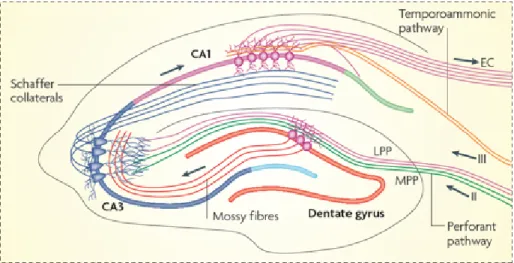
Figure 1.3.1. Hippocampal Circuitry of a Transverse Slice. Hip-pocampus has a very stereotypical structure. Hippocampal slices retain their cytoarchitecture and connections ex vivo which facilitates the stud-ies of synaptic plasticity mechanisms. Placing a stimulation electrode at one pathway allows the studies of specific types of synapses. (Image is modified from Deng, Aimone, & Gage, 2010)
The hippocampus is located in the medial temporal lobe and it repre-sents a part of the limbic system (Eichenbaum, 1997). In addition to being the site where synaptic plasticity was first described, is also a structure highly amenable to experimental manipulation. This is due to its highly laminar organization, which allows for the connections within the structure to be maintained when manipulated ex vivo. When sliced transversally, tri-synaptic pathway of the hippocampus can be preserved intact. Cortical projections enter the hippocampus via the perforant and the temporoam-monic pathways (Figure 1.3.1 (Deng, Aimone, & Gage, 2010)). Perforant pathway starts with dentate gyrus (DG) and DG granular cells send axons to CA3 area via mossy fibers, and the Schaffer collateral axons emerging from CA3 pyramidal neurons form synapses onto the apical dendrites of CA1 pyramidal neurons. On the other hand, temporoammonic pathway
axons form synapses onto the basal dendrites of CA1 pyramidal neurons. In this thesis, Schaffer collateral synapses have been studied.
Behavioral studies showed that CA1 pyramidal neurons fire condi-tional to the animal’s location (hence they are called place cells) (O’Keefe & Dostrovsky, 1971) and the Schaffer collateral synapses were shown to be modified during the formation of this place memory (McHugh, Blum, Tsien, Tonegawa, & Wilson, 1996; Mehta, Quirk, & Wilson, 2000). This long-term modifications on the transmission efficacy of a synapse is a func-tion of the converging activity and the subsequent firing caused by that activity. There has been two ways of studying plasticity electrically, re-peated high or low frequency activation of pre-synaptic terminals (Bliss & Lømo, 1973; Bienenstock et al., 1982), and coincident activation of post-synaptic action potentials (APs) and excitatory post-post-synaptic potentials
(EPSPs) (Markram, L¨ubke, Frotscher, & Sakmann, 1997; Bi & Poo, 1998;
Lisman & Spruston, 2010).
Recent advancements in the field of optics in combination with photo-activatable compounds have made it possible to study plasticity at the level of single synapses.
1.4
Dendritic
Spines
and
Structure-Function
Coupling
Although focus in Hebbs postulate (see section 1.2) is often placed on the need for coincident activity in order to effect changes in efficacy be-tween synapses, an integral part of the theory is the requirement for ”some growth process or metabolic change” to accompany the changes in efficacy. Therefore, we will discuss the structural changes that occur during synap-tic plassynap-ticity below.
Dendritic spines are the post-synaptic structures on which the major-ity of excitatory synapses of pyramidal neurons in the brain are located.
Their morphology is highly regulated by incoming activity. As described above, the induction of plasticity requires CaMKII and Ras/MAPK sig-naling pathways. However, the activation pattern and the morphology of the spine determines the level at which these interactions happen. This occurs due to filtering and compartmentalization of electro-chemical in-teractions within the volume of the spine head (Harris & Stevens, 1989;
Tonnesen, Katona, R´ozsa, Nagerl, et al., 2014). The post-synaptic density
(PSD), the region at the tip of a spine, is the region where the majority of receptors are clustered and interact, and the size of this specializa-tion is strongly correlated with the volume of the spine head (Harris & Stevens, 1989; Arellano, Benavides-Piccione, DeFelipe, & Yuste, 2007). As early as 1975, it was shown that the size of a spine changes immedi-ately following a stimulation (Van Harreveld & Fifkova, 1975) in dentate gyrus granular cells. In a follow up study, this spine enlargement was sup-pressed when a protein synthesis inhibitor, anisomycin, was applied prior
to the stimulation (Fifkov´a, Anderson, Young, & Van Harreveld, 1982).
Further evidence supporting the connection between functional and struc-tural coupling was seen when in response to brief bursts of high frequency stimulation of Schaffer collateral-commisural projections, the number of shaft synapses increases and the variability of dendritic spines decreases (Lee, Schottler, Oliver, & Lynch, 1980). More recently, an electron mi-croscopy study showed that when an axon makes more than one synapse with multiple spines, those spines have similar volumes, indicating that activity is critical for determining spine size (Bartol et al., 2015).
A major advance in our understanding of the relationship between spine size and synapse function accompanied the technological develop-ment which allowed for precise stimulation of single spines. Technical developments in the field of microscopy enabled researches to monitor
structural changes at single-synapse level in vivo. Invention of caged
of laser beams advanced the field one step further in terms of the speci-ficity of the targeted synapses (Ellis-Davies, 2007). Caged compounds are light-sensitive precursors of ligands that are inactive in the absence of light. Application of light pulses onto these compounds breaks the cage by a process called photolysis and frees the ligand from the cage, enabling spatio-temporally controlled manipulations of targeted processes. Schiller et al. used UV excitable caged glutamate to study NMDA-dependent
Ca2+ dynamics at single spines (Schiller, Schiller, & Clapham, 1998) and
they showed that, when paired with AP firing, uncaging controlled
NM-DAR activation caused a supralinear increase in Ca2+ through these
re-ceptors. Patterned two-photon uncaging was used to understand dendritic
integration mechanism at cortical (Branco, Clark, & H¨ausser, 2010) and
CA1 pyramidal neurons (Smith, Ellis-Davies, & Magee, 2003; Losonczy & Magee, 2006; Losonczy, Makara, & Magee, 2008). Using two-photon glu-tamate uncaging in combination with somatic and dendritic patch clamp recordings, significant contributions were made by showing that pseudo-synchroneous multisite glutamate uncaging leads to the non-linear inte-gration of EPSCs at hippocampal CA1 pyramidal neurons (Smith et al., 2003; Losonczy & Magee, 2006; Losonczy et al., 2008) and spatio-temporal properties of the multisite uncaging patterns determine the level of non-linearity of dendritic calcium and somatic EPSC integration (Branco et al., 2010).
This technology allowed for the careful assessment of spine volume in relation to the amount of current that the synapse conducts (Matsuzaki et al., 2004). A tight positive correlation between synaptic current and spine head volume has been demonstrated (Matsuzaki et al., 2004; Asrican et al., 2007; Harvey & Svoboda, 2007). This findings allowed researchers to use spine head volume as a proxy for plasticity.
1.5
Single Spine Plasticity
Matsuzaki et al. used two-photon glutamate uncaging and imaging to stimulate single dendritic spines and monitor their structural responses regarding activity (Matsuzaki et al., 2004). They developed a glutamate uncaging protocol by comparing normalized volume responses of dendritic spines to two different electrical stimulation condition. First, they stimu-lated Schaffer collaterals with 100 Hz, 1 sec electrical stimulation with the
presence of 1 mM M g2+ as they imaged a CA1 dendritic branch.
Simi-lar spine volume change was also induced by with 2 Hz, 60 sec electrical
stimulation without M g2+. Therefore, this [M g2+] dependent frequency
mapping allowed them to come up with the uncaging pattern they used for the study (60 uncaging pulses (each 0.6 msec) in 1 min, see Table 1.1). Glutamate uncaging induced similar spine enlargement as well, and they also showed that this form of LTP is NMDAR and CaMKII dependent ((Matsuzaki et al., 2004), Figure 1f and Figure 2e, respectively).
In a following study (Harvey & Svoboda, 2007), another regular uncaging pattern was found to evoke similar single-spine volume dynam-ics using 30 uncaging pulses (each pulse 4 msec, instead of 0.6 msec in the previously described study) for 1 min. Additionally, they showed that the stimulation of multiple spines with two different uncaging protocols (30 pulses,1 min, 4msec vs 30 pulses, 1 min, 1msec), did not lead to the potentiation of spines stimulated with shorter pulse lengths, hence this protocol is referred to as sub-threshold.
Govindarajan et al. using the same stimulation protocol in combina-tion with forskolin in order to raise the intracellular levels of cyclic-AMP (cAMP) in the stimulated neurons. cAMP is a second messenger that acts on protein kinase A (PKA) pathway, hence boosting the protein transla-tion in the cell. This study showed that the uncaging induced LTP under
AMPAR NMDAR Caged Glutamate bpAP uEP SC
Dend
ri
te
Spine
Head
Uncaging LaserFigure 1.5.1. Pre-synaptic Neurotransmitter Release can be Mim-icked by Glutamate Uncaging. Two-photon glutamate uncaging lows glutamate to be released in a spatially specific, controlled way, al-lowing single spines to be studied. Studies addressing NMDA dependent
plasticity are performed in the absence of M g2+
the influence of forskolin extended the longevity of the structural spine plasticity (Govindarajan et al., 2011).
Table 1.1 summarizes the stimulation protocols that has been used to induce LTP at single dendritic spines using electrical and uncaging laser pulses.
Table 1.1. Requirements of Inducing Plasticity at Single Den-dritic Spines. Plasticity can be induced at single spines using electrical stimulation of glutamate uncaging. Glutamate uncaging under the
ab-sence of [M g2+] induces plasticity that is conditional to the number of
uncaging pulses and pulse width.
Study Stim. Type Freq. (Hz) Stim. Len. (sec) # of Pulses Mg (mM) Uncaging Pulse Len. (msec) Matsuzaki et al. (2004) Electrical 100 1 100 1 -Electrical 2 60 120 0 -Uncaging 1 60 60 0 0.6 Harvey & Svoboda (2007) Uncaging 0.5 60 30 0 4 Israely et al. (2011) Uncaging 0.5 60 30 0 4
1.6
Spike-Timing Dependent Plasticity
It has been shown that relative firing times between a pair of feed-forward connected neurons determines the direction and the level of plasticity at
the synapse connecting these neurons. This phenomenon is known as
spike-timing dependent plasticity (STDP) (Markram et al., 1997; Bi & Poo, 1998). STDP is proposed to be the mechanism governing the synaptic plasticity in vivo (Paulsen & Sejnowski, 2000). However, this proposal is highly debated (Lisman & Spruston, 2005, 2010).
There has been various working models proposed to be the induction
mechanism of STDP (Markram, Gerstner, & Sj¨ostr¨om, 2012). Original
model was based on the timing difference between one pre-synaptic and one post-synaptic spike. Repeated activation of this pre→post firing or-der induced LTP at excitatory synapses of pyramidal neurons as long as
timing difference was smaller 50 msec (Bi & Poo, 1998). This finding was in strong agreement with Hebb’s original proposal (see section 1.2). Ad-ditionally, reversing the order of pre→post firing induced LTD (Markram et al., 1997; Bi & Poo, 1998). This simple two-spike interaction model shown be explaining cortical development and remapping of visual cor-tical maps through competition (Song, Miller, & Abbott, 2000; Song & Abbott, 2001) and hippocampal receptive fields (Mehta et al., 2000).
Further research showed that this two-spike model is not sufficient to explain a large body of the experimental data which lead to the develop-ment of three- and four- spike models (Froemke & Dan, 2002; Froemke, Tsay, Raad, Long, & Dan, 2006; Pfister & Gerstner, 2006). In these mod-els instead of pre→post or post→pre spike interactions, higher order sta-tistical interactions between multiple inter-spike intervals were taken into consideration such as pre→post→pre, post→pre→post (Pfister & Gerst-ner, 2006), pre→post→pre→post or post→pre→post→pre (Froemke & Dan, 2002). These models were shown to be better at explaining the experimental data which shows the importance of complex spike-timing interactions.
1.7
Naturalistic Patterns, Synaptic Responses
and Plasticity
The induction of LTP was discovered by high frequency electrical stim-ulation (HFS) and variations of HFS trains are used to induce LTP in the hippocampus and other brain regions. Although these constant high frequency protocols are effective at inducing potentiation, they do not represent the breadth of activity patterns that can be observed in vivo. The existence of both temporal and rate coding in neural networks has been well established (Ferster & Spruston, 1995; Christopher deCharms & Merzenich, 1996; Prut, Slovin, & Aertsen, 1995; Bienenstock et al., 1982;
Tsodyks & Markram, 1997) yet how such parameters influence plasticity at single inputs has not been addressed yet. A number of computational and experimental studies have been conducted, investigating the effects of naturalistic stimulation patterns on the induction of plasticity. Here, we are going to review these studies briefly and try to summarize how different groups approached this problem.
Migliore and Lanski (Migliore & Lansky, 1999) made a computational model of synaptic transmission to test to what extent the temporal vari-ations at the stimulation train can change the state of a synapse. Their results showed that, even if the mean stimulation frequency is maintained constant, the probability of inducing LTP and LTD can be a function of the temporal variation of the stimulation train. Such temporal variations of the input train has not taken into account in the experiments discussed earlier.
The first study to use naturalistic stimulus patterns (NSP) to study short and long term plasticity showed that NSPs induce LTP in hippocam-pal slices (Dobrunz & Stevens, 1999), whereas they found no relation-ship between the instantaneous frequency and the response magnitude for NSPs. The NSPs were taken from the timing of action potentials recorded in vivo from hippocampal place cells of awake, freely moving rats. They showed that NSPs show highly variable timing. The interspike intervals measured in vivo were multiplied by 3 to account for the temperature
difference between the in vivo measurements (37 oC) and slice recordings
(24oC) (Dobrunz & Stevens, 1999).
This study mostly focused on short term plasticity, such as the re-sponse size variations with respect to stimulus number and inter-stimulus intervals (ISI) within the stimulation train. The authors investigated LTP using natural stimulus pattern, and they tested field EPSP slopes for two independent pathways in the same hippocampal slice. A 256-point natu-ral stimulus pattern was applied for 12 min, which caused a long lasting
potentiation (1.5 times more compared with the baseline) of the response to constant frequency (0.17 Hz) stimulation. The control pathway, which was not stimulated with the natural pattern, was not potentiated.
Another study investigated the plasticity concequences of naturalistic patterns in spike-timing dependent plasticity (STDP) setting (Froemke & Dan, 2002). Using the firing patterns of two cortical pyramidal neurons that have overlapping visual fields as pre- and post-synaptic trains, it has been shown that the activity-induced synaptic modifications do not depend only on the relative spike timing between the neurons, but also on the inter-spike intervals within each neuron (Froemke & Dan, 2002).
In neocortical slices, stimulation patterns derived from slow wave sleep (SWS) induced LTP (Rosanova & Ulrich, 2005) or LTD (Czarnecki, Bir-toli, & Ulrich, 2007) depending on the pattern of stimulation (Rhyth-mic burst or spindle-like trains, respectively). deKay et al. compared naturalistic patterns with constant frequency stimulation in a compara-tive study between young adult (P28-P35) and juvenile (P12-P18) rats (deKay, Chang, Mills, Speed, & Dobrunz, 2006). They showed that the average responses to naturalistic stimuli and constant frequency stimu-lus both showed modest depression in young adults, but juveniles showed facilitation for NSPs but short-term depression after constant frequency stimulation. Tunstall et al. (Tunstall, Agnew, Panzeri, & Gigg, 2010) used two different naturalistic stimulus patterns to stimulate neurons in subiculum. First pattern included bursts of activity, whereas second pat-tern was spaced more evenly. Differences in the short term responses to two different patterns were reported, and they concluded that dynamic interactions between rate and temporal coding exist between input spikes and stimulus response but they have not observed any LTP for either class. Gundfinger et al. used irregular stimulus trains resembling the natural spike statistics from DG neurons to study short and long term plasticity mechanisms at mossy fiber synapses in acute hippocampal slice
prepa-rations (Gundlfinger et al., 2007; Gundlfinger, Breustedt, Sullivan, & Schmitz, 2010). They used in vitro electrophysiology and computational modeling to study the interactions between LTP and STP at mossy fiber synapses and showed that LTP occluded the frequency facilitation by re-ducing the dynamic range of the synapse. They concluded that phenomena such as alterations in the place field size and speed could be explained by mechanism related to experience-dependent changes in the properties of short-term facilitation.
In another study combining electrophysiological recordings with real-istic modeling of STP in excitatory hippocampal synapses, Kandaswamy et al. showed that STP increases the information transfer in a time and frequency dependent fashion. The study showed that STP in Schaffer col-lateral synapses increased the information transfer in a wide range of input frequencies from 2 to 40 Hz. Moreover, time dependent analysis of mu-tual information predicted that in low-release probability synapses, STP acts to maximize information transfer specifically for short high frequency bursts. They concluded that since many types of synapses are not likely to experience extensive periods of high-frequency activity under natural conditions, these synapses do not reach a steady state in vivo.
In summary, the studies discussed above found that the short term responses are very sensitive to the temporal variations of the input pat-terns. Transient bursting patterns are necessary for LTP to occur in the hippocampus (Pike, Meredith, Olding, & Paulsen, 1999) but not sufficient, since not all naturalistic patterns induce LTP. Finally, in the light of these experiments, a biophysical modeling study, Migliore et al. concluded that the state of the synapse at the time of plasticity induction is a key factor in determining whether a pattern will lead to the induction of LTP or LTD (Migliore, De Simone, & Migliore, 2015).
1.8
Contribution of this Dissertation
The work in this dissertation investigates the functional and structural consequences of non-regular patterns of activity at single inputs. The aim of the study was to understand how more naturalistic stimulation scenarios encode information at single synapses, and is built around the idea that the transmission of information does not occur through regularly spaced neurotransmitter release in vivo (Zador & Dobrunz, 1997).
In chapter 2 we listed the materials that we used in our experiments and explained the methods that have been used.
We introduce a dendritic spine analysis toolbox named SpineS in chap-ter 3. Studies addressing structural dendritic modifications require the collection of large scale dendritic spine images which requires laborious manual analysis. Manual analysis of vast number of dendritic spines is not only time consuming and tiresome, it also is prone to subjective blun-ders. Therefore, we developed SpineS for the automatic quantification of dendritic features. SpineS is Matlab based and open-source.
In chapters 4 and 5, we present the investigations of long- and short-term structural spine dynamics comparing regular and Poisson distributed stimulation patterns. This is the first study using naturalistic patterns to understand structural plasticity mechanisms at single dendritic spines.
In chapter 6 we discuss the implications of the findings and consider potential mechanisms underlying the obtained results. Further, we will reflect upon the implications of these findings to the study of synaptic plasticity and how this work may contribute to the understanding of plas-ticity within neural circuits.
Chapter 2
2.1
Materials
2.1.1 Dissection Solution
First, we prepared 10× concentrated Krebs-Ringer solution (Table 2.1)
and we kept this stock solution at 4oC. Later, we prepared 1× solution
(Table 2.2) by adding CaCl2 and M gCl2 while the solution was bubbled
using 95% O2 and 5% CO2 for 30 min. The solution was afforated using
distilled water to 250 mL, sterilized with vacuum filter, aliquoted in 40
mL volumes and stored at −20oC until dissection.
Table 2.1. 10× Krebs Ringer Dissection Solution.
10× Krebs Ringer mM M.W. in 250 mL
KCl 25 74.55 0.465 g
N aHCO3 260 84.01 5.460 g
N aH2P O4 11.5 mM 119.98 0.344 g
D − Glucose 110 mM 180.16 4.95 g
Table 2.2. 1× Krebs-Ringer Dissection Solution.
1× Krebs-Ringer mM in 250 mL 10× Krebs-Ringer 100 25 mL M iliQW ater - 300 mL Sucrose 238 20.36 g CaCl2 1 0.25 mL (1M CaCl2) M gCl2 5 1.25 mL (1M MgCl2) 2.1.2 Dissection Equipment
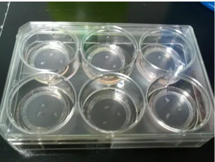
Brains were dissected using surgical tools (Figure 2.1.1). During the pro-cedure, the extracted brain and hippocampal slices were kept in ice-cold Krebs-Ringer solution. Following the extraction, hippocampi were placed on a filter paper with Krebs-Ringer solution and fixed on the chopper
(Figure 2.1.2) plate. Afterward, they were sliced 350µm-thick transverse slices and kept in an incubator in six-well plates (Figure 2.1.3).
Figure 2.1.1. Surgical Dissection Set. Brain is extracted out of skull using the surgical set to be sliced using tissue slicer.
Figure 2.1.2. Tissue Slicer. Hippocampi were sliced into 350 µm thick slices for culturing.
Figure 2.1.3. Cultures in Six-Well Plate. Hippocampal slices are incubated in six well plates in the incubator. We placed three to four slices per well sitting on inserts with the culture media underneath them. Medium was changed every two to three days.
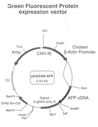
2.1.3 Biolistic Gene Transfer
Hippocampal neurons from organotypic slice cultures were transfected us-ing a Helios gene gun (Bio-Rad) after four to seven days in vitro (DIV). Gold beads (10 mg, 1.6 µm diameter, Bio-Rad) were coated with 100 µg of pCAGGS-AFP (Fig. 2.1.4) plasmid DNA (Ogawa & Umesono, 1998),
according to the Bio-Rad protocol1, and delivered biolistically into the
slices at 180-200 psi (Woods & Zito, 2008).
1
Figure 2.1.4. pCAGGS-AFP Vector. pCAGGS-AFP plasmid was used for the sparse transfection of hippocampal neurons. Plasmid size: 5.54 Kb, constructed by Hidesato Ogawa (Ogawa & Umesono, 1998).
2.1.4 Internal Solution for Patch Pipette
Whole cell recordings were performed at room temperature, with glass pipettes (access resistance 6 to 10 MΩ) filled with internal solution
con-Table 2.3. Artificial Cerebrospinal Fluid (ACSF). ACSF (2X) mM M.W. in 1 L N aCl 127 58.44 14.84 g KCl 2.5 74.55 0.372 g N aHCO3 25 84.01 4.2 g N aH2P O4 1.25 119.98 0.299 g
taining: 115 mM potassium gluconate, 10 mM HEPES, 20 mM KCl, 4 mM Mg-ATP, 0.3 mM Na-GTP, 10 mM Na-phosphocreatine and 1 µM
CaCl2. pH was adjusted to 7.4 using KOH. In some recordings, ALEXA
488 Fluor (30µM, Invitrogen, Germany) was added to the internal solution (Edelmann & Lessmann, 2011).
2.1.5 Artificial Cerebro-Spinal Fluid (ACSF)
During the experiments, the slices were perfused with carbogenated (95%
O2 and 5% CO2) artificial cerebral spinal fluid (ACSF) containing the
following (in mM): 127 N aCl, 25 N aHCO3, 25 D-glucose, 2.5 KCl, 1
M gCl2, 2 CaCl2 and 1.25 N aH2P O4 and TTX, delivered with a
peri-staltic pump at 1.5 ml/min (Table 2.3). Uncaging ACSF (uACSF) was
the same as ACSF except for 4 mM CaCl2, 0 mM M gCl2, MNI-glutamate
and 0 TTX. Anisomycin (50 mM), cycloheximide (60 mM), APV (50 µM )
or different [M g2+] was added to the solution when specified as well
Table 2.4. Organotypic Slice Culture Media. mM in 500 mL N aHCO3 6 0.252 g HEPES 30 3.57 g D-Glucose 27 2.43 g 1X MEM 394 mL Horse Serum 100 mL CaCl2 1 mM M gSO4 1 mM Ascorbic Acid 25% 24 µL Insulin (10 mg/mL) 50 µL GlutaMax 2.5 mL
2.2
Methods
2.2.1 Organotypic Slice Cultures
Cultured hippocampal slices were prepared from C57BL/6J mice
(postna-tal day 7 to 10) (G¨ahwiler, 1981; Stoppini, Buchs, & Muller, 1991). Briefly,
350 µm thick slices were obtained with a chopper in ice-cold ACSF
con-taining 2.5 mM KCl, 26 mM N aHCO3, 1.15 mM N aH2P O4, 11 mM
D-glucose, 24 mM sucrose, 1 mM CaCl2 and 5 mM M gCl2, and cultured
on membranes (Millipore). The slices were maintained in an interface configuration with the following media: 1× MEM (Invitrogen), 20% horse serum (Invitrogen), GlutaMAX 1 mM (Invitrogen), 27 mM D-glucose, 30
mM HEPES, 6 mM N aHCO3, 1 M CaCl2, 1 M M gSO4, 1.2% ascorbic
acid, and 1 µg per mL insulin (Table 2.4). The pH was adjusted to 7.3, and osmolarity adjusted to 300-310 mOsm. All chemicals were purchased from Sigma unless otherwise indicated.
2.2.2 Two Photon Laser Scanning Microscopy
Two photon excitation microscopy is an imaging technique that was de-veloped by Winfried Denk as a side project during his doctorate (Denk, Strickler, & Webb, 1990; Svoboda & Yasuda, 2006). This technique uses femto-second laser pulses to excite fluorophores. Femto-second pulses en-able pseudo-synchronous excitation of a fluorophore by two-near coinci-dent photons hitting them so that the second photon creates a non-linear jump at the energy level of the excited particles, allowing fluorophores to be imaged using lower laser pulses than in one-photon based techniques.
In our experiments, two-photon imaging and uncaging was performed using a galvanometer-based scanning system (Prairie Technologies, ac-quired by Bruker recently) on a BX61WI Olympus microscope, using a Ti:sapphire laser (Coherent) controlled by PrairieView software.
Imaging
Slices were perfused with oxygenated ACSF (Section 2.1.5. Imaging was started 15 to 30 min after the initiation of slice incubation. Secondary or tertiary dendrites of CA1 neurons were imaged using a water immersion objective (60×, 0.9 NA, Olympus) with a zoom of 10×. Image stacks (0.3 µm per section) were collected once every 5 min for up to 4 h at a resolution of 1024 × 1024 pixels, resulting in a field of view measuring approximately 19.8µm × 19.8µm. Z-stacks were used to quantify spine volumes (see Chapter 3 for details) in all experimental conditions, and all images in one experiment were acquired under the same imaging conditions maintaining equal laser power and PMT gain settings. We monitored imaging laser power fluctuations throughout experiments using a laser power meter (Thor Labs).
Figure 2.2.1. Two Photon Imaging and Uncaging of Single Den-dritic Spines. Left: A CA1 pyramidal neuron expression AFP, Right Top: Branch of Interest, Right Bottom: Stimulated Spine. Scale Bar: 1µm
Glutamate Uncaging
Glutamate is the major neurotransmitter operating at excitatory synapses. MNI-caged-L-glutamate (4-methoxy-7-nitroindolinyl-caged-L-glutamate)
is a compound that is inert in cells in the initial form. Two photon
laser pulses at 720 nm wavelength are able to break the cage hence enabling spatio-temporally controlled simulation patterns for the mimicry of synaptic activation.
Objective
60X / 0.9NA
Pump
1.5ml/minACSF
2Ca/1MguACSF
4Ca/0Mg TrashChamber
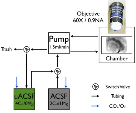
Switch Valve Tubing CO2/O2Figure 2.2.2. ACSF Circulation. During imaging and uncaging, slices were perfused using artificial cerebro-spinal fluid (ACSF). Two different
ACSFs were used with different concentrations of Ca2+, M g2+ and
MNI-L-Glutamate. Normal ACSF was used during incubation, baseline, and post-stimulus periods, whereas uncaging-ACSF (uACSF) was used only during the stimulation period which starts with the completion of the last baseline image stack and ends with stimulus delivery (6 min in total).
Before using a new batch of MNI-L-glutamate (Tocris), we performed whole-cell patch-clamp recordings of pyramidal neurons to monitor
uncaging-evoked mini EPSCs (mEPCSs). We located the laser pulse
approximately 0.5 µm away from the spine head perimeter (Figure 2.2.1). First, we recorded spontaneous mEPSCs for 5 to 10 min and attempted the application of different laser power values under physiological Ca and
Mg concentrations (2 mM CaCl2, 1 mM M gCl2) to obtain an uncaging-evoked mEPSC similar in size to the spontaneously occurring mEPSCs on average.
Experimental Design
We incubated slices 20 to 30 min as they are perfused with ACSF (0.5µM TTX, 2 mM Ca, 1 mM Mg) at room temperature before the beginning of the experiment. Experiments are initiated with baseline Z-stack imaging of a secondary or tertiary dendritic branch of a CA1 neuron (Figure 2.2.1). The dendritic branch was imaged every 5 min for 15 to 30 min before glutamate uncaging. After the collection of baseline images, we switched to uncaging-ACSF (uACSF), containing 2.5 mM MNI-Glutamate, 0 mM Mg and 4 mM Ca. Uncaging Pattern (Figure 2.2.3) delivered 0.5µm away from the tip of the spine.
We switched back to the use of normal ACSF after uncaging, and take the first image was obtained 2 min after the stimulation and, afterward, every 5 min during the following 4 h.
2.2.3 Pulse Train Modeling using a Poisson Process
We designed uncaging pulse trains using a homogeneous Poisson process to generate irregular uncaging patterns that we call naturalistic trains to stimulate single dendritic spines.
There are two ways of generating homogeneous Poisson spike trains. The first approach is based on subdividing total spike train length into a series of non-overlapping time intervals, each of duration δt. Afterward, a sequence of uniformly distributed random numbers between 0 and 1 can be used to generate a spike for each interval as long as the random number x ≤ rδt. Here r is the instantaneous firing rate, which is constant over time for homogeneous Poisson train. This means that the probability
Baseline Images
Stimulation Train
2’
4h
uACSF
Caged Glutamate
TTX
Post Stimulation Images
Regular
NT-Uniform
NT-Beginning
NT-End
0 60 sec
Figure 2.2.3. Schematic Illustration of the Experiment. Following the incubation, baseline images were collected. uACSF circulation started
immediately after the acquisition of last baseline image stack. At the 5th
min of uACSF circulation, one of four uncaging patterns was delivered for 1 min and structural imaging was continued up to 4 h post-stimulation in order to follow uncaging-evoked spine volume changes.
of a spike occurring during a time interval δt is equal to the value of the instantaneous firing rate during that interval times the length of the interval (Equation 2.1).
P{1 spike during (t − δt, t + δt)} = rδt (2.1)
In the second approach exponential distribution is used to derive in-terspike intervals for a Poisson spike train. Poisson process provides a description of the number of events in a given time period (Equation 2.2).
P(n spikes during ∆t) = e
−r∆t(r∆t)n
The exponential distribution, which can be obtained by taking the derivative of the cumulative distribution function of the Poisson distribu-tion, will provide the length of time between events (Equation 2.3).
f(∆t) = r∆te−r∆t (2.3)
Once the exponentially distributed random spike times are generated, successive spike times can be obtained by adding the previous spike time with the randomly drawn interspike interval.
2.2.4 Statistical Analysis
All statistical analyses were performed using custom code written in Mat-lab. Permutation (Shuffle) test was used for the analysis presented in Figure 5.3.7. Nonparametric MannWhitney-U test was used to compare spine volumes at any time bin versus baseline or different condition. Time series were compared using repeated-measures ANOVA.
In order to compare the error between different volume estimation methods and volume differences that might introduced due to fluorescence fluctuations over time, we used a symmetric mean absolute percentage er-ror (sMAPE) based similarity score (SS). The sMAPE is a common mea-sure for trend comparisons between time series data (Makridakis, 1993).
sM AP Emethodspine 1−method2 = 100 ×
1 n n X i=1 |method1 i − method2i| |method1 i + method2i| (2.4)
Here, n is the number of time points for the analyzed spine. sMAPE is used in sections 3.3 and 3.7.
Chapter 3
Spines: A Tool for
Automatic Dendritic Spine
Analysis
Contributions: Ali ¨Ozg¨ur Argun¸sah, Devrim ¨Unay and Inbal Israely
conceived the study. Ali ¨Ozg¨ur Argun¸sah, Ertun¸c Erdil, Muhammad
Usman Ghani, Arif Murat Ya˘gcı, S¨umeyra Demir Kanık and Devrim
¨
Unay wrote the code. Ali ¨Ozg¨ur Argun¸sah, Anna Felicity Hobbiss and
Yazm´ın Ramiro Cort´es provided experimental data.
Affiliations:
1Champalimaud Neuroscience Programme, Lisbon, Portugal.
2Sabancı University, Istanbul, Turkey.
3Bahce¸sehir University, Istanbul, Turkey.
4Bo˘gazi¸ci University, Istanbul, Turkey.
5Universidad Nacional Autonoma de Mexico, Mexico City, Mexico.
6Izmir University of Economics, Izmir, Turkey.
Support: This work is supported by Funda¸c˜ao para a Ciˆencia e a
Techologia (FCT), Funda¸c˜ao Champalimaud (FC) and Instituto
Gulbenkian de Ciˆencia (IGC) and The Scientific and Technological
3.1
Abstract
Two photon-imaging experiments have begun to elucidate the dynamic na-ture of dendritic spines, showing that they undergo changes in shape both during development and in response to synaptic stimulation. The experi-ments which track such changes require the collection of multi-dimensional data over prolonged periods of time, generating large amounts of informa-tion which requires tedious manual labor in order to be analyzed. In addi-tion to involving lengthy analysis periods, manual analysis may introduce operant bias which may alter the accuracy of quantification. Therefore, we developed an open source image-processing toolbox called SpineS for the automatic quantification of dendritic features such as spine head volume, spine neck length, and inter spine distances, from imaging data collected with confocal and two-photon fluorescence microscopy. This toolbox al-lows for the rapid quantification of many spines within the field of view, as it increases estimation precision and eliminates inter-operant estimation differences.
3.2
Introduction
The efficacy of excitatory synapses changes with activity (Bliss & Lømo, 1973) as well as during learning (Whitlock et al., 2006), and these changes correlate with the morphological alterations of dendritic spines (Asrican et al., 2007; Matsuzaki et al., 2004). In particular, the linear relationship between spine volume and current amplitude of a spine (as discussed in section 1.4), and bidirectional changes in spine volume correspond to the induced plasticity (Asrican et al., 2007; Matsuzaki et al., 2004;
Ramiro-Cort´es & Israely, 2013; Tonnesen et al., 2014). These changes in efficacy
and structure reflect activity at a synapse, and can impact subsequent in-formation transmission between inputs across the dendritic arbor (Magee,
2000; London & H¨ausser, 2005; Bartol et al., 2015). Understanding how such changes are physically maintained in the cell is key to elucidating the mechanisms whereby information is stored in the brain. Activity-dependent structural changes at spines can last from several minutes to hours, and are visualized through multi time point sampling of Z-stack im-ages, often collected for many hours. For example, in an experiment that addresses structural LTP or LTD mechanisms at a single dendritic spine using two photon glutamate uncaging and imaging, researchers image a dendritic branch every 5 min up to 4 h (48 Z-stacks). Given the image acquisition conditions and the type of neuron that dendritic segment im-ages are collected from, one branch may have up to 50 spines. Analyzing 2400 spines is not only laborious and time-consuming but also prone to operant subjectivity.
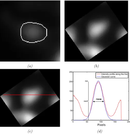
Therefore, we developed a Matlab based toolbox called SpineS for rapid and robust quantification of spine head volumes and neck lengths. Z-stacks from multiple time points can be analyzed using SpineS. The process starts with importing Z-stacks and registering them over time. Maximum inten-sity projections (MIPs) of Z-stacks are computed after the registration and filtered using a median filter. Filtering is followed by segmentation of head of dendritic spines and spine volumes are estimated using integrated fluorescence intensity (IFI) method. Afterward, neck paths are computed using a fast-marching algorithm for the estimation of spine neck lengths. Detailed description of these steps is presented below.
The first step performed by the SpineS package is to load data. Since each lab uses a different imaging system, and data formats and specifica-tion can be very different, we used bio-formats library provided by Open Microscopy Environment (OME) Project (Goldberg et al., 2005). Bio-formats library provides tools for importing various image Bio-formats. The algorithm computes maximum intensity projected (MIP) images (Figures 3.2.1 and 3.2.3), for each time point as it loads image stacks and performs
MxNxT Filtering, Z-projection, Registration Spine Seg. t=1 t=7 t=22 t=36 t=49 Dendrite Segmentation 3D Spine Neck Path Finding using Fast Marching MxNxT Use 3D z-stack ROI of the Spine
of Interest
Compute 3D Skeleton from binary segmented dendrite to
find candidate points to end the shortest path algorithm
Compute 3D Spine Head Center from binary segmented spine head and use this point to start the shortest path algorithm MxNxZxT t=1 t=2 t=T Volume Neck Measure
Figure 3.2.1. Workflow of SpineS. Z-stack from multiple time points are analyzed. Each Z-stack is imported and registered after Z-projections computed and filtered using median filtering. Dendritic spines are seg-mented using a watershed based algorithm and each spine volume is es-timated using IFI method and normalized with the median fluorescence intensity of the dendrite at the corresponding time point. Neck paths are computed using a fast-marching algorithm from spine head center to the closest geodesic point on the dendrite by imposing some constraints.
an initial translation correction (Figure 3.4.1). Users select the spines to be analyzed by clicking on the center of the spine head at the first time point (Figure 3.2.2). Next, the registered MIP images are filtered using a simple median filter during the segmentation process. The me-dian filter has just one parameter in order to determine the number of neighboring pixels that are used for the calculations of the median value,
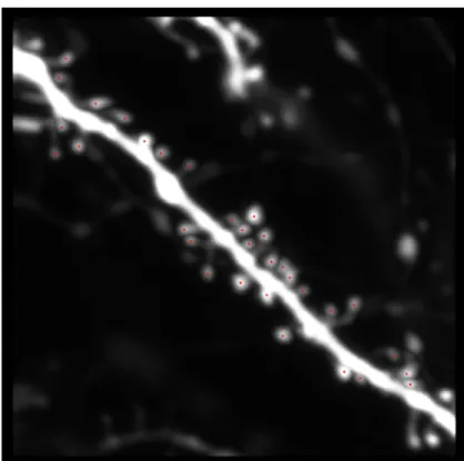
Figure 3.2.2. Spine Selection for Analysis. User clicks at the center of every spine at the first time point. These spines will be tracked in the next time points by the algorithm.
which is set by the user. The filtered image is then binarized using Otsu thresholding (Otsu, 1975), which results in a rough segmentation of the dendritic branch including spines. Further, the medial axis of the dendrite is computed by applying a fast marching distance transform (Kimmel & Sethian, 1996) on the dendritic segment, then we apply a locally adaptive sized disk-shaped structuring element around the medial axis of the den-drite to remove spines for denden-drite segmentation. To further refine the segmentation, we use the assumption that the dendrite diameter remains consistent in the local field of view after the initial registration. We
com-pute the diameter of the dendrite at all locations and consider the median value to be the true dendrite diameter and remove all pixels beyond the diameter. This gives us a clear segmentation of the dendrite.
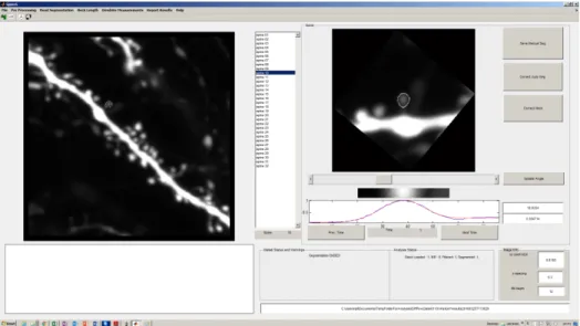
Figure 3.2.3. SpineS Graphical User Interface (GUI). GUI provides filtering, automatic segmentation, segmentation correction, manual seg-mentation and manual FWHM estimation tools. The big plot on the left is the MIP image of the analyzed dendrite. Smaller plot on the right show one of the 32 spines that are analyzed in this example.
In order to define the spine head, we use a multilevel segmentation algorithm. First, we obtain a coarse segmentation of the spine-head using a watershed-based technique. Since the spine head boundaries found in this step are generally larger than the expected boundaries, we segment the interior of this region for refinement. A graph-based image segmentation algorithm followed by hierarchical agglomerative clustering is applied to obtain refined spine head segmentation (Figure 3.5.1). Spine volumes can be computed using IFI of the segmented spine head image. Once the spine of interest has been segmented, a fast marching algorithm computes the