RESUMO.- [A dosagem sérica de CPK-MB em cães com desvio ST por quimiluminescência.] Embora frequente em humanos, as doenças hipóxicas e isquêmicas do coração são pouco relatadas em cães, com poucos relatos de infarto agudo do miocárdio (IAM) nesta espécie. Alguns achados no eletrocardiograma podem sugerir hipóxia/isquemia miocárdica, como a elevação ou depressão do segmento ST,
mas não há estudos que mostram se os desvios do segmen-to ST estão associados a lesões miocárdicas e aumensegmen-to sé-rico da creatinafosfoquinase (CPK-MB). A fim de investigar possíveis lesões nas células miocárdicas em condições de má perfusão, 38 cães foram estudados, 20 com eletrocar-diograma normal e 18 com elevação ou depressão do seg-mento ST, registrados em papel, na derivação II, velocidade de 50 mm/s e sensibilidade N (1mV = 1cm). A mensuração da creatinafosfoquinase isoenzima MB (CPK-MB) em cães normais (grupo 1) determinou os valores controle (em ng/ ml), que foram comparados com os obtidos a partir de cães com desvio (grupo 2), permitindo a confirmação ou não da lesão miocárdica. Os valores médios de CPK-MB obtidos de cães nos grupos 1 e 2 foram 0,540ng/ml (DP±0,890) e 0,440ng / ml (DP ± 1.106), respectivamente. A um nível de significância de 5%, a relação de CPK-MB com a idade, mas -sa e creatinofosfoquinase total (CPK-T) não foi significativa
Serum dosage of CPK-MB in dogs with ST deviation by
chemiluminescence
1André L.F. Santos2*, Guilherme G. Pereira3, Mário M. Santos3, Vanda C.R. Gutierrez4, Priscila C. Calyjur2 and Maria Helena M.A. Larsson3
ABSTRACT.- Santos A.L.F., Pereira G.G., Santos M.M., Gutierrez V.C.R., Calyjur P.C. & Larsson M.H.M.A. 2014. Serum dosage of CPK-MB in dogs with ST deviation by chemilumines-cence. Pesquisa Veterinária Brasileira 34(Supl.1):74-78. Setor de Patologia Veterinária, Fa-culdade de Medicina Veterinária e Zootecnia, Universidade de São Paulo, Av. Orlando Mar-ques de Paiva 87, São Paulo, SP 05508-270, Brazil. E-mail: [email protected]
Although frequently in humans, hypoxic and ischemic heart diseases are poorly docu-mented in dogs, with only few reports of acute myocardial infarction (AMI) in this species. Some electrocardiographic findings might suggest myocardium hypoxia/ischemia, like ST segment elevation or depression, but there are no studies showing whether deviations in ST segment are associated to myocardial injury and serum increase of creatine phosphoki -nase (CPK-MB). In order to investigate possible myocardial cells injury in poor perfusion conditions, 38 dogs were studied, 20 with normal electrocardiogram and 18 with ST seg-ment elevation or depression, recorded in lead II, at a paper speed of 50 mm/sec and N sensibility (1mV=1cm). Serum measurement of creatine phosphokinase isoenzyme MB (CPK-MB) in normal dogs (group 1) determined control values (in ng/mL), which were compared to those obtained from dogs with deviation (group 2), which allowed confir -mation or not of myocardial injury. CPK-MB mean values obtained from dogs in groups 1 and 2 were 0.540ng/ml (SD±0.890)ng/mL and 0.440ng/mL (SD±1.106), respectively. At a significance level of 5%, the relation of CPK-MB with age, mass and total creatine phos -phokinase (CPK-T) was not significant in groups 1 and 2. CPK-MB showed no difference, at 5% level, between groups 1 and 2. In conclusion, it is possible to use the human chemilumi -nescent immunometric assay kit in canine species and that hypoxia/ischemia revealed by ST segment deviation does not mean significant myocardium injury.
INDEX TERMS: Creatinephosphokinase, CPK-MB, dogs, ST segment.
1 Received on September 5, 2014.
Accepted for publication on January 6, 2015.
2 Centro de Pesquisas sobre o Genoma Humano e Células-Tronco, Rua do
Matão 106, São Paulo, SP 05508-090. *Corresponding author: andrvet@ usp.br
3 Departamento de Clínica Médica, Faculdade de Medicina Veterinária e
Zootecnia, Universidade de São Paulo (USP), Av. Orlando Marques de Paiva 87, São Paulo, SP 05508-270, Brazil.
4 Hospital Universitário, USP, Av. Orlando Marques de Paiva 87, São Paulo,
nos grupos 1 e 2. Não houve diferenças na CPK-MB, ao ní-vel de 5%, entre os grupos 1 e 2. Conclui-se que é possíní-vel utilizar o kit de ensaio imunométrico por quimiolumines-cência humano na espécie canina e que a hipoxia/isquemia revelada pelos desvios do segmento ST, não significa lesão miocárdica.
TERMOS DE INFDEXAÇÃO: Creatinafosfoquinase, CPK-MB, cães, segmento ST.
INTRODUCTION
Acute myocardial infarction is a common affection in hu-mans, having as a typical symptom the “chest pain” irradia-ting to the back and arms. Individuals with these symptoms are monitored by electrocardiogram and by dosing myo-cardial hypoxia-ischemia markers (Bonow et al. 2009, Tha -rwat 2013). Myocardial hypoxia is the insufficient cardiac muscle oxygenation (Robbins et al. 2001), in spite of the adequate perfusion (Bonow et al. 2009), while ischemia is the oxygen deprivation followed by inadequate metabolites removal.
During hypoxia, enzymes are liberated in the blood cir-culation (Loegering & Critz 1971) and, in case of hypoxia/ myocardial infarction, creatine phosphokinase (CPK) ca -talyses the reversible reaction: creatine + ATP > phospho-creatine + ADP, named Lohman Reaction (Aktas et al. 1993, Bottomley & Weiss 2001).
CPK is a dimeric molecule of 381 amino acids that con-sists of two subunits, (M and B, which have the same wei-ght but are immunologically distinct and synthesized by two different genes) and presents 282 cysteine residues that are necessary for its catalytic activity. There are three isoenzymes: CPK-MM, present in striated muscle; CPK-BB, present predominantly in the brain and digestive system and CPK-MB, present in the cardiac tissue, kidneys, intesti -nes and lungs (Aktas et al. 1993).
CPK synthesis control is not well understood. Studies show that, in dogs myocardium, short term anoxia causes an increase in the concentration of the mRNA for the B su-bunit synthesis, resulting in an increase of 35% to 100% of the CPK-MB serum activity, therefore it is used for the diagnosis of experimentally induced acute myocardial in-farction in dogs (Aktas et al. 1993).
In dogs, the increase in CPK-MB serum activity occurs in myocardial involvement following parvovirosis, heart-wor-ms, endocarditis, hypertrophic cardiomyopathy (Aktas et al. 1993), racing (Tharwat 2013) and the opposite is ob-served in dilated cardiomyopathy in Dobermans (O’Brien 1997). Although being considered as gold standard in myocardial injury diagnosis, it cannot be ignored that CPK is present in other organs, diminishing its specificity (Ra -mos& Magalhães 2002).
CPK variant isoenzymes, named macro-CPK types I and II, are the common reason of false positives in CPK-MB serum activity measures. Macro-CPK type I is frequent in elderly women, while macro-CPK type II is frequent in he-patic cirrhosis and metastatic disease in human patients (Camarozano & Henriques 1996, Ricchiuti et al. 1998, Ra-mos & Magalhães, 2002).
In human infarction patients, CPK-MB levels increase in 4 to 12 hours from the start of “angor pectoris” and serum levels return to base values in 48 to 72 hours (Ramos & Ma-galhães 2002). According to Ricchiuti et al. (1998), serum CPK-MB release in dogs with experimental coronary occlu-sion is similar to this enzyme release during acute myocar-dial infarction in humans. Activity or weight determination of CPK-MB can be conducted in serum or heparinized plas-ma (Aktas et al. 1994, Tharwat 2013), although hemolisys or hyperbilirubinemia may result in false serum CK-MB in-creases, what does not happen in lipemic serum (Aktas et al. 1993).
Artherosclerosis is rare in dogs and when it occurs, it is associated to uncontrolled hypothyroidism, but hypoxia--ischemia, with consequent infarction is common due to coronary embolism, – infectious endocarditis, particularly of the aortic valve – septicemia and pulmonary neoplasia (Thomas 1987). Dogs presenting gastric dilatation-volvu-lus syndrome frequently develop ventricular arrhythmia – extrasystolic and ventricular tachycardia – that may cause focal ischemia and myocardial necrosis, and the same fact occurs in acute pancreatitis (Herndon et al. 2002, Burgener et al. 2006).
ST segment increase/decrease are common in Veteri-nary Cardiology and these electrocardiographic findings agree with myocardial hypoxia and ischemia respectively. However, there are very few papers in veterinary medicine that associate this electrocardiographic alteration to myo-cardial injury and serum CPK-MB levels increase.
Electrolitic disturbances, especially those related to potassium (K+), can cause alterations in the ST segment morphology in the absence of myocardial hypoxia/ische-mia, hence the need to dose serum K+ when ST segment deviations are present (Tilley 1992). Hypopotassemia oc-curs when the serum potassium level is under 3,0mEq/L, causing an increase in the intracellular/extracellular K+ relation, making the myocardium less excitable. The cau -ses are chronic renal insufficiency, excessive administra -tion of mineralocorticoids, emesis, severe diarrhea and prolonged administration of loop diuretics in high doses (Tilley & Junior 2003). In hyperpotassemia, the serum K+ concentration is over 6.5mEq/L and there is, consequen-tly, a decrease in the intracellular/extracellular K+ rela-tion and myocardial hyper excitability, having as causes acute renal insufficiency, vesicle rupture, urethral obs -truction, muscle trauma, digitalis intoxication, high doses of potassium-sparing diuretics, among others (Tilley & Junior, 2003).
The objectives of this study are to investigate possible myocardial injury and increase in serum CPK-MB in dogs presenting ST segment deviations.
MATERIALS AND METHODS
We used 38 dogs, males and females, known breeds or not, va -riable body weight and ages, distributed in two groups: group 1 consisted of 20 healthy dogs, with normal electrocardiograms and group 2 consisted of 18 dogs presenting uneven ST segments.
by the College of Veterinary Medicine and Zootechnics of USP Bio-ethics Commission, protocol nº 238/2002. Dogs presenting neo-plasias, gastroenteritis, dermopathies and that frequented coastal
areas - because of the risk of contracting Dirofilaria immitis – were not included in the study. Physical examination constituted in de-termining the cardiac and respiratory frequencies, measuring the rectal temperature, inspection of the oral, ocular and penile-vagi-nal mucous membranes, abdomipenile-vagi-nal and linfonodes palpation and cardio respiratory auscultation.
The radiographic examinations were made in the CGR radio-diagnostic equipment, model Chenonceaux, of 600mA and 130Kv, with radiologic table with grid and Potter-Bucky recipromatic
system type Par Speed, rotating anode X-ray tube. The films were developed in the automatic processor RP-OMAT Processor Kodak.
The radiographic examination was made in three projections: ri-ght latero-lateral, left latero-lateral and dorsoventral.
Electrocardiograms were made in Ecafix ECG machine, model
ECG-6 or Schiller model AT-1v, with the animals positioned in ri-ght lateral decubitus on isolating surface. Bipolar derivations DI,
DII and DIII were taken and the augmented unipolar aVR, aVL and
aVF, besides the pre cordial derivation CV5RL, CV6LL, CV6LU and V10, according to Tilley & Junior (2003).
Ecocardiograms were made in ultrasonography machine Hi-tachi, model EUB 515-A, with 5MHz micro convex transducer. A Sony printer, model UP 890-CE was used. The following para-meters were measured and calculated in M mode, according to
the specifications and values Boon (2011): dimension of the left ventricle cavity in the end of systole (DSVEs); thickness of the left ventricular free wall in diastole (Epd), thickness of the inter -ventricular septum in diastole (ESd); shortening fraction (FE), calculated by the formula (DVEd-DVEs)x100/DVEd; aortic root (Ao), diameter of the left atrium (AE) and relation between Ao and AE.
Blood samples were collected by puncturing the radial, sa-phena or jugular veins – either right or left – using disposable needles and syringes. We used tubes containing EDTA-K2 with anticoagulant to perform the hemogram and tubes without an-ticoagulant for the serum biochemistry and CPK-MB tests. After collection, the blood was centrifuged at 1500g for 10 minutes. Blood serum for dosing CPK-MB (mass) and K+ was kept at -20°C
for 24 hours. Hemogram, renal and hepatic profiles and serum K+
determination were conducted at the Clinical Laboratory of the Veterinary Hospital of FMVZ-USP, while CPK-MB measure was conducted at the University Hospital Clinical
Laboratory-HU--USP. Total count of erithrocytes and leukocytes and hemoglobin
determination were performed using the automated system Se-rono, model System 9020AX. The hematocrit was determined by the microhematocrit technique in a Celm centrifuge, model MH
and the differential count of leukocytes was performed under
Carl Zeiss Jenamed optical microscope in in natura blood smears
stained by Rosenfield.
The evaluation of the renal and hepatic profiles and total CPK
(CPK-T) was conducted by Technicon-Bayer automated
biochemi-cal analyser, model RA-100. The renal profile was evaluated by se -rum urea levels – urease colorimetric assay – and creatinine levels,
by the kinetic method. The hepatic profile was evaluated by the determination of serum alkaline phosphatase (kinetic colorime
-tric assay), aspartate and alanine aminotransferase (kinetic assay
in ultraviolet), total protein (biuret assay) and albumin (bromo-cresol green assay). CPK-T was dosed by chemical chain reactions
involving hexokinase and glucose-6-phosphate dehydrogenase as
from the speed of formation of NADPH, measured at 340nm. Serum potassium dosage was performed by the ion-selective electrode assay, using the OMNI-4 analyser.
CPK-MB was calculated by chemiluminescence according to
the following procedure: 1) test unit: anti-ligand (Avidin) coated
pearl. Patient serum is added (50ul) and incubated at 37°C for 30
minutes under intermittent agitation; 2) 6.5 ml of reagent is ad-ded. It constitutes of two types of mouse monoclonal antibodies: biotin labeled anti-CPK-MB which will bind to the avidin in the
pearl and a buffered anti-CPK-BB labeled with alkaline phospha -tase; 3) serum CPK-MB and CPK-BB bind to the respective antibo-dies; 4) chemiluminescent substrate (ester phosphate adamantly dioxetane) is added and incubated for 10 minutes, followed by reading in luminometer (CPK-BB is measured and CPK-MB is indirectly measured through mathematical equations); 5) some assay characteristics: a) calibration: up to 500ng/mL; b)
analyti-cal sensitivity: 0.42ng/ mL; specificity: CPK-MB specific antibody
with low cross reaction with other isoenzymes; c) bilirubin cau-ses depression in the measured values; d) hemolysis: eritrocytes presence in the concentration of up to 30ug/mL does not alter the assay precision.
RESULTS
The animals in group 1 presented hemogram, potassium, renal and hepatic profiles and thoracic radiological exams inside the normal standards (Tilley & Junior 2003).The ob-tained results of CPK-T were within the normal reference values, between 20 to 235 U/L.
Groups 1 and 2 presented disparities concerning age and body weight. The dogs in group 1 were younger – 19 dogs less than 6 years old. In group 2, the majority of the dogs were older – 15 animals with more than 7 years. The dogs in group 1 presented more body mass and more ho-mogeneously distributed in comparison to group 2. In rela-tion to CPK-MB, group 1 dogs presented a mean of 0,540ng/ mL and standard deviation of 0.890ng/mL and group 2 presented mean of 0.440ng/mL and standard deviation of 1.106ng/mL. Fisher exact test, in 5% significance level, did not detect any association between the groups and the se-rum CK-MB (Table 1 and 2).
Table1. Identification and serum CPK-MB and CPK-T in
normal dogs (group 1)
Identification Racial Sex Age Mass CPK-MB CPK-T
definition (years) (g) (ng/mL) (U/mL)
1 WRD F 2 14,70 0,85 83,74
2 WRD M 2 6,20 0,86 142,85
3 WRD F 3 20,40 0,39 169,38
4 WRD F 2 10,30 <0,05 88,90
5 WRD M 6 21,30 0,76 471,66
6 Poodle Toy F 6 7,00 1,20 190,07 7 Poodle Toy F 6 4,20 1,90 312,91
8 WRD M 2 10,20 0,04 88,68
9 Poodle Toy M 6 8,20 3,60 203,50 10 Labrador Retriever F 3 31,80 <0,05 60,21 11 Belgian Sheperd M 7 24,20 <0,05 159,55
12 Teckel M 2 12,40 0,25 88,01
13 WRD F 3 24,70 <0,05 72,68
14 WRD M 5 19,00 0,75 125,95
15 WRD F 2 21,80 0,16 68,79
16 WRD M 3 16,60 <0,05 140,82 17 WRD M 3 19,00 <0,05 7,99 18 WRD M 2 12,00 <0,05 113,81 19 WRD M 2 17,40 <0,05 164,91 20 WRD M 3 15,40 <0,05 101,92
DISCUSSION
Veterinary literature still lacks studies that contemplate cardiac markers, however, in recent years papers were pu -blished measuring CPK-MB and troponins T and I in heal-thy dogs and in some specific cases, like myocardiopathies, arrhythmia, dyspnea, gastric dilatation-volvulus syndrome and after prolonged exercise (Fonfara et al. 2010, Tharwat et al. 2013).
Regarding CPK-MB, group 1 dogs presented mean of 0.54ng/mL and standard deviation of 0.89ng/mL and group 2 presented mean of 0.44ng/mL and standard devia-tion of 1.106ng/mL. Tharwat et al. (2013) dosed CPK-MB in Greyhounds before, 2 hours later and 24 hours after a 7 km race and got a mean of 0.35ng/mL by Elisa assay. Animals in group 2 presented a more homogeneous body weight distribution, which did not occur in group 1. That can be explained by the predominance of Poodles (seven animals) and Cocker Spaniels (three animais), that together consti -tuted 55% of the sample (table 2). Regarding CPK-MB, both groups showed high frequence of null values that weresi-milar to those found by Tharwat et al. (2013), consisting of values varying between 0.1 and 0.2mg/mL, considered very low.
There were evidences of the association between CPK--MB and body weight in both groups, since the lower the body weight, the higher the serum CPK-MB concentration. Small dogs present higher cardiac frequencies and are ge-nerally more susceptible to stress, and consequently to phi-siological miocardial hipoxya durind systole (Bonow et al. 1991).
Although higher levels of CPK-MB were expected in group 2, statistical tests did not reveal significative differen -ces in levels in both groups. There are few studies in dogs associating CPK-MB levels with ST segment deviations. One of the stimuli for the formation of collateral vascula-ture deriving from the coronary trunk is chronic hipoxya/
ischemia (Braunwald & Sobel 1991). With this information, it could be considered that in group 2, deficient myocardial oxygenation would be a stimulus to the formation of enou-gh collateral vasculature which could keep myocyte integri -ty or minimize the injuries, but that would not avoid the re-polarization disturbance. CPK-MB is not a good marker for small injuries, once its serum levels increase slowly, what can explain so many null values (Ramos &Magalhães 2002, Tharwat 2013).
CONCLUSIONS
The human chemiluminescent immunometric assay kit for serum CK-MB measure, can be used in dogs to moni-tor possible myocardial damage, in experimental or clinical conditions.
The mean CPK-MB values obtained in healthy animals cannot be considered referential because of the sample size, but they confirm the results found by Tharwat et al. (2013). If there is no difference among the animals, it is suggested that CPK-MB is not sensitive in the detection of myocardial lesions, or that these lesions are insignificant.
Study limitations.- As this is a clinic study and thus depends on the
ani-mals owners willingness and because said aniani-mals are healthy, it was not possible to perform the electrocardiographic monitoring and establish the CPK-MB curve for, at least, 6 hours.
REFERÊNCIAS
Aktas M., August D., Lefebure H.P., Toutain P.L. & Braun J.P. 1993. Creatine kinase in the dog: a review. Vet. Res. Commun. 17:353-369.
Aktas M., August D., Concordet D., Vinclair P., Lefebvre H., Tountain P.L. & Braun J.P. 1994. Creatine kinase in dog plasma: preanalytical factors of variation, reference values and diagnostic significance. Res. Vet. Sci.
56:30-36.
Bonow R.O., Libby P. & Zipes D.P. 2009. Braunwald - Tratado de Doenças Cardiovasculares. 8ª ed. Guanabara Koogan, Rio de Janeiro. 4864p. Boon J.A. 2011. Veterinary Ecocardiography. 2ª ed. Wiley Blackwell, West
Sussex. 632p.
Bottomley P.A. & Weiss R.G. 2001. Noninvasive localized MR quantification of creatine kinase metabolities in normal and infracted canine myocar -dium. Radiology 219:411-418.
Camarozano A.C.A & Henriques L.M.G. 1996. Uma macromolécula capaz de alterar o resultado do CK-MB e induzir ao erro diagnóstico de infarto do miocárdio. Arqs Bras. Cardiol. 66:1-9.
DPC 2002. CK-MB. DPC, São Paulo. 25p.
Loegering D.J. & Critz J.B. 1971. Effect of hypoxia and muscukar activity on
plasma enzyme levels in dogs. Am. J. Physiol. 220:100-103.
O’brien P.J. 1997. Deficiencies of myocardial troponin-T and creatine ki -nase MB isoenzyme in dogs with idiopathic dilated cardiomyopathy. Am. J. Vet. Res. 58:11-16.
Ramos R.F. & Magalhães H.M. 2000. Infarto agudo do miocárdio: novos métodos de diagnóstico e prognóstico, p.147-151. In: Timerman A. & César L.A.M. (Eds), Manual de Cardiologia (SOCESP). Atheneu, São Pau-lo, SP.
Ricchiuti V., Sharkey S.W., Murakami M.M., Voss E.M. & Apple F.S. 1998.
Cardiac troponin I and T alteration in dog hearts with myocardial infarc-tion. Am. J. Clin. Pathol. 110:241-247.
Rishniw M., Barr S.C., Simpson K.W., Winand N.J. & Wootton J.A.M. 2004. Cloning and sequencing of the canine and feline cardiac troponin I ge-nes. Am. J. Vet. Res. 65:53-58.
Robbins S.L., Kumar V. & Cotran R.S. 2001. Patologia Estrutural e Funcio-nal. 6ª ed. Guanabara Koogan, Rio de Janeiro. 780p.
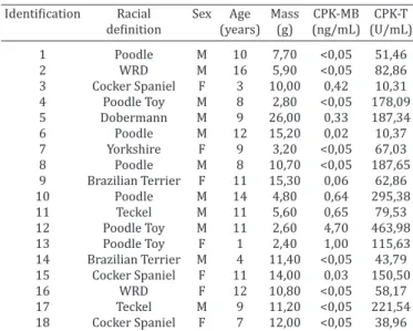
Table 2. Identification and serum CPK-MB and CPK-T in dogs
with ST-segment deviations (group 2)
Identification Racial Sex Age Mass CPK-MB CPK-T
definition (years) (g) (ng/mL) (U/mL)
1 Poodle M 10 7,70 <0,05 51,46 2 WRD M 16 5,90 <0,05 82,86
3 Cocker Spaniel F 3 10,00 0,42 10,31
4 Poodle Toy M 8 2,80 <0,05 178,09 5 Dobermann M 9 26,00 0,33 187,34 6 Poodle M 12 15,20 0,02 10,37
7 Yorkshire F 9 3,20 <0,05 67,03
8 Poodle M 8 10,70 <0,05 187,65 9 Brazilian Terrier F 11 15,30 0,06 62,86 10 Poodle M 14 4,80 0,64 295,38
11 Teckel M 11 5,60 0,65 79,53
12 Poodle Toy M 11 2,60 4,70 463,98 13 Poodle Toy F 1 2,40 1,00 115,63 14 Brazilian Terrier M 4 11,40 <0,05 43,79
15 Cocker Spaniel F 11 14,00 0,03 150,50
16 WRD F 12 10,80 <0,05 58,17
17 Teckel M 9 11,20 <0,05 221,54
18 Cocker Spaniel F 7 12,00 <0,05 38,96
Schobber K., Kirbach B., Cornand C. & Oechtering G. 2002. Diagnostische
und differenzialdiagnostische Wertigkeit zirkulierender kardialer Tro -ponine bei Hund und Katze. Tierärztl. Praxis 30:327-331.
Tharwat M., Al-Sobayil F. & Buczinski S. 2013. Influence of racing on the serum concentrations of the cardiac biomarkers troponin I and creatine kinase myocardial band (CK-MB) in racing greyhounds. Vet. J. 197:900-902.
Thomas W.P. 1987. Cardiology, p.117-156. In: Bonagura J.D. (Ed.),
Myocar-dial Disease of the Dog. Churchill Livingstone, New York.
Tilley L.P. & Junior F.W.K.S. 2003. Consulta Veterinária em 5 minutos. 2ª ed. Manole, São Paulo. 1423p.