Micropilar and embryonic events during hydration of
Melanoxylon brauna
Schott seeds
1Eduardo Euclydes de Lima e Borges
2, Glauciana da Mata Ataíde
3*,
Antônio César Batista Matos
2ABSTRACT – Germination is a complex process that involves molecules properties that make up the cell walls, hydrolytic enzymes that break the bonds between the polymers and action of reactive oxygen substance. Melanoxylon brauna is a forest species of high economic value. In order to evaluate the physiological and biochemical changes that occur in the embryonic axis during germination, fresh matter, length, activities of the enzymes pectin methylesterase, polygalacturonase, superoxide dismutase, catalase, peroxidase and hydrogen peroxide levels were quantified in the embryonic axis. Furthermore, in the micropyle area the composition of carbohydrates and micropyle physical resistance were evaluated with and without drying. During soaking, if there are increases in fresh matter and length of the embryonic axis, there is the same trend of polygalacturonase and pectin methylesterase enzymes. The hydrogen peroxide content was reduced during the soaking, as well as the puncture force of the micropylar area. It is concluded that the seed coat and the cotyledons are responsible for 90% of the water soaked by the seeds. The events in the micropyle and embryonic axis occur independently in the first 16 hours. The weakening of the micropyle features an elastic step and a plastic one. Enzymes pectin methylesterase and polygalacturonase act in cellular expansion of the embryonic axis.
Index terms: puncture force, hydrogen peroxide, cell wall, germination.
Eventos micropilar e embrionário na hidratação de
sementes de Melanoxylon brauna Schott
RESUMO – A germinação é um processo complexo que envolve propriedades das moléculas que compõem a parede celular, enzimas hidrolíticas que quebram ligações entre polímeros da pectina e da hemicelulose e ação das substâncias reativas de oxigênio. Melanoxylon brauna é uma espécie florestal de alto valor econômico. Com o objetivo de avaliar alterações fisiológicas e bioquímicas no eixo embrionário durante a germinação, foram quantificados a massa fresca, o comprimento, as atividades das enzimas pectina metilesterase, poligalacturonase, superóxido dismutase, catalase, peroxidase e os teores de peróxido de hidrogênio. Foram avaliados na região micropilar a composição dos carboidratos e a resistência da micrópila seca e sem secagem. Durante a embebição, ocorrem aumentos na massa fresca e comprimento dos eixos embrionários, mesma tendência das enzimas poligalacturonase e pectina metilesterase, que participam da expansão celular. O conteúdo de peróxido de hidrogênio reduziu durante a embebição, assim como a força de ruptura da região micropilar. Conclui-se que o tegumento e os cotilédones são responsáveis por 90% da água embebida pelas sementes. Os eventos na micrópila e eixo ocorrem independentes nas primeiras 16 horas. O enfraquecimento da micrópila apresenta uma etapa elástica e outra plástica. As enzimas pectina metilesterase e a poligalacturonase atuam na expansão celular do eixo embrionário.
Termos para indexação: força de ruptura, peróxido hidrogênio, parede celular, germinação.
1Submitted on 03/30/2015. Accepted for publication on 8/17/2015. 2Departamento de Engenharia Florestal, UFV, 36570-000 – Viçosa, MG, Brasil.
3Departamento de Silvicultura, UFRRJ, 23895-000 – Seropédica, RJ, Brasil. *Corresponding author <[email protected]>
Introduction
Germination is the result of expansion of the embryonic axis due to the embryo potential growth and cell wall weakening of the micropyle, which is the limiting component of the radicle growth. According to Nonogaki (2014), germination will occur when the active embryo radicle is
able to break the strength exerted by tissue wraps. Different enzymes are involved in the germination process, both in the embryonic axis and in the micropyle.
detected by Scheler et al. (2015) in Lepidium sativum seeds, with the penetration of the radicle in the endosperm being dependent of cells cohesion loss in place, resulting from the action of the enzyme. Moreover, the growth of the embryonic axis of Dalbergia nigra seeds by polygalacturonase was observed, which facilitates expansion of the axis by effect of hydration, according to Ataíde et al. (2013). Reactive oxygen species (ROS) are considered toxic molecules formed during normal metabolic functions and induced when plants are exposed to environmental stimuli (Éaux and Toledano, 2007). However, they also act as signaling molecules in response to different stresses during germination (Gomes and Garcia, 2013; Tenhaken, 2015) or even germination under normal conditions, as occurs in Dalbergia nigra seeds (Matos et al., 2014). Reactions catalyzed by peroxidase also result in stimulation of sprouting, especially in the weakening of the cell wall, since they produce the hydroxyl radical (Richards et al., 2015).
To avoid irreversible cell damage, the antioxidant system enzymes come into play when ROS levels exceed certain levels,
as verified for Dalbergia nigra seeds (Matos et al., 2014). The first
line of defense in plants against oxidative stress is the superoxide dismutase (SOD), an enzyme that catalyzes the conversion of superoxide radicals (O2●–) at H2O2 (Alscher et al., 2002). The H2O2 formed in turn can be removed from the cells by enzymes such as peroxidase (POX), and catalase (CAT) which, in general, use it as a substrate, reducing it to water.
Among the forest native species of ecological and economic importance for use in plantations, there is the Melanoxylon brauna, popularly known as braúna, naturally occurring in
the Atlantic Forest in Northeast and Southeast Brazil, being
the species known for the quality and great economic value of its wood (Lorenzi, 2009). Currently, it is in the Brazilian List
of Endangered Flora Species (MMA, 2008). Despite the great
economic and environmental value and studies involving aspects
related to the seed species physiology (Flores et al., 2014a; Flores et al., 2014b; Borges et al., 2015), more scientific research about
the spread of M. brauna is needed, particularly with an emphasis on dehydration and germination steps.
Thus, it is proposed to quantify the physiological and biochemical changes which occur in the embryonic axis and in the micropyle during the germination of M. brauna seeds, such as the antioxidant system enzymes activity, polygalacturonase and pectin methylesterase and the mechanical resistance of the micropyle, with a view to understanding the germination process of the species.
Material and Methods
Melanoxylon brauna seeds were collected from ripe
fruit in five trees from natural regeneration in the Brazilian
district of Cataguases, Minas Gerais state (23K 0721180 and
7701850, UTM, 609 m of altitude), and taken to Forest Seed Analysis Laboratory at University Federal of Viçosa. After
drying in the sun, at up to about 12-15% moisture, the seeds were processed and mixed, forming a single lot. The seeds were stored in a refrigerator (about 5 °C) in cardboard drums until the beginning of the experiments.
The fresh matter of seeds parts were quantified (integument,
cotyledons and embryos) and of isolated embryos during soaking. To this end, whole seeds were kept on two sheets of germitest-type paper moistened with water at 30 °C, with
constant light from four 40 W daylight-type fluorescent lamps,
and sampled at times zero, 2, 4, 16 and 24 hours, when the seeds were dissected in integument, cotyledons and embryonic
axis, superficially dried on paper towels and each part weighed
in scales of down to a hundredth precision. In the fresh matter evaluations of the embryonic axes isolated from these seeds, they were removed with a scalpel and placed to soak under the same temperature and light conditions above outlined, and the samples were withdrawn at times zero, 2, 4, 16 and 24 hours.
The determinations done in isolated embryos at times zero, 2, 4, 16 and 24 hours were:
Fresh matter – Five replications of ten embryonic axes were
used by means of weighing the sample on a precision balance. Length of the embryonic axes – To calculate the average length of the embryonic axis, these were individually measured using a photographic enlarger and a millimeter
ruler. Five replicates of ten embryonic axes were used.
Pectin methylesterase enzyme – The extraction of the pectin methylesterase enzyme was performed according to the
method described by Pinto et al. (2011), with modifications. Four replications of 0.1 g of embryonic axes removed at times
zero, 2, 4, 16 and 24 hours of soaking were used, which were homogenized in 4.0 mL of solution of NaCl 1.0 M, pH 7.5, containing 1.0% (m/v) of insoluble polyvinylpolypyrrolidone
(PVPP). This solution was centrifuged at 15,000 g for
30 minutes in a centrifuge refrigerated at 4 °C, and the supernatant (enzyme extract) was used for determination of
enzyme activity. The activity was quantified according to
Grsic-Rausch and Rausch (2004), being considered that a pectin methylesterase unit corresponds to 1 µM of NADPH formed per minute in pH 7.5 and 25 ºC.
Polygalacturonase Enzyme – The enzyme extracts were
obtained as described by Guimarães et al. (2001). Four
according to Miller (1959). The enzyme unit was defined as
the amount of protein required to produce the equivalent of 1.0 µmol of glucose per minute.
Hydrogen peroxide (H2O2) – The extraction and production of hydrogen peroxide were conducted according to Gay and Gebicki (2000). Three replicates of 0.1 g for each sample withdrawal time were used.
Superoxide dismutase (SOD) and catalase (CAT) enzymes – The crude enzyme extract used were obtained
from the method described by Flores et al. (2014a). Three
replicates of 0.1 g for each sample withdrawal time were used for both enzymes. The superoxide dismutase activity was determined according to Del Longo et al. (1993) and
Beauchamp and Fridovich (1971). The activity of catalase
was determined according to Hodges et al. (1997). The enzyme activity was calculated using the molar extinction
coefficient of 36 M.cm-1, according to Anderson et al. (1995).
Peroxidase enzyme – Enzymatic extracts were obtained according to Peixoto et al. (1999). The activity was determined according to Kar and Mishra (1976) using the molar extinction
coefficient of 2.47 mM-1.cm-1 (Chance and Maehley, 1995) and
expressed in µmol-1.min-1.mg-1 of protein. Three replicates of
0.1 g per hour of sample withdrawal were used.
The protein contents of the enzymatic extracts were determined by the method by Bradford (1976), using BSA (bovine serum albumin) as a standard.
To quantify changes in seeds micropyle, whole seeds samples were germinated under the same conditions described above, removing the seeds micropylar area for the following assessments:
Micropyle resistance in whole seeds – The resistance of
the micropyle area was quantified by the puncture force in
whole seeds in the soaking times of 0 (control), 24, 48 and 72 hours of soaking after removal of the embryonic axis in each of these times.
Resistance of the isolated micropyle, wet and after drying in the soaking times of 0, 2, 4, 16, 24, 48 and 72 hours – In these times, the puncture force was measured in the wet seeds and after drying at room temperature. Independent samples were used for the measurement of wet and dry seeds. The
micropyle resistance was also quantified after subjecting them
to a temperature of 80 °C for 24 hours in order to kill the seed and subsequent denaturation of the enzymes present in the micropyle, and the puncture force was measured after this period, whether or not followed by drying.
All quantifications of alterations in the cell wall strength
were done by the strength for tissues rupture using a texture analyzer (Stable Micro Systems Texture Analyzer). The micropyle area (about 2 mm) of the dry or hydrated seeds
was cut using a multi-tool kit with a circular saw. Then it was positioned on the probe and drilled. The required force to puncture the endosperm expressed in Newton (N) was used as a strength parameter of the micropylar endosperm to the embryo elongation during germination. The resistance data were
obtained from the average of five replicates of 10 individual
seeds, taken at random after each aforementioned range.
Extraction of the cell wall and carbohydrates quantification– The micropyle cell wall was purified according to Borges et al. (2000), with modifications: 300 mg of micropylar material
were homogenized in a tris-HCl buffer, 50 mM, pH 8.0, at 5 ºC.
After filtration in a nylon filter, two more washings and filtering
were done. The residue was washed in deionized water at 5 °C and kept in suspension, being considered as a crude extract of the cell wall.
The extraction of carbohydrates of the micropyle area was performed as described by Carpita and Gilbeaut (1993), and alditol acetates were prepared for monosaccharide analysis in gas chromatography, according to Englyst and Cummings
(1984). Four replications were done in the soaking times of 0,
2, 4, 16, 24, 48 and 72 hours.
Results and Discussion
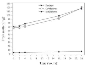
The hydration of M. brauna seed embryos began slowly after four hours in water, while the cotyledons and
the integument started in the first two hours of soaking, continuously reaching higher values for 24 hours (Figure 1).
Two aspects must be highlighted in the hydration of the three compartments. In the case of cotyledons, the size of both is clearly superior to the embryo, besides having reserves with hygroscopic properties, such as starch, soluble sugars and
proteins, and thus, being capable to hydrate more efficiently.
The integument, by having a high concentration of galactomannan (unpublished data) in a thin inner layer, acts in the same way, since this reserve tissue is highly hygroscopic. Due to the reduced size of the embryonic axis with respect to the integument and the cotyledons and being wrapped by both, it can be stated that the root protrusion is determined in part by the greater time to achieve water content needed to swell and increase the potential growth, acting against the integument tissues that are opposed to this expansion.
This is corroborated by the data in Figure 2, which represent
Thus, it is clear that once having access to water, axis expansion starts. Possibly by the activation of enzyme activities that weaken the connections that maintain the cell wall cohesion of the embryonic axis cells in the soaking exponential phase, with small increases in expansion of the embryonic axis in the stationary phase. These stages correspond to soaking phases I and II, respectively, according to Bewley et al. (2013). Alternatively, physical expansion of the wall may have occurred due to the polymers mechanical properties such as xyloglucan, for example, which comprises it (Park and Cosgrove, 2012). Other elements are involved for axis new growth to occur, corresponding to soaking phase III, which corresponds to the phase of root protrusion (Bewley et al., 2013).
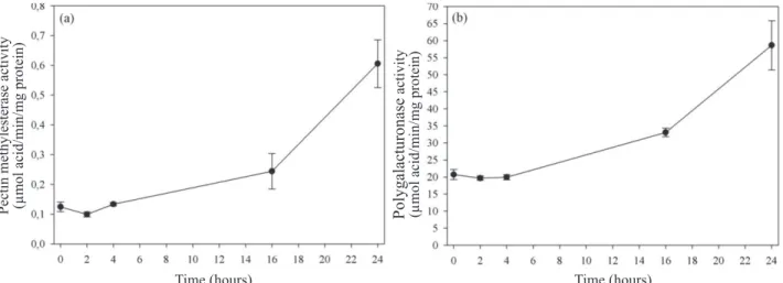
By the results obtained with enzymes pectin methylesterase
(Figure 3a) and polygalacturonase (Figure 3b) in embryonic
axis, it is possible to see that both are pre-formed and their activities have greatly increased in 16 hours and are more pronounced in 24 hours. The polygalacturonase activity is high at the beginning of hydration, which may imply that demethylation and calcium bridges formation have already taken place during the seed formation phase. The expansion in the second phase would be conditioned to the presence of both enzymes. Together they would act more strongly in the weakening or extensibility of cell wall from four hours, more
specifically in pectin, allowing its expansion with the water
inlet. Thus, enzyme participation in the initial expansion of the embryonic axis is clear.
Factors that increase the wall extensibility influence
hydration, which in turn allows cell growth. Pectin is formed by homogalacturonan or rhamnogalacturonans I and II. According to Peaucelle et al. (2012), the degree of methyl
esterification of the homogalacturonan defines porosity,
elasticity and compressibility of the wall. The action of pectin methylesterase allows increased wall susceptibility to the action of polygalacturonase, resulting in changes in the wall biomechanical properties (Wakabayashi et al., 2003). Thus both act, allowing cell expansion upon hydration.
Moreover, the observed increase in fresh matter of the
isolated embryonic axes in the first four hours (Figure 2a)
also appears to be related to the presence of reactive oxygen substances. A high concentration of hydrogen peroxide up to four hours of hydration was observed, stabilizing at 16 and with a marked reduction in 24 hours, when it reached
minimum values (Figure 4).
Hydrogen peroxide breaks bonds between polysaccharides in non-enzymatic reactions, causing weakening of the wall (Schopfer, 2001). Thus the weakening of the cell wall by non-enzyme reactions may have reduced the wall potential, allowing cell expansion by the hydration resulting from
Figure 1. Fresh matter of the parts of seeds of Melanoxylon
brauna (embryonic axis, cotyledons and integument) during soaking at a temperature of 30 °C. +– Standard error.
Figure 2. Fresh matter (a) and medium length (b) of embryonic
axes isolated from Melanoxylon brauna seeds during soaking at a temperature of 30 °C. +– Standard error.
Time (hours)
Time (hours)
Time (hours)
Fresh matter (mg)
Fresh matter (mg)
the water potential difference between the medium and the axis cells in phase I. This possibility is in accordance with
the proposal that the hydration in phase I is purely physical, according to several authors cited by Weitbrecht et al. (2011).
Figure 3. Pectin methylesterase activity (a) and polygalacturonase (b) in embryonic axes isolated from Melanoxylon brauna
seeds during soaking at a temperature of 30 °C. +– Standard error.
Figure 4. Hydrogen peroxide content (µmol/g FM) in
embryonic axes isolated from Melanoxylon brauna seeds during soaking at a temperature of 30 °C. +– Standard error.
The conformation of the two curves (Figure 2a and Figure 4)
is clear, where it is possible to observe inverse behaviors between them. The concentration of hydrogen peroxide also has the three-phase conformation with a decrease in 2 hours, followed by a stabilization phase between 4 and 16 hours, and decreasing again in 24 hours. The reduction in the peroxide concentration over the hydration period has eliminated the chance of possible damage caused by the presence of the reactive oxygen substance.
The production of hydrogen peroxide can have a different source from that usually cited (mitochondrion or peroxisome),
being produced by the action of NADPH peroxidase which, according to Bedard et al. (2007), is the enzyme producing ROS in plants and animals. Moreover, the production and maintenance of hydrogen peroxide can also be by the action of NADPH oxidase, according to Karmer et al. (2010). According to them, hydrogen peroxide production outside the cell could return in the form of hydrogen peroxide, formed by the effect of extracellular pH 5.0.
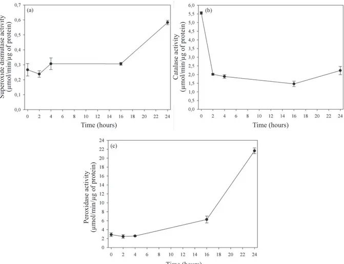
In Figure 5a, it is seen that the enzyme superoxide dismutase activity was stable in the first 16 hours and increased by 24 hours, and it was not possible to implicate it in the first
phase of action in the reduction of reactive oxygen species such as superoxide. It is possible that this concentration is low at this stage, with no need for dismutation by the enzyme. It is noteworthy that the presence of the substance under the conditions of this study not detected.
On the other hand, the enzyme catalase (Figure 5b) had high activity at first and then decreased until 16 hours of
soaking, returning to the same level of two hours. Unlike the
latter, peroxidase (Figure 5c) showed low activity in the first
four hours, with successive increases in up to 24 hours. It is noticed that both enzymes have fairly similar activities in the control embryos, with more pronounced decrease of catalase, which approached the peroxidase values in up to four hours of hydration. Subsequently, there was a substantial increase in the latter, which can indicate greater effectiveness of peroxidase to eliminate excess hydrogen peroxide produced by the increase in metabolic activity during hydration.
Considering that the production of ROS occurs during seed
Time (hours) Time (hours)
Time (hours)
Pectin methylesterase activity (µmol acid/min/mg protein) Polygalacturonase
activity
(µmol acid/min/mg protein)
formation due to the synthesis metabolism (Liu et al., 2014), catalase activity would act to reduce the level of these substances during germination so they do not cause damage to the cells, but keeping
them in a concentration that would act in the wall flexibility. Thus, hydration occurring in phase I would be possible by the Fenton
reaction. Subsequently, with the hydration increases and metabolic activation, the increase of ROS induced the increase of peroxidase
activity. It is noteworthy that, according to Weitbrecht et al. (2011), phase III is typically metabolic and therefore the production of ROS is high, especially by the mitochondrion, one of the producers
of these substances (Vanlerberghe, 2013). Thus, without the ROS
reduction in phase III there would be inactivation of enzymes
catalase and peroxidase, without which the flexibility of the wall
and subsequent increase in size would not have occurred.
Figure 5. Activity of enzymes superoxide dismutase (a), catalase (b), and peroxidase (c) in embryonic axes isolated from
Melanoxylon brauna seeds during soaking at a temperature of 30 °C. +– Standard error.
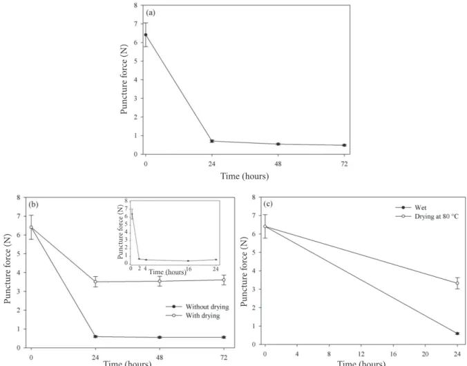
In assessing the change in the micropyle wall strength in
whole seeds, shown in Figure 6a, it was possible to observe
its marked reduction in 24 hours, in the same manner as in
the micropyle isolated in two hours (Figure 6b). The puncture
force values found during these times near 1 N are similar. In
Figure 6b it is seen that the weakening of the wall occurs in
two hours and remains relatively constant for 72 hours. Such similarity could not be otherwise, unless the embryo inhibited any activity at that location, which does not seem to be the case.
Drying the micropyle resulted in increasing its resistance.
For permanent deformation of the wall to occur, there would
be the need for changes in both components. The wall elastic element could be credited to the mechanical action of the polymers (Suslov et al., 2010), thus keeping the wall expansion property. As for the plastic change, it would result from the action of enzymes attached to the wall or exuded from the radicle to that location, which would degrade the wall component, such as, for example, of pectin.
After submission to the temperature of 80 °C and subsequent drying of the micropyle, the puncture force increased compared to the force required to puncture the wet
seeds, as shown in Figure 6c. Thus, the possible action of
Time (hours) Time (hours)
Time (hours)
Superoxide dismutase activity
(µmol/min/µg of protein)
Catalase activity
(µmol/min/µg of protein)
Peroxidase activity
soluble enzymes in the micropyle is discarded in view of their deterioration by heat.
Thus, the presence of proteins in the cell wall can be an actual fact, in case of its synthesis in a similar manner to Arabidopsis seeds, which, according to Dekkers et al. (2013),
the expansin gene is expressed exclusively in the micropylar endosperm. According to the authors, although there are variations in cell wall architecture among different species,
the weakening of the wall by specific proteins is widely
conserved in the seed germination mechanism.
Figure 6. Required puncture force to rupture (N) the micropylar area of whole seeds (a) the isolated micropyle (b) and after
subjection to a temperature of 80 °C (c) in Melanoxylon brauna seeds during soaking at a temperature of 30 ºC. +–
Standard error. The detail in Figure 6b is the puncture force in the micropyle isolated within the first 24 hours.
Considering the results obtained in the initial hydration phase, expansin would be one of the candidates to work in the elastic component of the micropyle wall. According to Cosgrove (2000), one of the features of expansin is the immediate extension of the wall, which begins within seconds after the extract containing expansin being placed in contact with the substrate, besides not causing continuous wall weakening.
The analysis of the micropyle cell wall composition
(Figure 7) shows the possible formation of galactomannan,
a cell wall reserve material, and rhamnogalacturonan, characteristic polysaccharide of pectin, responsible for wall stability by forming bonds with calcium or boron (Peaucelle
et al., 2012). Apparently, the presence of rhamnogalacturonan, consisting in rhamnose and galactose, is small, which allows the expansion of the wall due to increased turgor being easier. Moreover, the absence of xylose and glucose eliminates the possibility of xyloglucan-cellulose intersection, which would give greater resistance to the turgor wall.
It is evident that the micropyle cell wall is not structured as in other composite tissues of cellulose-hemicellulose and pectin. There were no changes in the sugars concentrations, assuming that there was no participation of hydrolytic enzymes, with the initial expansion of the wall being due to reduction in strength.
Time (hours)
Time (hours) Time (hours)
Pun
ct
ur
e f
o
rce (N)
Pun
ct
ur
e f
o
rce (N)
Pun
ct
ur
e f
o
rce (N)
Time (hours)
Pun
ct
ur
e f
o
rce (N)
8 7 6 5 4 3 2 1 0
According to Prietruszka and Lewicka (2007), the growth results from increase and irreversible deformation of the cell wall, while the elastic extension is not permanent and is reversed when the force is removed. According to the authors, the polymers, as the cell wall components are inert and have a stable elastic behavior at a wide temperature range, suggest that the elastic behavior is purely physical. Thus, the micropylar wall plastic component resistance of the M. brauna seeds is overcome by metabolic action, such as, for example, enzymes attached to the micropyle wall or exuded from the embryo axis.
Conclusions
The integument and cotyledons are responsible for 90% of the water soaked by the Melanoxylon brauna seeds within 72 hours;
The events in the embryonic axis and in the micropyle
occur independently in the first 16 hours;
The weakening of the micropyle occurs in two stages, one plastic and another elastic;
Enzymes polygalacturonase and pectin methylesterase are involved in cell expansion of the embryonic axis;
Germination of M. brauna seeds comprises the change in plasticity of the micropyle wall by a physical effect (phase I), followed by permanent alteration of the wall (plastic element) by the action of enzymes or proteins (phases II-III).
Acknowledgments
The authors thank CNPq [Conselho Nacional de
Desenvolvimento Científico e Tecnológico (National Counsel
of Technological and Scientific Development)] for the productivity scholarship for the first author and Professor Lúcio Alberto de Miranda Gomide, of Universidade Federal de Viçosa, for making available the texture analyzer.
References
ALSCHER, R.G.; ERTURK, N.; HEATH, L.S. Role of superoxide dismutase (SODs) in controlling oxidative stress. Journal of Experimental Botany, v.53,
p.1331-1341, 2002.http://jxb-oxfordjournals-org.ez35.periodicos.capes.gov.
br/content/53/372/1331.full.pdf+html
ANDERSON, M.D.; PRASAD, T.K.; STEWART, C.R. Changes in isozyme
profiles of catalase, peroxidase, and glutathione reductase during acclimation
to chilling in mesocotyls of maize seedlings. Plant Physiology, v.109, p.1247-1257, 1995. http://www.ncbi.nlm.nih.gov/pubmed/12228666
ATAÍDE, G.M.; BORGES, E.E.L.; GONÇALVES, J.F.C.; GUIMARÃES, V.M.; FLORES, A.V.; BICALHO, E.M. Activities of alfa-galactosidase
and poligalacturonase during hydratation of Dalbergia nigra (Vell.) Fr All. Ex Benth) seed at different temperatures. Journal of Seed Science, v.35, n.1, p.92-98, 2013.http://www.scielo.br/scielo.php?pid=S2317-15372013000100013&script=sci_arttext
BEAUCHAMP, C.; FRIDOVICH, I. Superoxide dismutase improved assays
and assay applicable to acrylamide gels. Analytical Biochemistry, v.44, p.276-287, 1971. http://www.ncbi.nlm.nih.gov/pubmed/4943714
BEDARD, K.; LARDY, B.; KRAUSE, K.H. NOX family NADPH oxidases:
Not just in mammals. Biochimie, v.89, p.1107-1112, 2007.http://www.ncbi. nlm.nih.gov/pubmed/17400358
BEWLEY, J.D.; BRADFORD, K.J.; HILHORST, H.W.M.; NONOGAKI, H.
Seeds: physiology of development, germination and dormancy. Nova York:
Springer, 2013. 392 p.
BORGES, E.E.L.; BORGES, R.C.G.; BUCKERIDGE, M.S. Alterações nas
composições de carboidratos e de ácido graxos em sementes de
jacarandá-da-bahia osmocondicionadas. Revista Brasileira de Fisiologia Vegetal, v.12, n.1, p.10-16, 2000. http://www.cnpdia.embrapa.br/rbfv/pdfs/v12n1p10.pdf
BORGES, E.E.L.; FLORES, A.V.; ATAÍDE, G.M.; MATOS, A.C.B. Alterações fisiológicas e atividade enzimática em sementes armazenadas de
Melanoxylon brauna Schott. Cerne, v.21, n.1, p.75-81, 2015. http://www.
scielo.br/pdf/cerne/v21n1/2317-6342-cerne-21-01-00075.pdf
BRADFORD, M.M. A rapid and sensitive method for the quantification
of microgram quantities of proteins utilizing the principle of protein-dye binding. Analytical Biochemistry, v.72, p.248-254, 1976. http://www.ncbi. nlm.nih.gov/pubmed/942051
CARPITA, N.G.; GILBEAUT, D.M. Structural models of primary cell walls
in flowering plants: consistency of molecular structure with the physical
properties of the walls during growth. Plant Physiology, v.3, p.1-3, 1993. http://www.ncbi.nlm.nih.gov/pubmed/8401598
CHANCE, B.; MAEHLEY, A.C. Assay of catalase and peroxidase.
Methods in Enzymology, v.2, p.764-775, 1995.http://www.ncbi.nlm.nih.gov/
pubmed/13193536
COSGROVE, D.J. Loosening of plant cell walls by expansins. Nature, v.407,
n.2, p.321-326, 2000. http://www.nature.com/nature/journal/v407/n6802/
pdf/407321a0.pdf
Figure 7. Cell wall of monosaccharides of the micropylar
area of Melanoxylon brauna seeds during soaking at a temperature of 30 ºC. +– Standard error.
Time (hours)
Monosaccharides
DEKKERS, B.J.W.; PEARCE, S.; BOLDEREN-VELDKAMP, R.P.; MARSHALL, A.; WIDERA, P.; GILBERT, J.; DROST, H.; BASSEL, G.W.; MÜLLER, K.; KING, J.R.; WOOD, A.T.A.; GROSSE, I.; QUINT, M.; KRASNOGOR, M.; LEUBNER-METZGER, G.; HOLDSWORTH, M.J.;
BENTSINK, L. Transcriptional dynamics of two seed compartments with opposing roles in Arabidopsis seed germination. Plant Physiology, v.163, n.1, p.205-215, 2013. http://www.ncbi.nlm.nih.gov/pubmed/23858430
DEL LONGO, O.T.; GONZÁLEZ, C.A.; PASTORI, G.M.; TRIPPI, V.S.
Antioxidant defenses under hyperoxygenic and hyperosmotic conditions in leaves of two lines of maize with differential to drought. Plant Cell
Physiology, v.37, n.7, p.1023-1028, 1993. http://pcp.oxfordjournals.org/
content/34/7/1023.full.pdf
ÉAUX, B.; TOLEDANO, M.B. Ros as signalling molecules: mechanisms
that generate specificity in ROS homeostasis. Nature Reviews Molecular
Cell Biology, v.8, p.813-824, 2007. http://www.ncbi.nlm.nih.gov/
pubmed/17848967
ENGLYST, H.N.; CUMMINGS, J.H. Simplified method for the measurement
of total non-starch polysaccharides by gas-liquid chromatography of constituent sugar as alditol acetates. Analyst, v.109, p.937-942, 1984. http:// pubs.rsc.org/en/Content/ArticleLanding/1984/AN/an9840900937
FLORES, A.V.; BORGES, E.E.L.; GUIMARÃES, V.M.; GONÇALVES, J.F.C.; ATAIDE, G.M.; BARROS, D.P. Atividade enzimática durante a
germinação de sementes de Melanoxylon brauna Schott sob diferentes temperaturas. Cerne, v.20, n.3, p.401-408, 2014a. http://www.redalyc.org/ articulo.oa?id=74432265009
FLORES, A.V.; BORGES, E.E.L.; GUIMARÃES, V.M.; ATAIDE, G.M.; CASTRO, R.V.O. Germinação de sementes de Melanoxylon brauna Schott em diferentes temperaturas. Revista Árvore, v.38, n.6, p.1147-1154, 2014b. http://www.scielo.br/pdf/rarv/v38n6/a19v38n6.pdf
GAY, C.; GEBICKI, J.M. A critical evaluation of the effect of sorbitol on the
ferric-xylenol orange hydroperoxide assay. Analytical Biochemistry, v.284, p.217-220, 2000. http://www.ncbi.nlm.nih.gov/pubmed/10964403
GOMES, M.G.; GARCIA, Q.S. Reactive oxygen species and seed
germination. Biologia, v.68, p.351-357, 2013. http://link.springer.com/
article/10.2478%2Fs11756-013-0161
GRSIC-RAUSCH, S.; RAUSCH, T. A coupled spectrophotometric enzyme assay for the determination of pectin methylesterase activity and its inhibition by proteinaceous inhibitors. Analytical Biochemistry, v.333, n.1, p.14-18, 2004. http://www.sciencedirect.com/science/article/pii/ S000326970400404X
GUIMARÃES, V.M.; REZENDE, S.T.; MOREIRA, M.A.; BARROS, E.G.; FELIX, C.R. Characterization of α-galactosidases from germinating soybean
seed and their use for hydrolysis of oligosaccharides. Phytochemistry, v.58, n.1, p.67-73, 2001. http://www.ncbi.nlm.nih.gov/pubmed/11524115
HODGES, D.M.; ANDREWS, C.J.; JOHNSON, D.A.; HAMILTON, R.I.
Antioxidant enzyme responses to chilling stress in differentially sensitive inbred maize lines. Journal of Experimental Botany, v.48, n.310,
p.1105-1113, 1997. http://jxb.oxfordjournals.org/content/48/5/1105.full.pdf
KAR, M.; MISHRA, D. Catalase, peroxidase, and polyphenolxidase activities during rice leaf senescence. Plant Physiology, v.57, p.315-319, 1976. http:// www.ncbi.nlm.nih.gov/pmc/articles/PMC542015/
KARMER, I.; ROACH, T.; BECKETT, R.P.; WHITAKER, C.;
MINIBAYEVA, F.V. Extracellular production of reactive oxygen species
during seed germination and early seedling growth in Pisum sativum. Journal
of Plant Physiology, v.167, p.805-811, 2010. http://www.sciencedirect.com/
science/article/pii/S0176161710000945
LIU, N.; LIN, Z.; GUAN, L.; GAUGHAN, G.; LIN, G. Antioxidant enzymes regulate reactive oxygen species during pod elongation in Pisum sativum and Brassica chinensis. PLoS ONE, v.9, n.2, p.1-9, 2014.http://journals.plos.org/ plosone/article?id=10.1371/journal.pone.0087588
LORENZI, H. Árvores brasileiras: manual de identificação e cultivo de plantas arbóreas nativas do Brasil. Nova Odessa: Plantarum, 2009. 384p.
MATOS, A.C.B.; BORGES, E.E.L.; SEKITA, M.C. Produção de espécies reativas de oxigênio em sementes de Dalbergia nigra sob estresse térmico.
Journal of Seed Science, v.36, n.3, p.282-289, 2014. http://www.scielo.br/
scielo.php?pid=S2317-15372014000300002&script=sci_arttext
MILLER, G.L. Use of dinitrosalicylic acid reagent for determination of reducing sugar. Analytical Chemistry, v.31, n.3, p.426-428, 1959. http://pubs. acs.org/doi/abs/10.1021/ac60147a030
MMA – Ministério do Meio Ambiente. Instrução Normativa nº 6, de 23 de dezembro de 2008. Lista Oficial das Espécies da Flora Brasileira Ameaçada de Extinção. 2008. http://www.mma.gov.br/estruturas/ascom_boletins/_ arquivos/83_19092008034949.pdf
NONOGAKI, H. Seed dormancy and germination-emerging mechanism and new hypotheses. Frontier Plant Science, v.5, n.233, p.1-14, 2014. http:// www.ncbi.nlm.nih.gov/pubmed/24904627
PARK, Y.B.; COSGROVE, D.J. Changes in cell wall biomechanical properties in the xyloglucan-deficient xxt1/xxt2 mutant of Arabidopsis.
Plant Physiology, v.158, n.1, p.465-475, 2012. http://www.ncbi.nlm.nih.gov/
pubmed/22108526
PEAUCELLE, A.; BRAYBROOK, S.; HÖFTE, H. Cell wall mechanics
and growth control in plant: the role of pectins revisited. Frontier Plant
Science, v.3, n.121, 2012. http://journal.frontiersin.org/Journal/10.3389/
fpls.2012.00121/abstract
PEIXOTO, P.H.P.; CAMBRAIA, J.; SANTANA, R.; MOSQUIM, P.R.;
MOREIRA, M.A. Aluminum effects on lipid peroxidation and on the activities of enzymes of oxidative metabolism in sorghum. Revista Brasileira
de Fisiologia Vegetal, v.11, p.137-143, 1999. http://www.cnpdia.embrapa.br/
rbfv/pdfs/v11n3p137.pdf
PIETRUSZKA, M.; LEWICKA, S. Effect of temperature on plant elongation and cell wall extensibility. General Physiology and Biophysics, v.26, n.1, p.40-47, 2007. http://www.ncbi.nlm.nih.gov/pubmed/17579253#
PINTO, L.K.A.; MARTINS, M.L.L.; RESENDE, E.D.; THIEBAUT,
J.T.L. Atividade de pectina metilesterase e da β-galactosidase durante o
amadurecimento do mamão cv golden. Revista Brasileira de Fruticultura, v.33, n.3, p.713-722, 2011. http://www.scielo.br/scielo.php?pid=S0100-29452011000300004&script=sci_arttext
RICHARDS, S.L.; WILKINS, K.A.; SWARBRECK, S.M.; ANDERSON,
A.A.; HABIB, N.; SMITEH, A.G.; McANISH, M.; DAVIES, J.M. The hydroxyl
radical in plants: from seed to seed. Journal Experimental Botany, v.66, n.1,
SCHELER, C.; WEITBRECHT, K.; PEARCE, S.P.; HAMPSTEAD, A.
BÜTTNER-MAINIK, A.; LEE, K.J.D.; VOEGELE, A.; ORACZ, K.; DEKKERS, B.J.W.; WANG, X.; WOOD, A.T.A.; BENTSINK, L.; KING, J.R.; KNOX, J.P.; HOLDSWORTH, M.J.; MÜLLER, K.;
LEUBNER-METZGER, G. Promotion of testa rupture during garden cress germination
involves seed compartment-specific expression and activity of pectin
methylesterases. Plant Physiology, v.167, p.200-215, 2015. http://www. plantphysiology.org/content/167/1/200.full.pdf+html
SCHOPFER, P. Hydroxyl radical-induced cell-wall loosening in vitro and in
vivo implications for the control of elongation growth. Plant Journal, v.28, p.678-688, 2001. http://www.ncbi.nlm.nih.gov/pubmed/11851914
SUSLOV, D.; VERBELEN, J.P.; VISSEMBERG, K. Is acid-induced extension
in seed plants only protein mediated? Plant signaling & Behavior, v.5, n.6, p.757-759, 2010. http://www.ncbi.nlm.nih.gov/pmc/articles/PMC3001582/ TENHAKEN, R. Cell wall remodeling under abiotic stress. Frontiers in Plant Science, v.5, article 771, p.1-9, 2015. http://journal.frontiersin.org/ article/10.3389/fpls.2014.00771/full
VANLERBERGHE, G.C. Alternative oxidase: a mitochondrial respiratory
pathway to maintain metabolic and signaling homeostasis during abiotic and biotic stress in plant. International Journal of Molecular Science, v.14, n.4, p.6805-6847, 2013. http://www.mdpi.com/1422-0067/14/4/6805
WAKABAYASHI, K.; HOSON, T.; HUBER, D.J. Methyl de-esterification as a major factor regulating the extent of pectin depolimerization during
fruit ripening: a comparasion of the action of avocado (Persea americana) and tomato (Lycopersicon esculentum). Journal of Plant Physiology, v.160, n.6, p.667-673, 2003. http://ac.els-cdn.com/S0176161704704514/1- s2.0-S0176161704704514-main.pdf?_tid=a17c87d0-dd61-11e4-aa23-00000aab0f26&acdnat=1428437391_a705745c8e2af0a89e5e67531ab31a41
WEITBRECHT, K.; MÜLLER, K.; LEUBNER-METZGER, G. First off the
mark: early seed germination. Journal of Experimental Botany, v.62, n.10,
p.3289–3309, 2011. http://jxb.oxfordjournals.org/content/62/10/3289.full.