Comunicação científica /
Scientific communication
horticultura horticultura
brasileirabrasileira
T
he bacterial species Pseudomonas cichorii, which was originally isolated from the herbaceous plants Cichorium intybus and Cichorium endivia, belongs to rRNA homology group I, together with P. syringae (Garrity et al., 2005). Recent taxonomic advances, based on biochemical and genotypic processes, demonstrated that the plant pathogenic species P. cichorii consists of a cluster of closely related genomic groups with genetic heterogeneity of the species (Trantas et al., 2013).This bacterium has already been reported causing leaf spot in many plants, and recently it was reported in Stevia rebaudiana in Florida (Strayer et al., 2012), associated with leaf blight in soybean in South Korea (Yu,
2012), a new symptom of okra bacterial leaf blight in a local variety of okra (Abelmoschus esculentus) in Japan (Inoue et al., 2013) and in Chengdu City in China (Li et al., 2014) causing leaf spot of vegetable sponge (Luffa cylindrical).
In Brazil it is considered an opportunistic pathogen in that it infects a wide range of dicotyledons and herbaceous plants, including vegetables and ornamental species. In lettuce, bacterial blight is not a limiting disease, varying with climatic and inoculum densities. Among crucifers, P. cichorri causes spots on the leaves and head of cabbage (Amorim et al., 2005). In
sunflower, this bacterium causes spot and blight on the stalks, first described
in 1981 (Robbs & Almeida, 1981).
It has also been found causing leaf spot in mint, Mentha arvensis (Maia et al., 1996). Among ornamentals, it mainly occurs in chrysanthemum, philodendron, and African violet. Silva Júnior et al. (2009) reported this
bacterium affecting the tomato crop in the state of São Paulo for the first time
in 2007 and also registered leaf blight on eucalyptus (Gonçalves et al., 2008).
Gerbera is an Asteracea that originates in South Africa and Asia (Radice & Marconi, 1998), and it is among the most extensively exploited
flowers in Brazil. In this plant, bacterial
blight caused by P. cichorii was first described in Apopka, Florida (Miller & Knauss, 1973) and subsequently in Greece (Alivizatos, 1986). Although its occurrence in Brazil has not been clearly MARQUES, E; BORGES, RCF; UESUGI, CH. 2016. Identification and pathogenicity of Pseudomonas cichorii associated with a bacterial blight of gerbera
in the Federal District. Horticultura Brasileira 34: 244-248. DOI - http://dx.doi.org/10.1590/S0102-053620160000200015
Identification and pathogenicity of
Pseudomonas cichorii
associated with
a bacterial blight of gerbera in the Federal District
Eder Marques; Rafaela CF Borges; Carlos H Uesugi
Universidade de Brasília, Depto. Fitopatologia, Brasília-DF; [email protected]; [email protected]; [email protected]
ABSTRACT
In 2013, leaf samples of gerbera plants showing symptoms of
bacterial blight were collected in cut-flower high tunnels, in the
settlement of Núcleo Rural Alexandre Gusmão, located in Brazlândia, Distrito Federal, Brazil. Seven isolates obtained were subjected to phenotypic and molecular characterization, including pathogenicity tests, LOPAT, and partial sequencing of the 16S rDNA gene. All isolates were gram-negative, aerobic, oxidase-positive, produced
fluorescent pigment, induced hypersensitivity in tobacco leaves, used sorbitol and glutamate and were pathogenic to 24 different plant
species. Results of these tests and analysis of the sequences of rDNA showed 100% identity with Pseudomonas cichorii. To our knowledge,
this is the first report of P. cichorii in gerbera in the Federal District.
Keywords:Gerbera jamesonii, leaf spot, LOPAT.
RESUMO
Identificação e patogenicidade de Pseudomonas cichorii associada ao crestamento bacteriano da gérbera no Distrito Federal
Em 2013, amostras foliares de plantas de gérbera, apresentando sintomas de crestamento, foram coletadas em telados de cultivo de corte, no Núcleo Rural Alexandre Gusmão, localizado em Brazlândia--DF. Sete isolados bacterianos obtidos foram submetidos à caracte-rização fenotípica e molecular, incluindo testes de patogenicidade, testes LOPAT e sequenciamento parcial do gene 16S rDNA. Todos os isolados foram gram-negativos, aeróbios, oxidase positivos,
produzi-ram pigmento fluorescente, induziproduzi-ram reação de hipersensibilidade
em folhas de fumo, utilizaram sorbitol e glutamato, e foram patogê-nicos a 24 diferentes espécies de plantas. Os resultados destes testes e análises de sequências do rDNA mostraram 100% de identidade com
Pseudomonas cichorii. Para o nosso conhecimento, este é o primeiro relato de P. cichorii, em gérbera, no Distrito Federal.
Palavras-chave:Gerbera jamesonii, mancha foliar, LOPAT.
established, it was reported in São Paulo (Malavolta Júnior et al., 1994). According to studies on the cataloguing of gerbera diseases in the state of Paraná from 2004 to 2006, leaf spot was noted when inspections were carried out in properties producing ornamental plants, but in only two commercial pot-plants (Ferronato et al., 2008). In the Federal District of Brazil, leaf spot has been observed in gerbera cultivars for some time. Therefore, the objective of this work was to investigate the causal agent associated with bacterial blight of gerbera and to study its range of hosts.
MATERIAL AND METHODS
In February 2013, leaves from
different plants showing symptoms of
leaf blight were collected in six high tunnels, approximately 8 m wide and 50 m long (8 x 50), from a field of gerbera (directly in the soil) situated at Núcleo Rural Alexandre Gusmão, located in Brazlândia, Federal District, Brazil. Overall, the plants showed 80% infection, especially in lower leaves.
The same region produces sunflower,
chrysanthemum, and goldenrod. Segments of symptomatic leaves were surface disinfected, macerated in sterile distilled water and then used for dilution plating on culture media 523 (Kado & Heskett, 1970). Single colonies were selected and submitted to standard morphological, physiological, and/or biochemical tests for bacterial
identification according to Garrity et al. (2005) and Schaad et al. (2001).
Hypersensitivity response (HR)
tests were carried out by infiltrating
a 109 cfu/mL (Scale 7 of McFarland)
bacterial suspension on tobacco leaves using a needle-less syringe. For this, the bacterial isolates were grown on 523 solid medium for 48 h and then diluted in sterile distilled water. Characteristic HR symptoms were scored at 24 h. Bacterial inoculation on gerbera plants (pathogenicity test) was carried out by spraying a 109 cfu/
mL bacterial suspension on the abaxial and adaxial surfaces of the leaves up
to the beginning of run-off; following
inoculation, plants were incubated for 24
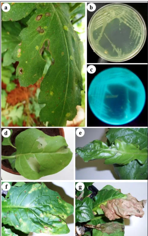
Figure 1. Pathogenicity tests of gerbera bacterial isolates. A) Gerbera plant with necrotic
lesions on leaf, observed in the field; b) Whitish colonies producing green pigment; in 523 culture medium; c) Observation of fluorescent pigments in King B medium, under UV light;
d) Hypersensitivity response (HR) test in tobacco leaf 16 hours after inoculation; e) Lesions on the edges of gerbera leaves; f) scattered necrotic spot-like lesions, with a depressed center, followed also by leaf chlorosis; and g) Coalescent lesions, leaf blight {teste de patogenicidade com isolados de gérbera. a) Planta de gérbera com lesões necróticas na folha, observada em campo; b) Colônias brancas produzindo pigmento verde, em meio 523; c) Observação do
pigmento fluorescente em meio B de King, sob luz UV; d) Reação de hipersensibilidade em
h at 28ºC in a wet chamber and then kept in the greenhouse for the remainder of
the experiment. In order to fulfill Koch’s
postulates, disease symptoms were observed and the pathogen re-isolated in culture medium.
Purification of the total DNA of the bacterial genome was carried out
using the Wizard® Genomic DNA
Purifications Kit (Promega, Madison, WI), and a partial sequence of the 16S rDNA gene was amplified using the universal primers for bacteria F984
5’-AACGCGAAGAACCTTAC-3’ a n d R 1 4 9 2 5 ’ - CTACGGYTACCTTGTTACGAC-3’
(Heuer et al., 1997). The obtained PCR
products were purified and sequenced
( M a c r o g e n I n c . ) . T h e r e s u l t i n g sequences were analyzed using the Basic Local Alignment Search Tool (BLAST; National Center for Biotechnology Information, http://www.ncbi.nlm.nih. gov). The contiguous sequences were assembled using the DNA Sequence Assembly BASER Software v.4.5.0 (http://www.dnabaser.com/index.html).
For phylogenetic analysis, the sequences were aligned using MAFFT (Katoh & Standley, 2013). The trees were determined using MrBayes v3.2.1 (Ronquist & Huelsenbeck, 2003), which uses the Markov chain Monte Carlo method (MCMC). The analysis was released using the default settings except the appropriate template, which was implemented automatically using “Reversible-jump” MCMC (Huelsenbeck et al., 2004). The program ran for 2,000,000 generations and the frequency separation stabilized below
0.008. The first 25% of the trees were
discarded (“burn-in”) before calculating the consensus tree.
RESULTS AND DISCUSSION
A total of seven gram-negative bacterial isolates (PC1, PC2, PC3, PC4, PC5, PC6, and PC7) were isolated from the symptomatic tissues of gerbera
plants collected in the field (Figure 1a).
The bacterial isolates were included in the Collection of Plant Pathogenic Bacteria of the Department of Plant Pathology (University of Brasília) with
the following designations: UnB 1379, UnB 1380, UnB 1381, UnB 1382, UnB 1383, UnB 1384, and UnB 1385.
All bacterial isolates had whitish colonies (Figure 1b); positive for
production of fluorescent pigments in King B medium (Figure 1c); HR positive (Figure 1d), gram negative, KOH positive, oxidative, negative for hydrolyses of gelatin and mucoid
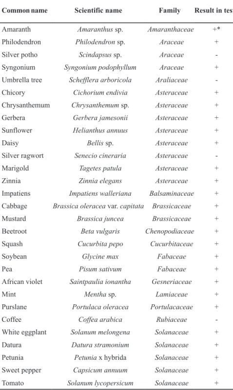
Table 1. Plants used in the pathogenicity and host tests, in reaction to artificial inoculation of
seven bacterial isolates obtained from gerbera in the Federal District (plantas utilizadas no
teste de patogenicidade e de hospedeiras, em reação a inoculação artificial com sete isolados
bacterianos, obtidos de gerbera no Distrito Federal). Brasília, UnB, 2013.
Common name Scientific name Family Result in test
Amaranth Amaranthus sp. Amaranthaceae +*
Philodendron Philodendron sp. Araceae +
Silver potho Scindapsus sp. Araceae
-Syngonium Syngonium podophyllum Araceae +
Umbrella tree Scheffleraarboricola Araliaceae
-Chicory Cichorium endivia Asteraceae +
Chrysanthemum Chrysanthemum sp. Asteraceae +
Gerbera Gerbera jamesonii Asteraceae +
Sunflower Helianthus annuus Asteraceae +
Daisy Bellis sp. Asteraceae +
Silver ragwort Senecio cineraria Asteraceae
-Marigold Tagetes patula Asteraceae +
Zinnia Zinnia elegans Asteraceae +
Impatiens Impatiens walleriana Balsaminaceae +
Cabbage Brassica oleracea var. capitata Brassicaceae +
Mustard Brassica juncea Brassicaceae +
Beetroot Beta vulgaris Chenopodiaceae +
Squash Cucurbita pepo Cucurbitaceae +
Soybean Glycine max Fabaceae +
Pea Pisum sativum Fabaceae +
African violet Saintpaulia ionantha Gesneriaceae +
Mint Mentha sp. Lamiaceae +
Purslane Portulaca oleracea Portulacaceae +
Coffee Coffea arabica Rubiaceae
-White eggplant Solanum melongena Solanaceae +
Datura Datura stramonium Solanaceae +
Petunia Petunia x hybrida Solanaceae +
Sweet pepper Capsicum annuum Solanaceae +
Tomato Solanum lycopersicum Solanaceae +
colonies in YDC medium. In the LOPAT test the isolates were: levan negative, oxidase positive, arginine dihydrolase negative and had no pectinolitic activity on potato discs. The reduction of nitrate was negative, as was the utilization of sorbitol, trehalose, cellobiose, D-arabinose, oxalate, tartrate, salicine, succinate, glycerol, saccharose and citrate. On the other hand, the isolates used mannitol and glutamate, in accordance with Garrity et al. (2005); and Schaad et al. (2001).
In inoculated gerbera plants, the typical symptoms of bacterial blight could be observed (Malavolta Júnior et al., 1994; Ferronato et al., 2008), three days after inoculation. Initially, dark lesions appeared on the edges of the leaves (Figure 1e). These evolved to become necrotic spot-like lesions, with a depressed center distributed across the leaf area. Leaves became
chlorotic (Figure 1f), coalescent and
dried out (Figure 1g). Stem and flower
rotting could also be observed. Plants exhibited irregular lesions on leaf edges with a depressed center (Figure 1f). The isolates were pathogenic to 24 other plants (Table 1). Taking into account
the fulfillment of Koch’s postulates, altogether, these results confirmed the
pathogenicity of the isolates.
Studies that report the occurrence of this bacteriosis in Brazil are scarce (Malavolta Júnior et al., 1994). In this article, most of the plants showed symptoms of infection by the bacterial isolates obtained from gerbera, although the symptoms were more pronounced in gerbera, in which lesions even coalesced and leaves died; however, the plants were still able to recover and bloom again. In the other plants the symptoms
were discrete and did not affect their
development, for example in tomato, in
which the lesions were small and sparse on the leaf. However, this was unlike the symptoms induced by isolates on tomato in the work of Silva Júnior et al. (2009), in which the disease appeared in the form of wet irregular lesions that evolved to irregular necrosis of the leaf area.
The biochemical oxidase test (positive) allowed the distinction of gerbera isolates from P. syringae group, P. congelans, P. rhizosphaerae and P. lutea. The arginine dihydrolase test (negative) also differentiated them from P. putida, P. mohnii and P. brassicacearum. Trehalose distinguished P. cichorii (used it) from P. migulae, which did not use it (Garryti et al., 2005; Schaad et al., 2001).
Heterogeneity within the species P. cichorii and its host range is already recognized (Trantas et al., 2013). Thus the amplification of the 16S rDNA
sequences using universal primers generated a product of approximately 545 bp for the seven isolates tested, with PCR products varying from 508 to 523 bp. The verification of the sequences in BLASTn showed identity with various Pseudomonas species. The non-fluorescent representatives were P. lutea; P. mohnii, P.ficuserectae, and P. rhizosphaerae. The fluorescent representatives were: P. syringae, P. migulae, P. brassicacearum, P. c o n g e l a n s , a n d P. p u t i d a. T h e phylogenetic tree that was generated
clarified the identity of isolates (Figure
2) which showed 100% similarity with accessions AB724282.1 (Inoue et al., 2013), JQ994483.1 (Strayer et al., 2012) and EF101305.1 (Gonçalves et al., 2008) of P. cichorii, whose original hosts are okra, stevia, and eucalyptus, respectively.
These results based on identification in different tests suggest that the causal
agent of bacterial blight of gerbera in the Federal District belongs to the Pseudomonas cichorii species. In addition, the isolates were pathogenic to the other 24 plants in artificial inoculations. This bacterium has a wide distribution and range of hosts, and this report is important for cataloging plant-pathogenic bacteria that occur throughout Brazilian territory and in special conditions may become epidemic.
REFERENCES
ALIVIZATOS, SA. 1986. Pseudomonas cichorii Stin Gerbera jamesonii Stin Ellada. Chronika Benaki Phytopathological Institute 15: 85-88.
AMORIM, L; BERGAMIN FILHO, A; KIMATI, H. 2005. Manual de Fitopatologia. São Paulo: Agronômica Ceres. 663p.
FERRONATO, LM; LIMA NETO, VC; TOMAZ, R. 2008. Doenças em cultivos de gérbera no Estado do Paraná. Scientia Agraria 9: 481-489. GARRITY, GM; STALEY, JT; BOONE, DR;
BRENNER, DJ; VOS, P; GOODFELLOW, M; KRIEG, NR; RAINEY, FA; SCHLEIFER, KH. 2005. Proteobacteria. In: BRENNER, DJ; KRIEG, NR; STALEY, JT. Bergey’s Manual of Systematic Bacteriology. New York: Michigan State University. p.575-623.
GONÇALVES, CR; LAU, D; OLIVEIRA, JR; MAFFIA, LA; CASCARDO, JCM; ALFENAS, AC. 2008. Etiology of bacterial leaf blight of eucalyptus in Brazil. Tropical Plant Pathology 33: 180-188.
HEUER, H; KRSEK, M; BAKER, P; SMALLA, K; WELLINGTON, EM. 1997. Analysis of actinomycete communities by specific amplification of genes encoding 16S rRNA and gel-electrophoretic separation in denaturing gradients. Applied and Environmental Microbiology 63: 3233-3241.
HUELSENBECK, JP; LARGET, B; ALFARO, ME. 2004. Bayesian phylogenetic model selection using reversible jump Markov chain Monte Carlo. Molecular Biology and Evolution 21: 1123-1133.
INOUE, T; KAJIHARA, H; MURAMOTO, K; YOSHIOKA, R; SAWADA, H. 2013. Fruit rot, a new symptom of okra bacterial leaf blight, caused by Pseudomonas cichorii. Japanese Journal of Phytopathology 79: 99-104.
KADO, CI; HESKETT, MG. 1970. Selective media for isolation of Agrobacterium, Corynebacterium, Erwinia, Pseudomonas and Xanthomonas. Phytopathology 60: 969-976.
KATOH, K; STANDLEY, DM. 2013. MAFFT Multiple Sequence Alignment Software Version 7: Improvements in Performance and Usability. Molecular Biology and Evolution 30: 772-780.
LI, BJ; LI, HL; SHI, YX; XIE, XW. 2014. First report of Pseudomonas cichorii causing leaf spot of vegetable sponge gourd in China. Plant Disease 98: 153.
MAIA, NB; MALAVOLTA JÚNIOR, VA;
CARVALHO, RV; FANCELLI, MI; CARMELLO, QAC. 1996. Ocorrência de Pseudomonas cichorii em Mentha arvensis. Summa Phytopathologica 22: 185-188. MALAVOLTA JÚNIOR, VA; ROBBS, CF;
VICTOR, O; RODRIGUES NETO, J. 1994. Crestamento bacteriano da gérbera. O Biológico 56: 1-2.
MILLER, JW; KNAUSS, JF. 1973. Bacterial blight of Gerbera jamesonii incited by Pseudomonas cichorii. Plant Disease Report 57: 504-505.
RADICE, S; MARCONI, PL. 1998. Clonación in vitro de diversas cultivares de Gerbera jamesonii a partir de capítulos florales. Revista de la Facultad de Agronomía 103: 111-118. R O B B S , C F ; A L M E I D A , A M R . 1 9 8 1 .
Crestamento bacteriano das folhas do girassol causado por Pseudomonas cichorii (Swingle) Stapp: primeira constatação no Brasil. Fitopatologia Brasileira 6: 127-130. RONQUIST, F; HUELSENBECK, JP. 2003.
MrBayes 3: Bayesian phylogenetic inference under mixed models. Bioinformatics 19: 1572-4.
SCHAAD, NW; JONES, JB; CHUN, W. 2001. Pseudomonas. In: SCHAAD, WM (ed).
Laboratory guide for identification of plant
pathogenic bacteria. 2.ed. St. Paul: The American Phytopathological Society. p.75-336.
SILVA JÚNIOR, TAF; BERIAM, LOS; AZEVEDO, SM; GIORIA, R; ALMEIDA, IMG; MARINGONI, AC. 2009. Ocorrência de Pseudomonas cichorii em tomateiro no Estado de São Paulo. Arquivos do Instituto Biológico 76: 285-290.
STRAYER, A; GARCIA-MARUNIAK, A; SUN, X; SCHUBERT, T; SUTTON, B. 2012. First report of Pseudomonas cichorii causing leaf spot of stevia detected in Florida. Plant Disease 96: 1690.
TRANTAS, EA; SARRIS, PF; MPALANTINAKI, EE; PENTARI, MG; VERVERIDIS, FN; GOUMAS, DE. 2013. A new genomovar of Pseudomonas cichorii, a causal agent of tomato pith necrosis. European Journal of Plant Pathology 137: 477-493.