Universidade de Lisboa
Faculdade de Farmácia
Analogues of the Antimicrobial Peptide
BP214
Catarina Rainho Coelho
Mestrado Integrado em Ciências Farmacêuticas
Universidade de Lisboa
Faculdade de Farmácia
Analogues of the Antimicrobial Peptide
BP214
Catarina Rainho Coelho
Monografia de Mestrado Integrado em Ciências Farmacêuticas apresentada à Universidade de Lisboa através da Faculdade de Farmácia
Orientador: Professor Paul Robert Hansen
Co-Orientador: Professora Francisca da Conceição Lopes
This report was carried out under the Erasmus program between the Department of Drug Design and Pharmacology of the University of Copenhagen and the Faculdade de Farmácia of the Universidade de Lisboa, with respectively Professor Paul Robert Hansen and Professor Francisca da Conceição Lopes as coordinators.
3
Resumo
O uso excessivo e descontrolado de antibióticos nas últimas décadas tem contribuído fortemente para o aumento do número de microrganismos resistentes e com isto, o aumento do número de mortes por infeções intratáveis e um aumento nas despesas para a saúde. Por questões económicas e regulamentares, o desenvolvimento de novos antibióticos tem também diminuído significativamente. Assim, a procura de novas alternativas terapêuticas aos antibióticos convencionais levou investigadores a focarem-se no estudo dos péptidos antimicrobianos. Os péptidos antimicrobianos são pequenas moléculas anfipáticas e catiónicas com elevada seletividade e especificidade, que existem naturalmente nos seres vivos e que contribuem para a defesa imunitária dos mesmos. Como a superfície das células bacterianas é carregada negativamente, os péptidos antimicrobianos, com carga positiva, são atraídos electrostaticamente, provocando a disrupção da membrana bacteriana e, assim, a morte celular. Atualmente, inúmeros péptidos antimicrobianos já foram identificados e alguns mesmo já se encontram aprovados como fármacos. Para além da atividade antimicrobiana, estes péptidos têm a capacidade de exibir atividades antibiofilme, imunomodulatória e anticancerígena. Contudo, a baixa biodisponibilidade, o curto tempo de semivida, a dificuldade na purificação e a toxicidade hemolítica que apresentam tornam essencial a síntese para a otimização destas estruturas.
Recentemente foi reportado o péptido BP214 (kklfkkilryl-NH2), um análogo do híbrido
cecropina-α-melitina BP100 (KKLFKKILKYL-NH2), que mostrou um elevado potencial
antimicrobiano contra estirpes de Acinetobacter baumannii resistentes à colistina e outros agentes bacterianos relevantes, para além de uma atividade hemolítica relativamente reduzida na presença de altas concentrações de péptido. Com o objetivo de identificar quais os aminoácidos mais importantes para a atividade do BP214, procedeu-se à realização de um scan de D-Alanina, ou seja, à substituição de cada um dos aminoácidos da sequência peptídica do BP214 pelo aminoácido D-Alanina. Assim, através da síntese de péptidos em fase sólida, foi então possível sintetizar onze novos análogos do péptido BP214.
A síntese em fase sólida é o método de escolha na síntese de péptidos, em particular a estratégia Fmoc/tBu, a mesma estratégia utilizada no presente estudo. Nesta síntese em fase sólida, um primeiro aminoácido, protegido pelo grupo Fmoc no grupo α-amina, é acoplado através do resíduo C-terminal a um suporte sólido polimérico, a resina. De
4
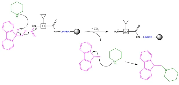
seguida, a desproteção da amina permite o ataque nucleofílico deste grupo ao ácido carboxílico do segundo aminoácido, com a formação de uma ligação amida. O grupo Fmoc é lábil a bases, por isso é removido através do tratamento com piperidina, uma base forte. Este ciclo repete-se até que todo o péptido desejado seja obtido.
Os péptidos sintetizados neste trabalho foram posteriormente analisados e purificados usando o HPLC em fase-reversa, de modo a obter uma purificação de pelo menos 95%. E, de forma a identificar e determinar a massa molecular dos mesmos, foi utilizada a espectrometria de massa MALDI-TOF. Em anexo, são apresentados os respetivos cromatogramas e espectros. Por fim, as suas atividades antimicrobiana e hemolítica foram determinadas.
A resistência antimicrobiana está sobretudo associada a bactérias Gram-negativas, por essa razão, a atividade antimicrobiana dos análogos do péptido BP214 foi testada contra estirpes de Escherichia coli, Pseudomonas aeruginosa, Klebsiella pneumoniae e
Acinetobacter baumannii, todas bactérias Gram-negativas clinicamente revelantes.
Utilizando a microdiluição em caldo, um dos métodos mais usados para avaliar o potencial antimicrobiano de fármacos, cada uma das bactérias selecionadas foi inoculada com diferentes concentrações dos péptidos em estudo e as suas concentrações mínimas inibitórias foram determinadas. Contudo, não basta apenas que os péptidos antimicrobianos apresentem elevada atividade bactericida para que possam ser aplicados na prática clínica. É também essencial que demonstrem baixa toxicidade contra as células hospedeiras, em particular, os eritrócitos. Para a determinação da atividade hemolítica, diferentes concentrações de péptido foram também incubadas com uma suspensão de glóbulos vermelhos e, a partir da hemoglobina libertada, a percentagem de hemólise foi calculada.
Nos análogos em que se procedeu à substituição de uma única lisina ou arginina pelo aminoácido alanina, verificou-se uma melhoria na atividade antimicrobiana contra a maioria das estirpes bacterianas, com concentrações mínimas inibitórias inferiores a 8 µg/mL. Por outro lado, com a substituição de uma única leucina, isoleucina, fenilalanina ou tirosina, os análogos demonstraram uma diminuição no potencial antimicrobiano em relação ao péptido BP214, em particular a substituição da leucina na posição 11, que revelou concentrações mínimas inibitórias superiores a 128 µg/mL na generalidade dos casos. Estes resultados sugerem que os aminoácidos lisina e arginina não são cruciais para a atividade do BP214 e que os aminoácidos leucina, isoleucina, fenilalanina e tirosina são importantes para a atividade deste mesmo péptido. Para além disso, todos os análogos
5
que revelaram concentrações mínimas inibitórias tão boas ou melhores que o péptido BP214, apresentaram também uma toxicidade hemolítica superior.
Também se observou que, de todas as estirpes bacterianas selecionadas, as piores concentrações mínimas inibitórias (≥ 34 µg/mL) dos análogos do péptido BP214 foram obtidas para a Pseudomonas aeruginosa. Este resultado já era de esperar, uma vez que a
Pseudomonas aeruginosa é um microrganismo que adquire facilmente resistências.
A partir da informação obtida no HPLC de fase reversa, foi ainda possível relacionar o tempo de retenção de cada péptido sintetizado com o seu caráter hidrofóbico. Deste modo, averiguou-se a influência da hidrofobicidade nas atividades antimicrobiana e hemolítica e consequentemente, na seletividade celular. Todos os análogos que mostraram um caráter hidrofóbico superior ao péptido BP214, foram também os que revelaram atividades antimicrobianas e hemolíticas superiores. Com isto concluiu-se que a hidrofobicidade contribui para o aumento da atividade antimicrobiana e da toxicidade hemolítica e assim, para a redução da seletividade celular.
Assim, apesar de alguns dos análogos do péptido BP214 terem-se revelado bastante promissores quanto ao seu potencial antimicrobiano, também mostraram uma toxicidade significativa que torna pouco provável a continuação dos seus estudos e a sua aplicação futura no tratamento de doenças infeciosas.
Antes da apresentação dos resultados foi também incluído, nesta dissertação, um procedimento detalhado de todos os métodos realizados para atingir os objetivos deste trabalho, com base nos protocolos fornecidos e utilizados pelo laboratório.
Palavras-chave: Péptidos antimicrobianos; Atividade antimicrobiana; Atividade
6
Abstract
The excessive and uncontrolled use of antibiotics over the last decades has contributed strongly to the increase in the number of resistant microorganisms and with this, the increase in the number of deaths due to intractable infections and an increase of the Healthcare expenditure. For economic and regulatory reasons, the development of new antibiotics has also decreased significantly. Thus, the search for new therapeutic alternatives to conventional antibiotics led researchers to focus on the study of antimicrobial peptides. In addition to antimicrobial activity, these peptides have the ability to exhibit antibiofilm, immunomodulatory and anticancer activities.
Recently a previous study reported BP214 peptide, an analogue of the cecropin-α-melittin hybrid BP100, which showed a high antimicrobial potential against colistin-resistant
Acinetobacter baumannii strains and other relevant bacterial agents and a relatively low
hemolytic activity. In order to identify the most important amino acids for BP214 activity, a D-Alanine scan was performed. Thus, eleven new analogues of the BP214 peptide were synthesized through the solid-phase peptide synthesis. Subsequently, the synthesized peptides were analysed and purified using MALDI-TOF mass spectrometry and reversed-phase HPLC. Finally, its antimicrobial and hemolytic activities were determined. In the analogues where a single lysine or arginine was replaced by alanine, an improvement in antimicrobial activity against most bacterial strains was verified. On the other hand, with the substitution of a single leucine, isoleucine, phenylalanine or tyrosine, the analogues showed a reduced antimicrobial potential compared to BP214. In addition, all analogues which showed antimicrobial activities as good or better than the BP214 peptide also showed higher hemolytic toxicities. From the information obtained with reversed-phase HPLC, it was still possible to relate the retention time of each peptide to its hydrophobic character. Thus, the influence of the hydrophobicity on antimicrobial and hemolytic activities and consequently on cell selectivity was ascertained. All analogues that showed a superior hydrophobic character to BP214 were also those that showed superior antimicrobial and hemolytic activities.
Before presenting the results, a detailed procedure of all the methods performed was also included in this dissertation.
Key words: Antimicrobial peptides; Antimicrobial activity; Hemolytic activity;
7
Acknowledgements
I want to thank my advisor Paul R. Hansen for accepting me in his department and for all the support, availability, trust and knowledge during the three months project in Copenhagen. I would also like to thank my advisor Francisca da Conceição Lopes for all the help she has provided, patience and advice throughout the elaboration of this work. I am also grateful to my colleagues in the Department of Drug Design and Pharmacology Abdullah Ben Naim Lone, Waffa Al-Mansour, Sabbah Ahmed and Bala Krishna Prabhala for all their support during laboratory work and for the good times we lived together. I am deeply thankful to Birgitte Simonsen, Thomas T. Thomsen and Natalia Molchanova for the availability, support and for teaching me during the distinct phases of my laboratory work, always concerned with my success and helping me achieve good results. I would like to thank my family, especially my mother, father and younger brother, for all the support, affection and strength, and because without them none of this was possible. To my dearest friends, who despite being far away, always worried about me. A special thanks to my friend and classmate Catarina Freire, who accompanied me on this adventure and who taught me a lot and put up with during these three months.
8
Abbreviations
ACCA – α-cyano-4-hydroxycinammic acid AMPs – Antimicrobial peptides
AMR – Antimicrobial resistance Boc – tert-butyloxycarbonyl CFU – Colony forming unit
CLSI – Clinical and Laboratory Standards Institute DIEA – N,N-diisopropylethylamine
EC10 – Effective concentration that causes 10% of erythrocyte lysis
EC50 – Effective concentration that causes 50% of erythrocyte lysis
EDTA – Ethylenediaminetetraacetic acid ELISA – Enzyme-linked immunosorbent assay
EUCAST – European Committee on Antimicrobial Susceptibility Testing Fmoc – 9-fluorenylmethyloxycarbonyl
HA – Hemolytic activity
HATU – (N-[(dimethylamino)-1H-1,2,3-triazole[4,5-b]pyridine-1-ylmethylene])-N-methylmethan-aminium hexafluorophosphate N-oxide
HF – Hydrogen fluoride
HOAt – 7-aza-1-hydroxybenzotriazol HOBt – 1-hydroxybenzotriazole LPS – Lipopolysaccharides
MALDI-TOF MS – Matrix assisted laser desorption/ionization-time-of-flight mass spectrometry
9 MHB-II – Cation-adjusted Mueller-Hinton broth MIC – Minimum inhibitory concentration
OD300 – Optical density at 300 nm
Pbf – 2,2,4,6,7-pentamethyl-dihydrobenzofuran-5-sulfony PBS - Phosphate buffered saline
PEG – Polyethylene glycol PTFE – Polytetrafluoroethylene RAM – Rink amide linker RBCs – Red blood cells
RP HPLC – Reversed-phase high performance liquid chromatography SPPS – Solid-phase peptide synthesis
tBu – tert-Butyl
TFA – Trifluoroacetic acid Trt – Trityl
10
Index
1. Introduction ... 13
2. Aims ... 18
3. Materials and Methods ... 22
3.1. Solid-Phase Peptide Synthesis: ... 22
3.1.1. Protecting groups ... 24 3.1.2. Resins ... 25 3.1.3. Linkers ... 26 3.1.4. Coupling Reagents ... 26 3.1.5. Cleavage Cocktails ... 28 3.2. Analysis ... 29 3.3. Purification ... 30
3.4. Determination of Antimicrobial Activity... 30
3.5. Hemolytic Activity ... 32
4. Procedure and Preparations ... 33
4.1. Solid-Phase Peptide Synthesis ... 33
4.1.1. Resin Swelling ... 33
4.1.2. Deprotection of Resin ... 33
4.1.3. Amino Acid Coupling ... 34
4.1.4. Fmoc-Group Removal ... 34
4.1.5. Coupling of Next Amino Acids ... 35
4.1.6. Final Fmoc-Group Removal ... 35
4.1.7. Cleavage ... 35
4.2. Analysis ... 36
4.2.1. Analytical HPLC ... 36
4.2.2. MALDI-TOF Mass Spectrometry ... 36
11
4.4. Determination of Antimicrobial Activity... 37
4.4.1. Peptide Solutions ... 37
4.4.2. Preparation of Bacterial Suspension ... 37
4.4.3. Broth Microdilution ... 38
4.5. Hemolytic Activity ... 39
4.5.1. Preparation of PBS Solution ... 39
4.5.2. Preparation of Melittin Solution ... 39
4.5.3. Peptide and Antibiotic Solutions ... 40
4.5.4. Preparation of RBC Suspension ... 40
4.5.5. Teste Procedure ... 40
4.5.6. Incubation and Reading Absorbance ... 41
5. Results ... 43
6. Discussion ... 44
7. Conclusions ... 47
References ... 48
Annexes ... 52
A1. Structure and Nomenclature of Amino Acids ... 52
A2. 3D Structures of BP214 and the analogues under study ... 53
A3. Amino acid sequences of Cecropin A, Cecropin B and Melittin ... 57
A4. Structures of Ciprofloxacin and Colistin ... 58
A5. MALDI-TOF MS ... 59
A6. Analytical HPLC ... 62
List of Figures
Figure 1.1 Number of deaths caused by antimicrobial resistance (AMR) each year in relation to other causes of death. ... 1312
Figure 1.3 Membrane disruption models. ... 15
Figure 2.1 Structure of BP214. ... 19
Figure 2.2 Structures of the analogues CC2-CC6. ... 20
Figure 2.3 Structures of the analogues CF1-CF6. ... 21
Figure 3.1 Scheme of solid-phase peptide synthesis. ... 23
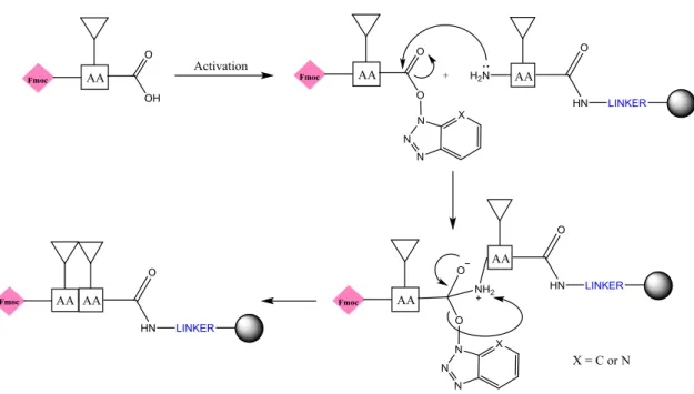
Figure 3.2 Mechanism of amide bond formation. ... 23
Figure 3.3 Boc group. ... 24
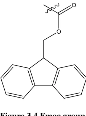
Figure 3.4 Fmoc group. ... 24
Figure 3.5 Mechanism of removal of the Fmoc group (pink) by treatment with piperidine (green). ... 25
Figure 3.6 Linkers most used in Fmoc SPPS. ... 26
Figure 3.7 Racemization of the C-α of the carboxylic acid group. ... 27
Figure 3.8 Coupling reagents used. ... 28
Figure 3.9 Mechanism of activation of the carboxylic acid through HATU/HOAt. ... 28
Figure 4.1 Scheme of broth microdilution... 39
Figure 4.2 Scheme of hemolytic activity. ... 41
List of Tables
Table 1.1 Antimicrobial activity, hemolytic activity and hydrophobic properties of the peptides under study. ... 4313
1. Introduction
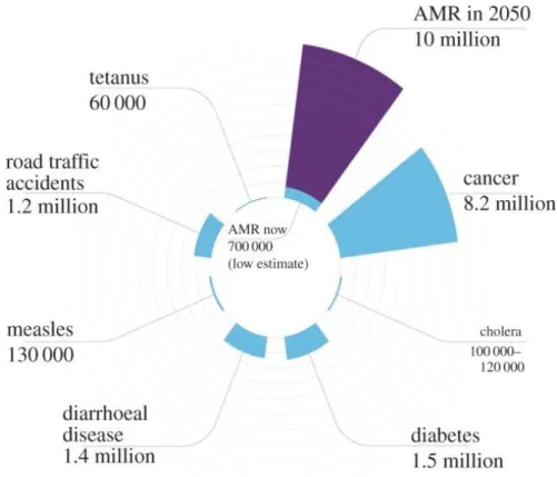
In the recent years, there has been a significant increase on the number of bacterial pathogens resistant to antibacterial agents. The World Health Organization (WHO) sees antibiotic resistance as one of the greatest threats to global health [1, 2]. This situation not only has an impact on human health (increased numbers of intractable infections and consequent mortality), but also has implications on health systems, with increased costs [2, 3] (Figure 1.1). The excessive and uncontrolled use of antibiotics in humans and animals is the major cause of the outbreak of strains resistant to antimicrobial agents [1]. Although some bacteria may be intrinsically resistant to certain antibiotics, exposure to these drugs contributes to the development of genetic mutations, target modifications and horizontal gene transfer, and thus to the selection of resistant pathogens [1, 3].
Figure 1.1 Number of deaths caused by antimicrobial resistance (AMR) each year in relation to other causes of death. It is estimated that in 2050, infections caused by
drug-resistant microorganisms will cause around 10 million deaths and will cost 100 trillion dollars. Source: Shallcross et al., 2015 [4]
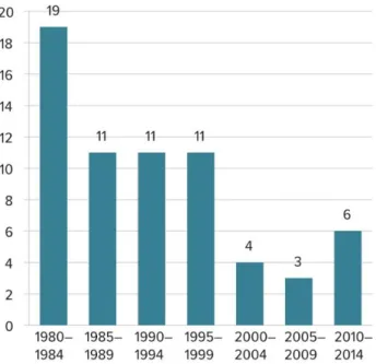
Also, the number of new antibiotics developed by the pharmaceutical industry in recent years has declined considerably due to economic and regulatory reasons (Figure 1.2).
14
Unlike the drugs used in the treatment of chronic diseases, antibiotics are usually used for short periods of time. On the other hand, the resistance associated with them makes recent antibiotics to be kept only as a last resort. This results in a low number of sales to pharmaceutical companies and consequently, a low return on investment. In addition, the very high costs of clinical trials and the complexity in obtaining regulatory approval compromise this development [5].
The urgent need to develop new strategies to combat this resistance has led many countries and organizations to make important decisions to optimize the use of antibacterial drugs and raise awareness of the population and promote research and development of new therapeutic alternatives [1, 2, 6].
Figure 1.2 Number of new antibiotics developed and approved in recent decades.
Source: Ventola, 2015 [5]
Antimicrobial peptides (AMPs) are a group of small molecules (composed by 12-100 amino acids) that exists naturally in living organisms (plants, fungi, bacteria, animals and humans) and contribute to the first line of defense against pathogens [7, 8]. These peptides are initially synthesized at ribosomes level on their inactive form and are then cleaved and processed to their active form, being able to acquire different conformations. The most predominant structures are α-helical, β-sheet, α-helix/β-sheet blending and extended [7]. Being α-helical and β-sheet the most common structures [9].
AMPs are amphipathic structures, meaning that on one side they have cationic (charged + 2 to +9) and polar residues and on the opposite side they have hydrophobic residues [7,
15
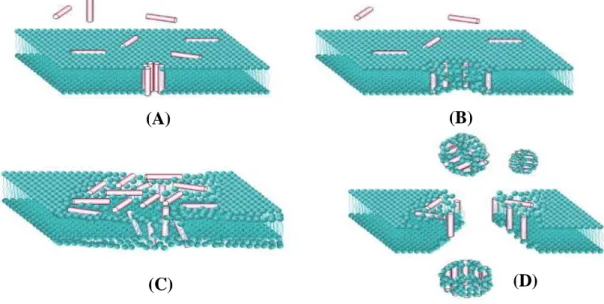
8]. Because of this feature, positively charged AMPs can interact with the surface of the negatively charged bacterial cell, leading to membrane modification and disruption and then, consequently, to cell death. Cationic residues are attracted electrostatically by anionic structures such as lipopolysaccharides (LPS) from the outer membrane of Gram-negative bacteria and cell wall lipoteichoic acids from Gram-positive bacteria [7]. The existence of a more negative surface potential in the membranes of prokaryotic cells than in eukaryotic cells explains the specificity of the peptides to these structures [10]. Currently, there are four proposed models that explain this mechanism by membrane disruption: barrel-stave, toroidal-pore, aggregate and carpet models (Figure 1.3); but none of these models is yet fully accepted [7]. However, it is known that the interaction of the AMPs with the membrane is influenced by the size, charge, secondary structure, amino acid composition, hydrophobicity and amphipathicity of the peptide [7, 11].
Figure 1.3 Membrane disruption models. (A) Barrel-stave model: formation of
barrel-like transmembrane pore by orientation of the hydrophobic residues of the AMPs in the lipid core bilayer - the hydrophobic region of the peptide is outwardly directed towards the membrane, while the hydrophilic region forms the coating of the pore; (B) Toroidal-pore model: AMPs insert inside the membrane and associate with the polar region of the phospholipids, affecting the curvature of the bilayer and consequently that the lipid region to bend and there is pore formation; (C) Carpet model: the peptides accumulate on the surface of the bilayer forming a carpet-type cover - when the concentration of AMPs becomes high, the membrane disintegrates (D) Aggregate model: AMPs are inserted into the membrane by the formation of peptide-lipid aggregates, allowing translocation of peptides across membranes [7, 12]. Source: Wimley, 2010 (adapted) [12]
(A) (B)
16
At certain concentrations, the AMPs are also able to translocate through the membrane and interact with different intracellular targets and other negatively charged molecules, such as DNA and RNA, inhibiting cell division and protein synthesis. Thus, the high affinity of AMPs for multiple targets hinders the appearance of resistant strains [7]. For the last few years, other biological activities have been revealed by AMPs. In addition to antimicrobial activity, the AMPs can exhibit antibiofilm, immunomodulatory and anticancer activities. Several studies have demonstrated that AMPs are capable of inhibit the formation of biofilms1 at concentrations below their MIC (minimum inhibitory concentration) therefore through mechanisms of action distinct from those used for antimicrobial activity [7]. AMPs have the ability to penetrate the matrix of biofilms, prevent their maturation, decrease bacterial cell adhesion and neutralize the effects of released bacterial endotoxins [11].
AMPs have also been shown to modulate the immunity response and pro- and anti-inflammatory responses by the capability to stimulate immune cell recruitment, promote cell differentiation, promote the expression of chemokines [7, 13] and prostaglandins and still regulate phenomes such as apoptosis, autophagy and phagocytosis [8, 13]. In mammals, AMPs are expressed by immune cells and epithelial cells of constitutive or induced form in the presence of infectious and inflammatory stimuli, for example [7, 8]. Other studies have shown that AMPs have a high specificity for cancer cells. This specificity is explained by the greater overall negative charge on the surface of malignant cells relatively to healthy cells. On the other hand, healthy cell membranes contain a higher amount of cholesterol and therefore are more rigid, preventing the entrance of peptides with anticancer activity [7].
Due to this diversity of functions, the study of AMPs has been a subject of great interest by researchers. Currently, more than 2000 AMPs have been identified [7] and more than 60 peptide drugs have already been approved by the US Food and Drug Administration (FDA) in the market. Also, many other peptides are already in clinical (> 140) and preclinical trials (> 500) and it is estimated that these numbers will continue to increase [14]. Although peptides have high effectiveness, specificity and selectivity, there are
1 Biofilms are aggregates of bacteria incorporated into extracellular polymer matrices produced by them,
highly resistant to antibiotics. The resistance of biofilms to antibiotics is due to the slow growth rate and the low metabolic activity of the bacterias that constitute it.
17
some limitations in their use as therapeutic alternatives, such as notably low bioavailability (oral), short half-life [14, 15], difficulty in purifying peptides from natural sources (requiring a large amount of biological material) [7] and hemolytic toxicity [16]. Thus, the easiness of peptide synthesis in the processes of modification and optimization of its sequences and functions is highlighted [7, 15].
18
2.
Aims
This work is based on the Oddo et al. paper2 which recently reported about BP214, kklfkkilryl-NH2, an analogue of the peptide BP100 (KKLFKKILKYL-NH2), a
cecropin-α-melittin hybrid, which showed rapid bactericidal properties, an hemolytic 50% effective concentration (EC50) of >150µM and a high activity against colistin-resistant
A. Baumannii strains featuring mutated lpxC, pmrA, and pmrB genes and other clinically
relevant human pathogens [16].
Cecropins are a family of broad spectrum antibacterial peptides that are mostly part of the insect’s immune system. These peptides are composed by 31-39 amino acids and have in their structure a strongly basic N-terminal domain and a hydrophobic C-terminal domain [10, 17]. Cecropins also have low toxicity against human erythrocytes and other eukaryotic cells. However, cecropins are susceptible to proteolytic degradation [18]. Melittin is also an antibacterial peptide (21 amino acids) present in bee venom, but is capable of causing erythrocytes lysis. Such as cecropin, melittin contains hydrophilic and hydrophobic domains, but with reverse polarity, i.e., a hydrophobic N-terminal domain and a basic C-terminal domain [10, 17].
Thus, several cecropin-melittin hybrid peptides have been synthesized and evaluated for their antibacterial and hemolytic activities [17], namely, the peptide BP100 (KKLFKKILKYL-NH2) [16]. Cecropin-melittin hybrids predominantly acquire α-helical
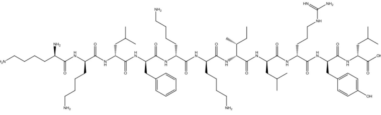
structures and are constructed by incorporating the hydrophilic and cationic N-terminal domain of cecropin with hydrophobic amino acids of the N-terminal domain of melittin [10]. BP100 demonstrated important bactericidal properties, low cytotoxicity and reduced susceptibility to proteases [16, 18]. From the peptide BP100, novel analogues have been developed, in particular, peptide BP214 (kklfkkilryl-NH2) (Figure 2.1), designed by
introducing novel elements and substituting L-amino acids (indicated by uppercase letters) with D-amino acids (indicated by lowercase letters) [16]. It is known that incorporation of D-amino acids increases proteolytic stability and decreases hemolytic activity by reduction of the helicity [9, 10].
2 Oddo A, et al. An Amphipathic Undecapeptide with All d-Amino Acids Shows Promising Activity against
Colistin-Resistant Strains of Acinetobacter baumannii and a Dual Mode of Action. Antimicrob Agents Chemother. 2015; 60:592-9
19
Figure 2.1 Structure of BP214.
In this report a D-Alanine scan was performed to identify the most important amino acids for BP214 activity. In addition to the synthesis of BP214 (kklfkkilryl-NH2), eleven novel
analogues were synthesized: aklfkkilryl-NH2 (CC2); kalfkkilryl-NH2 (CC3);
kkafkkilryl-NH2 (CC4); kklakkilryl-NH2 (CC5); kklfakilryl-NH2 (CC6) (Figure 2.2);
kklfkkilrya-NH2 (CF1); kklfkkilral-NH2 (CF2); kklfkkilayl-NH2 (CF3); kklfkkiaryl-NH2 (CF4);
kklfkkalryl-NH2 (CF5); kklfkailryl-NH2 (CF6) (Figure 2.3). Alanine is a non-polar amino
acid that was chosen due to the fact that, relative to other amino acids, it possesses a favourable degree of flexibility that prevents the loss of the secondary structure of the peptide sequences. Subsequently, their antibacterial and hemolytic activities were evaluated. However, only the procedures for the peptide BP214 and the analogues CC2, CC3, CC4, CC5 and CC6 are mentioned in this work. The other analogues are analysed on a different assay since the CF compounds were synthesized by another student. The results and the conclusions are presented at the end for all the under study peptides.
20
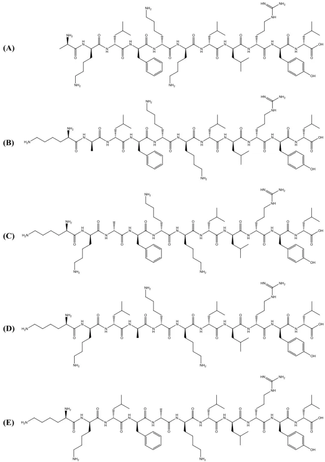
Figure 2.2 Structures of the analogues CC2-CC6. (A) CC2; (B) CC3; (C) CC4; (D) CC5; (E) CC6.
21
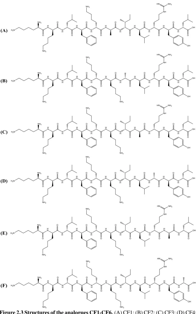
Figure 2.3 Structures of the analogues CF1-CF6.(A) CF1; (B) CF2; (C) CF3; (D) CF4; (E) CF5; (F) CF6
22
3.
Materials and Methods
3.1. Solid-Phase Peptide Synthesis:
Currently three methods can be used for the peptide synthesis: chemical synthesis, which includes solution phase and solid phase; enzymatic synthesis; and recombinant synthesis [19].
For the peptide synthesis was used the solid-phase peptide synthesis (SPPS). The SPPS is the method of choice that most researchers use because all reactions are carried out in one single container; large excesses of amino acid and reagents can be used to drive reactions to completion and subsequently can be removed by washing, not requiring the purification of the intermediates in each step; high yields can be obtained; the reaction cycles are shorter and faster than other methods; and uses repetitive steps making process automation possible [15, 20].
The SPPS (Figure 3.1) consists of the use of a polymeric support (resin), to which a first amino acid, protected with a protecting group in the α-amino group and, if necessary, in the side chains, is coupled through the C-α of the carboxylic acid group. The α-amino protecting groups provide only temporary protection, being removed after each coupling, while the side chain protecting groups are semi-permanent protecting groups, thus avoiding the occurrence of undesirable side reactions and the formation of by-products [15, 20]. The carboxylic acid group still needs to be activated by a coupling reagent to reduce the racemization of the chiral α-carbon [20, 21]. In addition, the resin has a linker which will facilitate the cleavage of the peptide after its synthesis is complete [20]. After the coupling of the first amino acid to the resin, the α-amino protecting group is then removed, being designated Nα-deprotection. The carboxylic acid of the second amino acid, upon activation, undergoes the nucleophilic attack of the α-amino group of the first amino acid resulting in the formation of an amide bond (Figure 3.2). The cycle is repeated between Nα-deprotection, activation and coupling until the desired peptide is obtained [15, 20]. In the end, the separation between the peptide and the resin is made with the aid of a cleavage cocktail, which enables the uptake of carbocations (highly reactive intermediates) formed during the synthesis, preventing them from reacting and causing undesirable by-products [20].
23
Figure 3.1 Scheme of solid-phase peptide synthesis.
Thus, for SPPS it is necessary to take into account six aspects: Nα-protecting group, side-chain protecting groups of amino acids, kind of resin, linker, coupling reagents and cleavage cocktails.
24
3.1.1. Protecting groups
The main strategies used in SPPS are Boc/benzyl strategy, developed by Merrifield in 1963, the only one used for many years [20], and the Fmoc/tBu strategy, which appeared later in the 1970’s [22].
The Boc/benzyl strategy consists of using the Boc (tert-butyloxycarbonyl) group (Figure
3.3) as the protecting group of the α-amino group and benzyl groups as the side chains
protecting groups. The Boc group is acid-labile, being removed with TFA (trifluoroacetic acid) in DCM (dichloromethane). The side chain protecting groups are removed by treatment with HF (hydrogen fluoride), a strong acid too [20].
Figure 3.3 Boc group.
The Fmoc/tBu strategy is based on the use of the Fmoc (9-fluorenylmethyloxycarbonyl) group (Figure 3.4) as the protecting group of the α-amino group and tBu (tert-Butyl) or trityl as the side chain protecting groups. Fmoc group is base-labile, so its subsequently removed by treatment with 20% piperidine in DMF (dimethylformamide) (Figure 3.5). The side chain protecting groups are acid-labile, so for their removal is used TFA [20].
Figure 3.4 Fmoc group.
Both strategies are equally valid, however the Fmoc/tBu strategy is the most advantageous and this was the strategy used in this work. The Fmoc/tBu strategy does not require the use of toxic and corrosive reagents, such as HF, and does not require the use of specialized conditions. In addition, this strategy allows an orthogonal protection and so a selective removal of the protecting groups, unlike the Boc/benzyl strategy [15, 20].
25
Figure 3.5 Mechanism of removal of the Fmoc group (pink) by treatment with piperidine (green).
The choice of side chain protecting groups depends on the amino acid and not all amino acids need protection. For example, for the amino acids Ser, Thr, Tyr, Glu and Asp is used the protecting group tert-Bu and for the amino acids Cys, Asn, Gln and His is used the Trt (trityl) group. The Pbf (2,2,4,6,7-pentamethyl-dihydrobenzofuran-5-sulfonyl) group is used to protect the amino acid Arg and the Boc group is used to protect the amino acids Lys and Trp [20]. In this work, the amino acids used were already protected in both the side chain and the α-amino group.
3.1.2. Resins
For adequate peptide synthesis, a number of requirements must be taken into account when selecting a resin: it must be insoluble in the solvents used in synthesis, be physically and chemically stable, have a good swelling capacity and exhibit low cross-linking for a good accessibility and diffusion of reagents [15, 21]. Based on the strategy used for solid phase synthesis of the peptides, TentaGel® resin, a PEG (polyethylene glycol) grafted polystyrene resin, was chosen. This resin is a copolymer consisting of a low cross-linked polystyrene matrix with 50-70% PEG and is the most commonly used resin in Fmoc SPPS [20]. As the peptide grows, it expands.
The resins also have functional groups that allow anchoring of the linker [15]. The number of functional groups present in the resin is represented by the loading value and is given in mmol per gram (mmol/g) of resin. There are low-load resins (≤0.25 mmol/g) and high-load resins (≥0.9 mmol/g). Although high high-loading resins are more cost-effective, they
26
favour peptide aggregation and the occurrence of undesirable intermolecular reactions. Therefore, a low load resin was used for this work [20].
3.1.3. Linkers
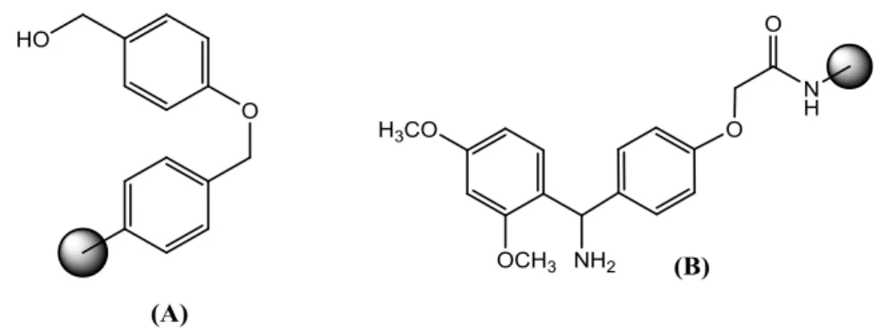
The linker represents the reversible bond between the peptide and the resin and will influence resin loading, peptide-resin spacing and C-terminal functionality of the synthesized peptide, i.e., it determines if the peptide is released as an acid peptide or an amide peptide [15, 20]. There are several linkers available, however the most used are Wang and Rink (Figure 3.6). The Wang linker is a peptide acid linker and the Rink linker is an amide linker; also, the resins are generally identified by their linker, there being the Wang Resin and the Rink Amide Resin. For PEG resins with polystyrene, as used on this work, a Rink amide linker (RAM) is often used [20].
Figure 3.6 Linkers most used in Fmoc SPPS. (A) Wang linker and (B) Rink amide linker.
3.1.4. Coupling Reagents
As discussed above, for the formation of a controlled amide bond between the N-terminus of one amino acid and the carboxylic acid of another amino acid it is essential that the C-α of the carboxylic acid group is activated for a more reactive specie [15, 20]. The formation of the amide bond can lead to loss of the chiral α-carbon integrity, allowing racemization. The activated species is most often a benzotriazyl ester, providing an efficient leaving group that facilitates the formation of the amide bond [20, 21]. The racemization of the α-carbon can occur via direct enolization or via 5(4H)-oxazolone formation (Figure 3.7) [21]. For this purpose, coupling reagents are used.
27
(A)
(B)
Figure 3.7 Racemization of the C-α of the carboxylic acid group. (A) Racemization via direct enolization and (B) Racemization via 5(4H)-oxazolone formation
Currently there are many coupling reagents available, however the most used coupling reagents are divided into three groups: carbodiimides, phosphonium salts and aminium salts [20, 21]. Thus, for this work, the following coupling reagents were chosen (Figure
3.8): 4 equivalents of HATU
((N-[(dimethylamino)-1H-1,2,3-triazole[4,5-b]pyridine-1-ylmethylene])-N-methylmethan-aminium hexafluorophosphate N-oxide), 4 equivalents of HOAt (7-aza-1-hydroxybenzotriazol) and 8 equivalents of DIEA (N,N-diisopropylethylamine) relative to resin loading (mmol/g) in DMF (Figure 3.9). HATU is an aminium salts. The aminium salts have a positive nitrogen atom in their structure and are typically used in combination with N-hydroxy derivatives, such as HOAt or HOBt (1-hydroxybenzotriazole), to improve the coupling reaction and reduce racemization [20, 21]. HOAt is preferable to HOBt because it has a nitrogen atom in the aromatic ring, making it more effective [20]. For the coupling reaction, addition of a base, such as DIEA, a non-nucleophilic tertiary amine, is still required [21]. The coupling reaction is still performed in DMF, a polar aprotic solvent, since Fmoc group protected amino acids and triazole based reagents are quite soluble in this solvent [20].
28
Figure 3.8 Coupling reagents used. (A) HATU, (B) HOAt and (C) DEIA
3.1.5. Cleavage Cocktails
In this case, Reagent B' TFA/H2O/triisopropylsilane (95:2.5:2.5), a variant of Reagent B3,
was used as cleavage cocktail [20]. In Fmoc SPPS an acid cocktail and scavengers are used to release the peptide from the resin. These scavengers are essential to capture the reactive species produced with the acidolytic treatment [21].
Figure 3.9 Mechanism of activation of the carboxylic acid through HATU/HOAt.
The base deprotonates the carboxylic acid, then the carboxylate anion attacks the electron deficient carbon of HATU (red). HOAt (blue) reacts with the formed intermediate resulting in an ester.
3 Reagent B: TFA/phenol/H
29
3.2. Analysis
Following synthesis, the peptides were analysed by analytical HPLC (high performance liquid chromatography) and MALDI-TOF MS (matrix assisted laser desorption/ionization-time-of-flight mass spectrometry).
HPLC is a method that allows the separation, identification and quantification of the different components of a sample. This separation is based on the different affinities of the constituents of the sample to a stationary phase and a mobile phase. The stationary phase consists of a porous column, in which the sample is eluted together with a solvent, the mobile phase, under the force of a high-pressure vacuum pump. Thus, the different interactions with the stationary phase and the mobile phase generate different migration times. In this case, reversed-phase HPLC (RP HPLC) was used. RP HPLC essentially depends on hydrophobic or van der Waals interactions. In reverse phase chromatography, the stationary phase is hydrophobic and the mobile phase is polar, where mostly water-based solutions are used. Therefore, substances with less affinity for the stationary phase, i.e. more polar, elute first and substances with a higher affinity for the stationary phase, i.e. non-polar, elute last [23]. In this work, analytical HPLC was performed on a C-18 silica column using 0.1 % aqueous TFA (buffer A) and CH3CN/H2O (9:1) (buffer B).
Analytical HPLC aims to identify and quantify the compound of interest and is used to obtain information about the retention time, not requiring sample collection [24].
MALDI-TOF mass spectrometry is an ionization method used for identification and characterization of peptides, proteins, polymers and other biological molecules [25]. MALDI-TOF mass spectrometry was used to identify and determine the molecular mass of the synthesized peptides. In this method, the compound in analysis is dissolved in a matrix on a metal plate. This matrix consists of a solution of small organic molecules with a high capacity for absorption of laser light. The mixture is dried, occurring the crystallization of the sample with the matrix. Thereafter, the metal plate is introduced into the spectrophotometer, where a laser is incised trough the sample/matrix mixture. The laser energy is absorbed by the matrix, triggering the transfer of protons from the matrix to the sample and simultaneously the desorption and ionization processes. The large accumulation of energy in the matrix and the rapid heating causes the passage of the compound into the gaseous state. The gas phase ions formed are accelerated by an electric field to a vacuum tube, the analyzer, where they are separated according of their
30
mass/charge ratio. The time the sample will take to reach the detector is directly proportional to its molecular mass [26]. In this work, the α-cyano-4-hydroxycinammic acid (ACCA) matrix was used for MALDI-TOF MS experiments. The obtained molecular weights were then compared with the theoretical masses calculated through the Peptide Mass Calculator4. In relation with other mass spectrometries, MALDI-TOF MS is a method of easy preparation, high sensitivity, rapid analysis, high contaminant tolerance and capable of analyzing large amounts of compounds and non-volatile and thermolabile compounds [25, 26]. In addition, the use of a matrix prevents the direct incidence of the laser on the sample and thus the destruction of the same [26].
3.3. Purification
For the purification of the peptides under study preparative HPLC was used. Preparative HPLC is based on the same methodology as the one presented by analytical HPLC (see
3.2.). However, preparative HPLC aims to isolate the compound of interest and collect
that fraction to a certain purity percentage [24]. In this case, the purity required for the synthesized peptides was ≥ 95%. Thus, preparative HPLC was also performed on a C-18 silica column using 0.1 % aqueous TFA (buffer A) and CH3CN/H2O (9:1) (buffer B).
3.4. Determination of Antimicrobial Activity
The broth microdilution in MHB (Mueller-Hinton broth) was used to determine the minimum inhibitory concentration (MIC) of the peptides synthetized according to the Clinical and Laboratory Standards Institute (CLSI) guidelines. MIC is defined as the lowest concentration of antimicrobial agent required to inhibit visible growth of a microorganism [27, 28] and its determination is considered the gold standard method for assessing antimicrobial activity [7]. The broth microdilution consists in the use of a liquid growth medium in which the bacteria are inoculated with decreasing concentrations of the antimicrobial agent. MHB is the medium most used for majority of microorganisms [28]. MICs were realized in triplicate using 96-well polypropylene microtiter plates and a final bacterial inoculum of approximately 5x10⁵ colony forming unit (CFU/mL). The
presence and absence of bacterial growth were evaluated visually by the presence or absence of turbidity, respectively. Bacterial inocula were prepared from the dilution of a pure culture in MHB and the number of colony forming units used was verified by
31
determination the optical density at 300 nm (OD300) of the inoculum through a
photometer. To verify the non-contamination of the wells of the plate, broth with bacterial inoculum without antimicrobial agent as control of growth (positive control) and only broth as control of sterility (negative control) was used.
The appearance of multi-drug resistant bacterial infections is mainly associated with gram negative bacteria due to the existence of an outer membrane that delays the penetration of the antibiotics into the cell by the ability to detect and repair the damages caused by them [29, 30]. Gram-negative bacteria are mostly associated with important nosocomial infections and their resistance to antibiotics such as fluoroquinolones, carbapenems, colistins (polymyxins) and cephalosporins, has increased significantly [29, 31]. BP214 peptide analogues were tested against Escherichia coli ATCC 25922, Pseudomonas
aeruginosa ATCC 27853, Klebsiella pneumoniae ATCC 13883 and Acinetobacter baumannii ATCC 19606, all Gram-negative bacteria. To verify the authenticity of the
assays and to compare results, antimicrobial agents whose MICs are known were further tested against the bacterial species selected. The antimicrobial agents were ciprofloxacin, colistin, peptide RW-BP100 (RRLFRRILRWL-NH2) and peptide BP214. Ciprofloxacin
is a broad-spectrum antibiotic that belongs to the class of fluoroquinolones [32]. Colistin (polymyxin E) is a potent cationic cyclic polypeptide antibiotic against multiresistant Gram-negative bacteria and sometimes the last active antibiotic available for these infections. However, the nephrotoxicity and neurotoxicity associated with this drug makes colistin a last-line therapeutic option, being replaced by less toxic drugs. Currently, strains resistant to colistin have been identified. Polymyxins act through disruption of the outer membrane of Gram-negative bacteria by interaction with the lipid A of LPS. Thus, resistance to colistin is mainly associated with changes in this same target [33, 34]. Peptides and antibiotics were tested at eleven different concentrations and the highest concentrations were defined based on breakpoints and data of the bacteria's behaviour already known. After inoculation of the bacterial suspension with the antimicrobial agents into the plate, a growth control aliquot diluted in PBS (phosphate buffered saline) is incubated on nutrient-rich agar plates. The cell count on the agar plates should be checked to verify that the correct number of CFU was used and to verify non-contamination of the bacterial inoculum used.
32
3.5. Hemolytic Activity
Another important feature to be considered in AMPs is their toxicity against host cells. Hemolytic activity (HA) testing is considered a fast and inexpensive method for evaluating the overall cellular toxicity of a compound. HA measures the lysis of red blood cells (RBCs) by measuring the hemoglobin released, through incubating different dilutions of the antimicrobial agent under study in PBS with a suspension of RBC in PBS. PBS is an isotonic buffer solution and does not damage most cells. The released hemoglobin is posteriorly evaluated through a spectrophotometer at 414 nm (since hemoglobin exhibits a peak at this wavelength) and the percentage of hemolysis is calculated. HA can also be represented by the concentration of compound that causes 10% (EC10) and 50% (EC50) of erythrocyte lysis. The assays were performed in triplicate
using 96-well microtiter plates and as positive and negative controls, melittin and PBS solution, respectively. Melittin is the gold standard of positive hemolytic controls because it exhibits high activity even at submicromolar concentrations and does not interfere with the spectrophotometric reading at 414 nm [35]. In addition, to ensure the accuracy of the results and for control purposes, the antibiotics ciprofloxacin and colistin, as well as the peptide BP214, whose hemolytic activity values are known, were used.
This method is associated with a noise of at least 4% related to inconsistencies in pipetting, damage to the RBC during its handling and the absorbance reader itself. HA also depends on the type of blood used, i.e., different human blood types and different blood types between different species exhibit different sensitivities to hemolysis and consequently different results can be obtained. Blood type 0 negative, as used in the present study, is most appropriate since it is absent from antigens that are found in blood groups A and B. Also, this test uses serum-free RBC, thus does not reflect in vivo conditions and therefore, peptide-protein interactions that may affect the concentration of free and active peptide in the bloodstream are not considered. Furthermore, although a hemolytic peptide can be considered toxic, a non-hemolytic peptide cannot be considered non-toxic only by this method, it can still be able to demonstrate toxicity against other cell types [35].
33
4. Procedure and Preparations
4.1. Solid-Phase Peptide Synthesis
4.1.1. Resin Swelling
1. Weigh 0.1 g of TentaGel® resin directly inside the reactor. In addition to the resin loading, the amount of resin required for the synthesis depends on the amount of peptide desired (Equation 1).
𝑔 𝑜𝑓 𝑅𝑒𝑠𝑖𝑛 = 𝑚𝑚𝑜𝑙 𝑜𝑓 𝑝𝑒𝑝𝑡𝑖𝑑𝑒 (𝑑𝑒𝑠𝑖𝑟𝑒𝑑)
𝑟𝑒𝑠𝑖𝑛 𝑙𝑜𝑎𝑑𝑖𝑛𝑔 (𝑚𝑚𝑜𝑙/𝑔) (1)
However, since the amount of peptide desired is only a theoretical value and does not account for impurities or losses, calculations should be made considering an excess. In order to facilitate the execution of the calculations presented, prepare a standard template for each peptide using Microsoft® Excel ™.
The reactor consists of a 5 mL syringe equipped with a PTFE (polytetrafluoroethylene) filter. Together with the reactor use pipette tips of 200 µL since they can be easily disposable.
2. Reinsert the piston into the syringe and push it down just above the resin. 3. Draw 3 mL of pure DMF into the syringe.
4. Discard the pipette tip and close the bottom syringe end with a pressure cap. 5. Let it swell in DMF for at least 2 hours or overnight.
4.1.2. Deprotection of Resin
1. Remove the piston and place a pipette tip.
2. Place the syringe on the suction plate and drain the solution.
3. Turn off the suction plate, transfer 3-4 mL of 20% piperidine in DMF into the syringe and leave standing for 4 min.
4. Drain the solution and wash three times with DMF.
5. Repeat steps 3 and 4 twice; thus, a total of three deprotection cycles. 6. Wash with DMF (x4), DCM (x3) and DMF (x4).
34
4.1.3. Amino Acid Coupling
1. Weigh the amount of each required protected amino acid into individual Eppendorf tubes (Equation 2).
𝑚𝑔 𝐴𝑚𝑖𝑛𝑜 𝑎𝑐𝑖𝑑 = 𝑀𝑜𝑙𝑒𝑐𝑢𝑙𝑎𝑟 𝑤𝑒𝑖𝑔ℎ𝑡 𝑜𝑓 𝐴𝑚𝑖𝑛𝑜 𝑎𝑐𝑖𝑑 𝑝𝑟𝑜𝑡𝑒𝑐𝑡𝑒𝑑 × 4 × [𝑠𝑦𝑛𝑡ℎ𝑒𝑠𝑖𝑠 𝑠𝑐𝑎𝑙𝑒(𝑚𝑚𝑜𝑙)] (2)
The synthesis scale refers to the amount of NH2 and depends on the size of the
peptide, i.e., the number of amino acids that constitute it.
2. Weigh the required amount of HOAt (4 eq.) to the amino acids into a tube and dissolve in DMF (Equation 3).
𝑚𝑔 𝐻𝑂𝐴𝑡 = 135.12 × 4 × [𝑠𝑦𝑛𝑡ℎ𝑒𝑠𝑖𝑠 𝑠𝑐𝑎𝑙𝑒 (𝑚𝑚𝑜𝑙)] × [𝑛𝑢𝑚𝑏𝑒𝑟 𝑜𝑓 𝑐𝑜𝑢𝑝𝑙𝑖𝑛𝑔𝑠] (3)
3. Weigh the required amount of HATU (4 eq.) to the amino acids into a tube and dissolve in DMF (Equation 4).
𝑚𝑔 𝐻𝐴𝑇𝑈 = 380.23 × 4 × [𝑠𝑦𝑛𝑡ℎ𝑒𝑠𝑖𝑠 𝑠𝑐𝑎𝑙𝑒 (𝑚𝑚𝑜𝑙)] × [𝑛𝑢𝑚𝑏𝑒𝑟 𝑜𝑓 𝑐𝑜𝑢𝑝𝑙𝑖𝑛𝑔𝑠] (4)
The solutions of HOAt and HATU are prepared to 0.4 M (Equation 5). 𝑚𝐿 𝐷𝑀𝐹 = 4 × [𝑠𝑦𝑛𝑡ℎ𝑒𝑠𝑖𝑠 𝑠𝑐𝑎𝑙𝑒 (𝑚𝑚𝑜𝑙)] × [𝑛𝑢𝑚𝑏𝑒𝑟 𝑜𝑓 𝑐𝑜𝑢𝑝𝑙𝑖𝑛𝑔𝑠]
0.4 𝑀 (5)
4. Divide the HATU solution into individual tubes according to the total number of couplings.
5. Add the HOAt solution to each amino acid in the Eppendorf tubes and shake. 6. Add the HATU solution to the HOAt/amino acid solution.
7. Add DIEA (8 eq.) to the previous solution and shake (Equation 6); the solution should turn yellow.
µ𝐿 𝐷𝐼𝐸𝐴 = 129.24 × 8 × [𝑠𝑦𝑛𝑡ℎ𝑒𝑠𝑖𝑠 𝑠𝑐𝑎𝑙𝑒 (𝑚𝑚𝑜𝑙)] × 0.742 (6) 8. Insert the piston, push it down just above the resin and change the pipette tip. 9. Draw the coupling solution into the syringe.
10. Cover the syringe with tin foil and leave it on a shaker for 2 hours or overnight.
4.1.4. Fmoc-Group Removal
1. Remove the piston and place the syringe on the suction plate. 2. Drain the coupling solution and wash six times with DMF.
35
3. Turn off the suction plate, transfer 3-4 mL of 20% piperidine in DMF into the syringe and leave standing for 4 min.
4. Drain the solution and wash three times with DMF.
5. Repeat steps 3 and 4 twice; thus, a total of three deprotection cycles. For peptides longer than ten amino acids, extend the time of deprotection cycles to 7 min each.
6. Wash ten times with DMF. 7. Turn off the suction plate.
4.1.5. Coupling of Next Amino Acids
1. For each next amino acid, repeat subheading 4.1.3., steps 5-9, followed by subheading 4.1.4., steps 1-7, until sequence is complete.
4.1.6. Final Fmoc-Group Removal
1. For the last amino acid remove the Fmoc-group and wash as described in subheading 4.1.4., steps 1-6.
2. Wash five times with ethanol and turn off the suction plate.
3. Insert the piston and push it down a little above the resin, then discard the pipette tip and leave the reactor in freeze-dry overnight.
4.1.7. Cleavage
1. Prepare at least 6 mL of TFA/H2O/triisopropylsilane (95:2.5:2.5) (v/v)
cleavage cocktail for each peptide. 2. Push the piston down above the resin.
3. Draw 3.5 mL of cleavage cocktail into the syringe.
4. Discard the pipette tip and insert the pressure cap into the bottom of the syringe.
5. Place the syringe on a shaker for at least 2 hours.
6. Remove the pressure cap and using the piston, push the cleavage solution into a 5 mL cryotube.
7. Remove the piston and wash the resin twice with approximately 1 mL of cleavage cocktail collecting the eluate in the cryotube.
36
9. When 0.2 mL or less are left, add 4 mL of cold (-20ºC) diethyl ether. Put the cap on and shake gently.
10. Centrifuge at 2000 rpm for 6 min.
11. Using a pipette, carefully remove and discard the supernatant. 12. Resuspend the solid into another 3-4 mL of cold ether.
13. Repeat steps 11-13 for a total of three washes. The last centrifuge run should be at 3500 rpm.
14. Leave the cryotube standing open overnight to let the residual ether evaporate. 15. Dissolve the crude product in 1-2 mL of 90% water and 10% acetonitrile +
0.1% TFA and freeze-dry to obtain fluffy white crystals overnight.
4.2. Analysis
4.2.1. Analytical HPLC
1. Weight approximately 0.5 mg of the crude product directly inside a glass vial and dissolve in H2O or acetonitrile till a concentration of 0.25-1 mg/mL.
2. Place the solution in an HPLC vial.
3. Run the sample on the analytical HPLC to obtain a chromatogram. Use water as blank.
4.2.2. MALDI-TOF Mass Spectrometry
1. Weight approximately 0.5 mg the crude product directly inside a glass vial and dissolve in H2O or acetonitrile till a concentration of 0.25-1 mg/mL.
2. Place 1 µL of the solution in the metal plate and let it dry. 3. Place 1 µL of the ACCA matrix in the metal plate and let it dry. 4. Place the metal plate in spectrophotometer to obtain the spectrum.
4.3. Purification
1. Dissolve the crude product in H2O or acetonitrile till a concentration of 15
mg/mL and shake.
2. Wash the HPLC needle with MeCN:H2O (1:1) and draw 200-300 µL of
previous solution into a 1 mL syringe.
3. Run the solution on the preparative HPLC and collect the fraction of the compound of interest into a glass tube. Use acetonitrile as blank.
37
5. Join the various fractions collected in a single glass tube and do the analysis of the sample purified as described in subheading 4.2.1., steps 2-3, and subheading 4.2.2., steps 2-4.
6. Place the purified sample in 5 mL cryotubes and freeze-dry.
4.4. Determination of Antimicrobial Activity
4.4.1. Peptide Solutions
1. Weight approximately 2 to 5 mg of each purified peptide in subheading 4.3. and dilute in H2O till a concentration of 10 mg/mL. Mix well in the vortex.
4.4.2. Preparation of Bacterial Suspension
1. Prepare nine glass tubes with 10 mL of sterile MHB-II5.
2. Select one colony from the pure culture of bacterial to be tested using a sterile loop and dilute (1:100) in one of the previous glass tubes.
3. Repeat step 2 for a total of three inoculated glass tubes.
4. Incubate the glass tubes overnight in a shaker at 37 °C and let grow.
5. Remove a 100 µL aliquot of the above bacterial suspensions and dilute (1:10) into three other glass tubes with pre-prepared 10 mL of MHB-II.
6. Incubate the glass tubes for 1h20 in a shaker at 37 ºC and let to grow.
7. Remove a 1 mL aliquot of the above bacterial suspensions and dilute (1:100) into three other glass tubes with pre-prepared 10 mL of MHB-II and let to grow again for 1h20 in a shaker at 37 °C.
8. Measure the OD300 of the above bacterial suspensions through the photometer
using plastic cuvettes. Use as white a cuvette only with sterile MHB-II. Considering the absorbance (A) equal to the concentration, then A = 1 = 1x10⁹. Thus, it is necessary that the OD300 corresponds to a value between 0.2-0.4 so
that the number of CFUs/mL is between 4x10⁵-7x10⁵.
9. Prepare three tubes with 10 mL of sterile MHB-II and three tubes with 18 mL of sterile MHB-II.
10. Based on the OD300 obtained, calculate the volume of each bacterial
suspension prepared in step 7 necessary to withdraw to dilute in the tubes with 10 mL of sterile MHB-II till a concentration of 0.02 mg/mL per tube.
38
11. Remove 2 mL of each of the above bacterial suspensions and dilute in the tubes with 18 mL of sterile MHB-II till a concentration of 0.002 mg/mL per tube.
4.4.3. Broth Microdilution
1. Dilute the solutions of peptides and antibiotics in sterile MHB-II in Eppendorf tubes to obtain the maximum desired concentrations. Dilutions are prepared at twice the highest concentration defined because the peptides and antibiotics will be diluted 50:50 with bacterial suspension. In this case, the dilutions were still prepared in triplicate using 200 µL of each dilution of antimicrobial agent per plate.
2. Mix well the solutions of peptides and antibiotics in the vortex.
3. Remove the 96-well polypropylene microtiter plates from their sterile package and label them using one line for each antimicrobial agent. Keep the lid closed while not handling the plate to prevent contamination.
4. Pipette 100 µL of sterile MHB-II into wells from column 2 to column 12. Pipette another 100 µL of sterile MHB-II into the sterility control wells (the first four wells of column 12).
5. Pipette 200 µL of each antimicrobial agent solution into each of the wells in column 1.
6. Dilute 50:50 solutions of antimicrobial agents from left to right at twice the desired final concentrations to obtain 100 µL in the wells from column 1 to column 11 (Figure 4.1).
7. Inoculate 100 μL of bacterial suspension prepared in subheading 4.4.2. in each well with antimicrobial agent solution and in the growth control wells (the last four wells of column 12).
8. Remove 10 μL of a growth control well from one plate and pipette into an Eppendorf tube with 990 µL PBS (1:1 000 dilution). Mix well in the vortex. 9. Repeat step 8 for two other plates; thus, a total of three Eppendorf tubes. 10. Dilute again (1:10 000) by pipetting 100 µL of each of the previous dilutions
into other Eppendorf tubes with 900 µL of PBS. Mix well in the vortex. 11. Place 100 µL of each of the dilutions from steps 8-9 on different nutrient-rich
agar plates to verify that the correct CFU number was used; resulting at the end three agar plates for each of the two dilutions.
39
12. Incubate the microtiter plates and the agar plates for 18-20h at 37 ° C.
13. Interpret the MICs obtained on the microtiter plates and count the colonies of the agar plates. About 50 colonies at the 1:10 000 dilution and 500 colonies at the 1: 1000 dilution are expected when using an inoculum density of 5x10⁵ CFU/mL.
Figure 4.1 Scheme of broth microdilution.
4.5. Hemolytic Activity
4.5.1. Preparation of PBS Solution
1. Dissolve one PBS tablet in 200 mL of deionized water. The PBS solution should be fresh and should be prepared on the day of the test or the day before.
4.5.2. Preparation of Melittin Solution
1. Remove the 96-well polypropylene microtiter plates from their sterile package.
2. The night before the test, add 150 µL of 5 µM melittin solution in PBS to the positive control wells (H1-H6). Each 1 mL of 5µM solution corresponds to 4 µL of melittin diluted in 1 mL of PBS. Melittin is very toxic and should be with care.
3. The day of the test, discard the solution of the wells and wash the wells with 150 µL of PBS.
40
5. The day of the teste, prepare 2.50 µM of melittin solution in PBS so as to have 500 µL for each plate. Each 1 mL of 2.50 µM solution corresponds to 2 µL of melittin diluted in 1 mL of PBS.
4.5.3. Peptide and Antibiotic Solutions
1. Weigh approximately 1 mg of each peptide and antibiotic into glass vials and dilute to approximately 500 µL of PBS. The antimicrobial agent solutions are prepared at twice the highest concentration defined since the peptides and antibiotics will be diluted 50:50 with RBC suspension. Mix well in the vortex.
4.5.4. Preparation of RBC Suspension
1. Transfer 1 mL of fresh 0 negative whole blood in EDTA into a 5 mL cryotube for centrifugation. RBC are fragile and need to be handle with care, so use a soft 3 mL Pastette.
2. Add 3 mL of PBS to previous blood, mix gently and centrifuge for 8 min at 3000 rpm.
3. Discard supernatant, add 4 mL of PBS, mix gently and centrifuge for 8 min at 3000 rpm.
4. Discard supernatant, add 4 mL of PBS, mix gently and centrifuge for 8 min at 4000 rpm. Plasma is associated with errors in the results, so plasma should not be present with the RBCs and the supernatant must be clear. Discard supernatant.
5. For each plate, prepare 8 mL of 0.5% v/v RBC suspension: using a 200 µL pipette tip cut into the tip, transfer 40 µL of RBC to 8 mL of PBS.
4.5.5. Teste Procedure
1. Pipette 75 µL of PBS to all the wells from B1 to G12 and to the negative control wells (H7-H12).
2. Pipette 150 µL of each peptide and antibiotic solution in row A using three columns for each antimicrobial agent. Each plate can accommodate four antimicrobial agents to be tested in triplicate. Remember to label each plate and note where each antimicrobial agent is inoculated.
41
4. Dilute 50:50 antimicrobial agent solutions from top to bottom, transferring 75 µl from row A to row G and mixing a couple of times. After mixing, discard 75 µL from row G.
5. Pipette 75 µL of RBC suspension into all wells (Figure 4.2).
6. Mix the antimicrobial agent solutions with the RBC suspension from row G to row A, i.e., towards the highest concentration wells, in order to reduce the impact of antimicrobial agents transfer from well to well. Mix positive and negative controls separately.
Figure 4.2 Scheme of hemolytic activity.
4.5.6. Incubation and Reading Absorbance
1. Cover each plate with a Biorad Microseal film and seal each well individually to prevent the solution from evaporating and consequently invalidating the test.
2. Place the plates in the incubator at 37 ºC for 1h. 3. Centrifuge the plates for 10 min at 4000 rpm.
4. Quickly and carefully transfer 60 µL of supernatant from each well to 96-well polystyrene ELISA plates, maintaining the respective positions of each well. These plates have transparent flat bottom wells, making it easier to read the absorbance. The supernatant will have the possible hemoglobin released by lysis of erythrocytes.
5. Place the plates on the spectrophotometer and read the absorbance at 414 nm. Positive controls represent 100% hemolysis and generally absorb in the range
42
of 0.78-0.9 nm. Negative controls represent 0% hemolysis and generally absorb in the range of 0.05-0.12 nm.
6. Calculate the percentage of hemolysis (Equation 7). % 𝐻𝑒𝑚𝑜𝑙𝑦𝑠𝑖𝑠 = 𝐴𝑏𝑠 (𝑠𝑎𝑚𝑝𝑙𝑒) − 𝐴𝑏𝑠 (𝑁𝑒𝑔𝐴𝑣𝑔)
𝐴𝑏𝑠 (𝑃𝑜𝑠𝐴𝑣𝑔) − 𝐴𝑏𝑠 (𝑁𝑒𝑔𝐴𝑣𝑔) × 100 (7) Abs (sample) = absorbance of the sample.
Abs (NegAvg) = averaged absorbance of the negative controls. Abs (PosAvg) = averaged absorbance of the positive controls.
43
5. Results
In Table 1.1 are presented the MIC and hemolytic activity values of the analogues under study, as well as those of the BP214 peptide and the other antimicrobial agents used for control and comparison purposes. In addition, this table shows the hydrophobic properties of the synthesized peptides obtained from analytical reversed-phase HPLC.
Table 1.1 Antimicrobial activity, hemolytic activity and hydrophobic properties of the peptides under study.
E. coli P. aeruginosa K. pneumoniae A. baumannii
CF1 kklfkkilrya-NH 2 64-128* 128 >128 64 >500 8 42.1 CF2 kklfkkilral-NH 2 16-32* 32 32-64* 8-16* 62.5 35 46.7 CF3 kklfkkilayl-NH 2 4-8* 64 4*-8 4*-8 35 56 49.6 CF4 kklfkkiaryl-NH 2 64 128*- >128 64-128* 64 >500 3 44.9 CF5 kklfkkalryl-NH 2 64*-128 64 128 16-32* >500 5 44.8 CF6 kklfkailryl-NH2 4 64*-128 4 4 <7.812 100 52.4 CC2 aklfkkilryl-NH2 8-4* 64 8 2-4* 15 65 51.8 CC3 kalfkkilryl-NH2 8 64*-128 8 2-4* <7.812 64 50.3 CC4 kkafkkilryl-NH2 32 64 32-64* 16 300 14 44.8 CC5 kklakkiryl-NH2 64 128 128 16-32* 500 10 44.4 CC6 kklfakiryl-NH2 4 64*-128 4-8* 4*-8 15.625 63 53.2 BP214 kklfkkilryl-NH2 8 16 4 8-16 35 29 47.7 RW-BP100 RRLFRRILRWL-NH2 4 8 4*-8 2-4 ND ND ND Ciprofloxacin 0.125 0.25 0.125 0.5*-1 0 1 ND Colistin 1 0.5 4*-8 0.5 0 1 ND Compound1 %B5 MIC (µg/mL) HA10 (µg/mL)3 %HA4 Sequence2
1 CF compounds were synthesized by another student
2 Lowercase letters indicate D-amino acids and uppercase letters indicate L-amino acids 3 HA
10, concentration of compound that causes 10% hemolysis
4 %HA, hemolytic activity at 500 µg/mL
5 %B, percentage of acetonitrile (buffer B) at the time of elution in analytical HPLC
*most common value ND, not determined