ABSTRACT
Peel bond strength of resilient liner modiied by
the addition of antimicrobial agents to denture
base acrylic resin
Cristiane S. ALCÂNTARA1, Allana F.C. de MACÊDO1, Bruno C.V. GURGEL2, Janaina H. JORGE3, Karin H.
NEPPELENBROEK4, Vanessa M. URBAN5
1- DDS, Faculty of Health and Life Sciences, Center of Superior Studies of Maceió - CESMAC, Maceió, AL, Brazil.
2- DDS, MSc, PhD, Assistant Professor, Department of Dentistry, Federal University of Rio Grande do Norte - UFRN, Natal, RN, Brazil.
3- DDS, MSc, PhD, Assistant Professor, Department of Dental Materials and Prosthodontics, Araraquara Dental School, Univ. Estadual Paulista - UNESP, Araraquara, SP, Brazil.
4- DDS, MSc, PhD, Assistant Professor, Bauru School of Dentistry, University of São Paulo - USP, Bauru, SP, Brazil.
5- DDS, MSc, PhD, Assistant Professor, Department of Dentistry, Ponta Grossa State University - UEPG, Ponta Grossa, PR, Brazil.
Corresponding address: Vanessa Migliorini Urban - Universidade Estadual de Ponta Grossa - Departamento de Odontologia - Campus de Uvaranas - Av. General Carlos Cavalcanti, 4748, 84030-900 - Ponta Grossa - PR - Brazil - Phone: 55-42-3220 3106 - e-mail: [email protected]
Received: December 3, 2010 - Modiication: August 14, 2011 - Accepted: September 18, 2011
I
n order to prolong the clinical longevity of resilient denture relining materials and reduce plaque accumulation, incorporation of antimicrobial agents into these materials has been proposed. However, this addition may affect their properties. Objective: This study evaluated the effect of the addition of antimicrobial agents into one soft liner (Soft Confort, Dencril) on its peel bond strength to one denture base (QC 20, Dentsply). Material and Methods: Acrylic specimens (n=9) were made (75x10x3 mm) and stored in distilled water at 37°C for 48 h. The drug powder concentrations (nystatin 500,000U - G2; nystatin 1,000,000U - G3; miconazole 125 mg - G4; miconazole 250 mg - G5; ketoconazole 100 mg - G6; ketoconazole 200 mg - G7; chlorhexidine diacetate 5% - G8; and 10% chlorhexidine diacetate - G9) were blended with the soft liner powder before the addition of the soft liner liquid. A group (G1) without any drug incorporation was used as control. Specimens (n=9) (75x10x6 mm) were plasticized according to the manufacturers’ instructions and stored in distilled water at 37°C for 24 h. Relined specimens were then submitted to a 180-degree peel test at a crosshead speed of 10 mm/min. Data (MPa) were analyzed by analysis of variance (α=0.05) and the failure modes were visually classiied. Results: Nosigniicant difference was found among experimental groups (p=0.148). Cohesive failure
located within the resilient material was predominantly observed in all tested groups.
Conclusions: Peel bond strength between the denture base and the modiied soft liner was
not affected by the addition of antimicrobial agents.
Key words: Antifungal agents. Tensile strength. Stomatitis. Denture bases.
INTRODUCTION
The oral candidiasis known as denture stomatitis is related to the use of removable dentures and is considered the most common oral lesion observed (65%)27 in patients wearing removable dentures.
Although the etiology of denture stomatitis is multifactorial, infection by Candida spp., especially
C. albicans, is considered the main etiologic factor. Local factors associated with the denture are also related to this pathology, such as: presence of
biofilm4,18, local trauma caused by dentures12,
xerostomia21, continuous use of the dentures and
alteration in salivary pH12.
Different treatments for denture stomatitis are available and may include topical antifungal and systemic therapy, care with oral hygiene, denture cleaning and disinfection procedures18,
replacement of old dentures, elimination of anatomic irregularities, re-establishment of atraumatic occlusion, and nutritional restitution3. Furthermore,
Materials Type Manufacturer Batch number
Composition Powder/
liquid ratio
Polymerization conditions
Soft Confort Resilient liner
Dencril Prod. Odontol., São Paulo, SP, Brazil
Powder (010068)
Poly(ethyl methacrylate), phtalate ester, ethyl alcohol
1.27 g/1 mL 5 min at room temperature
QC 20 Heat-curing acrylic resin
Dentsply Ind. Com. Ltda., Petrópolis, RJ,
Brazil
Liquid (503793)
Poly(methyl methacrylate),
methyl
methacrylate,n-butyl methacrylate
2.3 g/1 mL 1. Heat water to boiling point; 2. Turn off the water bath; 3.
Put the lask in and leave it for
20 min; 4. Turn on the water bath; 5. When the boiling point is reached, wait another
20 min.
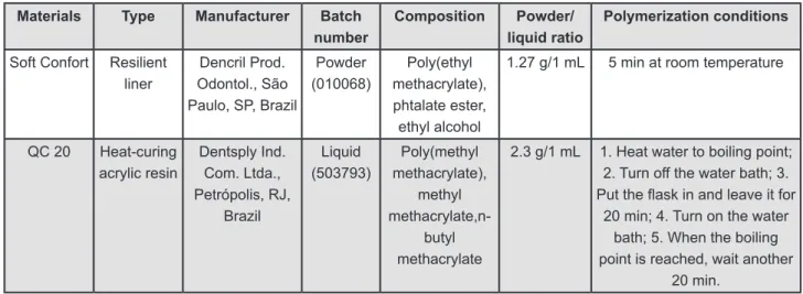
Figure 1- Materials selected for this study
Peel bond strength of resilient liner modiied by the addition of antimicrobial agents to denture base acrylic resin
the mucosal epithelium, patients should sleep without the dentures6. The choice of a treatment or
association of more than one treatment is an aspect to be individually considered. Re-infection of the treated oral mucosa may occur in up to two weeks post-treatment, and is attributed to the survival of Candida spp. due to insuficient concentration
of the antifungal agent on the denture surfaces16.
Therefore, it is crucial to adopt methods that reduce or preferably eliminate the microorganisms from denture surfaces.
In addition, resilient materials have been routinely used with the purpose of recovering tissues that are in contact with the denture base24. These materials
partially absorb chewing load on the denture during function, thus reducing the energy transmitted to the associated paraprosthetictissues17. However, these
materials are easily degradable and susceptible to microbial colonization14, which may cause different
degrees of denture stomatitis.
To prolong the clinical longevity of resilient materials and reduce plaque accumulation, incorporation of antimicrobial agents into these materials has been proposed20. This combination
may be a logical therapy in the treatment of denture stomatitis because of several factors: 1. reducing the trauma caused by the internal surface of removable dentures; 2. eliminating contact of the contaminated surface with the oral tissues and consequently, interrupting the cycle of re-infection, and 3. action of antimicrobial agents incorporated into the material on the infected tissues20. In
this context, denture stomatitis may be treated before fabricating new dentures, in a relatively short period. The reason is attributed to their gradual degradation and hardening, so it should not take longer than two weeks, which is a period similar to the one required for the treatment with conventional topical antifungal drugs20,22.
The incorporation of antimicrobial agents into resilient materials has shown to be effective and
feasible both in in vitro and in vivo studies6,20,22.
Despite these therapeutic advantages, the incorporation of drugs into polymeric materials, including tissue conditioners and resilient liners, may affect their properties. For the resilient liner to adequately perform its function of recovering the tissues injured by trauma, it should remain bonded to the acrylic base of the removable denture5. Peeling of the resilient material from the
denture base has been reported as the cause of clinical failure and the bond between the resilient materials and the denture base acrylic resins has been the object of previous investigations13,17. Thus,
the aim of this study was to evaluate the effect of the addition of antimicrobial agents (nystatin, miconazole, ketoconazole, and chlorhexidine diacetate) to a resilient liner on its peel bond strength to a denture base acrylic resin. The hypothesis investigated in this study was that the addition of antimicrobial agents to a resilient liner would result in changes in the peel bond strength to a denture base acrylic resin.
MATERIAL AND METhODS
The acrylic materials, manufacturers, batch numbers, compositions, powder/liquid ratios, and polymerization conditions selected for this study are listed in Figure 1. The selected antimicrobial agents were nystatin, miconazole, ketoconazole (Alonatu Farmácia de Manipulação e Cosméticos/Farmácia Dermatus, Maceí, AL, Brazil – Req. 119704-1), and 98% chlorhexidine diacetate (Acros Organics, Morris Plains, NJ, USA).
Specimen preparation
Specimens (n=9)17 measuring 75x10x3 mm13,17
Group Antimicrobial agent Amount of drug incorporated
G1 None (control) None
G2 Nystatin 500,000 U
G3 Nystatin 1,000,000 U
G4 Miconazole 125 mg
G5 Miconazole 250 mg
G6 Ketoconazole 100 mg
G7 Ketoconazole 200 mg
G8 Chlorhexidine diacetate 5%
G9 Chlorhexidine diacetate 10%
Figure 2- Drug dosages incorporated into the resilient liner powder in all experimental groups
(Zetalabor, Rovigo, Veneto, Italy) between two glass plates. The mold/matrix set was invested in
conventional metal dental lasks in Type III dental
stone (Herodent, Vigodent, Rio de Janeiro, RJ,
Brazil). The dental lasks were closed and remained
under pressure (500 kgf) in a hydraulic press during stone setting time. After this period, the dental
lasks were opened and the stainless steel matrixes
were removed.
QC 20 was proportioned, mixed according to the manufacturer’s instructions (Figure 1), and was inserted into the silicone matrix mold. The
dental lask was closed and kept under pressure
at room temperature (23±2°C) for 30 min. After this period, the test specimens were submitted to the polymerization cycle “B” recommended by resin manufacturer (Figure 1). When the polymerization
cycle ended, the dental lasks were bench cooled for
30 min and then under running water for 15 min. The specimens were removed from the molds and stored in distilled water at 37°C for 48 h10.
After this period, specimens were submitted to
surface preparation to receive the modiied resilient
liner. One of the specimen surfaces was abraded automatically in a polishing machine using #600 silica carbide abrasive paper (Norton Abrasivos, São Paulo, SP, Brazil). The abraded surface was cleaned with detergent for 20 s, washed under running water, and dried. The specimen was then placed in a hollow stainless steel mold with internal measurements of 75x10x6 mm. The specimen area (650 mm2) to not be bonded to the resilient material
was covered with a polyester strip.
The antimicrobial powders in each experimental group (Figure 2) were manually mixed with resilient lining powder with a spatula, until a homogenous mixture was obtained24,25. The resilient lining
liquid was added to this mixture and the material was mixed in accordance to the manufacturer’s
instructions (Figure 1). The modiied material was
inserted into the hollow mold containing the test specimen of the heat-curing acrylic resin prepared for the relining procedure. This set was covered with
glass slide and kept under inger pressure during
the resilient liner polymerization time recommended by the manufacturer (Figure 1). The excesses of
the modiied resilient liner were eliminated and the
specimen was removed from the mold. The relined specimens were then stored in distilled water at 37°C for 24 h prior to the peel test.
Peel test
A universal testing machine (Versat 2000, Panambra Ind. Tech. SA, São Paulo, SP, Brazil) was used to perform the peeling bond strength test of the relined test specimens at an angle of 180°. A
portion of modiied resilient material not bonded
to the resin base (65 mm) was folded upwards
and ixed onto the top hook of the equipment at
20 mm from the adhesive bond area of the test specimen. The other un-relined portion of the
heat-curing acrylic resin was ixed onto the bottom
hook of the equipment13,17 at the same distance
from the adhesive bond area. each test specimen was submitted to tension to promote peeling of the
modiied resilient liner from the heat-curing acrylic
resin base at a speed of 10 mm/min until failure occurred.
Bond failures were visually observed and
classiied into three categories: adhesive, when
peeling occurred between the modiied resilient
liner and the denture base acrylic resin; cohesive, when there was tearing (rupture of the resilient liner within the area bonded to the denture base) or snapping (resilient material had stretched and then ruptured away from the bonded area) within the
modiied resilient liner; and mixed, when regions with two types of failure were observed on the surface of the denture base material13,17.
The results of rupture force were initially obtained in N and transformed into peeling bond strength in MPa and then submitted to one-way
Experimental groups Mean (SD)* Mode of failure
G1 0.054 (0.017) 80% cohesive; 20% mixed
G2 0.049 (0.017) All cohesive
G3 0.043 (0.012) 80% cohesive; 20% mixed
G4 0.046 (0.013) 80% cohesive; 20% mixed
G5 0.042 (0.014) All cohesive
G6 0.046 (0.012) 40% cohesive; 40% mixed; 20% adhesive
G7 0.050 (0.010) 80% cohesive; 20% mixed
G8 0.060 (0.012) All cohesive
G9 0.049 (0.012) 80% cohesive; 20% mixed
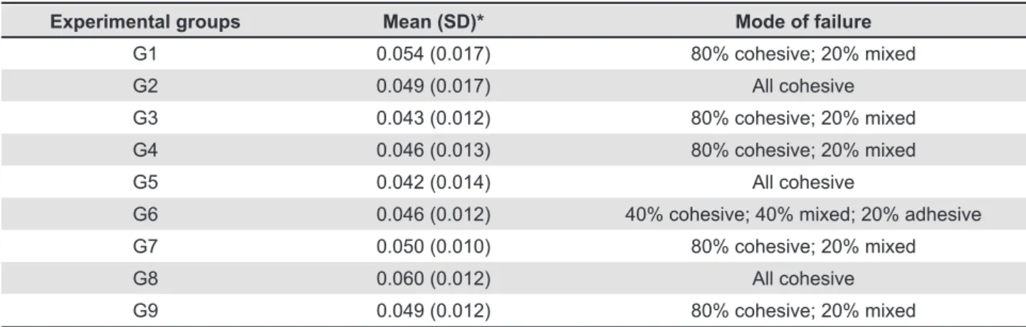
Table 1- Peel strength (MPa) at 24 h
* There was no statistical difference (p>0.05) among the experimental groups
Peel bond strength of resilient liner modiied by the addition of antimicrobial agents to denture base acrylic resin
RESULTS
The results of peel bond strength are shown
in Table 1. There was no signiicant difference
(p=0.148) among the experimental groups. Therefore, the incorporation of antimicrobial agents in the concentrations assessed did not affect the peeling bond strength between the resilient liner and the denture base resin after 24 h of immersion in distilled water.
The failure modes obtained after performing the tests are shown in Table 1. The majority of bond failures were cohesive (tearing and/or snapping) within the resilient liner. For the experimental groups G2 (nystatin at 500,000 U), G5 (miconazole at 250 mg), and G8 (5% chlorhexidine diacetate), a mixture of tearing and snapping was observed. Peeling away from the denture base was only observed for groups G6 (ketoconazole at 100 mg) and G9 (10% chlorhexidine diacetate). For the other groups, cohesive and mixed bond failures were observed.
DISCUSSION
The hypothesis investigated in this study that “the addition of antimicrobial agents to the resilient liner would result in alterations in the peeling bond strength to denture base resin” was rejected because there was no difference between the experimental groups assessed in comparison with the control group without the addition of drugs.
During clinical use, the resilient materials are highly subjected to degradation and susceptible to the colonization by microorganisms. If these materials are not regularly replaced, they may act as microorganism reservoirs, causing systemic complications23. An example of this is the presence
in the oral cavity of Staphylococcus aureus, a microorganism responsible for respiratory infections15. The combination between resilient
materials and antimicrobial agents seems to be a logical therapeutic modality for denture stomatitis. This method results in a reduction of the trauma caused by the old denture and tissue reconditioning associated with antimicrobial therapy; important etiologic factors in triggering infection by Candida
spp. are simultaneously eliminated. In addition, this method favors a relined denture that can more easily be kept clean by the patient20.
Several drugs have shown reduced water solubility, so maximum dose is required to have the effectiveness required for a certain medication8. Among the antimicrobial agents
assessed, chlorhexidine shows higher solubility in water, followed by nystatin, miconazole, and ketoconazole8. Although these medications are
soluble in water, they are insoluble in monomers and plasticizers1.Thus, they could not interfere with
the polymerization or plasticization1 process of these
materials. However, their physical presence within the polymer matrix could interrupt the structure of the polymerized materials21. Resilient materials
containing nystatin showed increased water sorption, and for these materials, this resulted in breaking their morphological structure7. According
to Addy and Handley2 (1981), change in material
properties may be consistent with the incorporation pattern of the medication into the polymer matrix. A previous study24 assessed the incorporation pattern
already existent dentures2, without necessarily
reducing their strength.
Although some soft materials are submitted only to compression and shear, tensile strength is used to measure the quality of the material. The ability of the material to resist tearing is of practical importance. In clinical use, including the cleaning and disinfection procedures, the soft materials are submitted to conditions that start the tearing process. Adequate bonding between denture base resin and soft material is therefore essential. Clinical failure of these materials is frequently attributed to the rupture of this bond, and the measurements of this bond are clinically relevant. Reduced bond between the soft liner and the denture base resin effectively negates any other property considered adequate for this material26. In
the peel bond strength test, the stress is conined
to a line restricted to the end of the bond, and is considered the most clinically representative of the failure modes26. This is the only method in which
the failure proceeds at controlled speed and it is a direct measure of peeling, while it also represents the elastic deformation of the material9. The peeling
test simulates the lining procedure more precisely, with a uniform and constant distribution of force throughout the bond area26.
The results of this study demonstrated that the addition of antimicrobial agents in all the assessed concentrations did not affect the peeling bond strength of the resilient liner to denture base resin. However, the bond strength values were considered low, since they were approximately 10 times lower than the acceptable value for the clinical use of resilient liners (0.44 MPa)11.
While the methodology in this study was
performed, some modiications were made, such as
the reduction in bond area and surface roughness of the denture base, to ensure that the methodology evaluated the bonding between materials rather than the cohesive strength of the liner material. If the bond failures observed in this study were predominantly cohesive within the liner material, the peeling bond strength would not be measured9.
The failure mode of the cohesive type provides information related to the material itself and not to the bond between materials19. emmer, et al.8 (1995)
suggested the term “strength failure” instead of “bond failure” when cohesive failures occur. Predominant cohesive failures, such as those that occurred in this study, indicate poor resistance to tearing of the resilient material. However, mixed and adhesive failures were observed in some samples, indicating that the cohesive strength values of the resilient liner and bond strength values to base resin were similar.
A previous study25 observed that a tissue
conditioner (Duraconditioner, Reliance Manufacturing
Co., Worth, IL, USA) modiied by the addition of
nystatin showed cohesive strength values similar to those of the control group. These values were close, if not similar, to the ones obtained in this study. Therefore, the cohesive strength of the resilient material tested in this study is equivalent to its bond strength to the denture base material. Thus, the material will snap or tear at the bond interface at forces lower than those necessary to cause bond failures.
One of the limitations of this study was that only one brand of the resilient liner was assessed. Moreover, the peeling bond strength could have been assessed after other storage periods. This assessment is also important to observe a possible
reduction in bond strength of the modiied liner
to the denture base material, since it has been reported that plasticizers and alcohols are released from resilient materials after periods of storage in water and this release is responsible for the decrease in the bond strength values between the materials13. However, these analyses are object of
future investigations.
CONCLUSIONS
Within the limitations of this in vitro study, it can be concluded that it is possible to incorporate any of the antimicrobial agents assessed in the selected concentrations into a resilient liner without changing the bond strength of this material to denture base resin. A clinical study is still needed to determine the therapeutic validity of this alternative treatment modality.
REFERENCES
1- Addy M. In vitro studies into the use of denture base and soft liner materials as carriers for drugs in the mouth. J Oral Rehabil. 1981;8:131-42.
2- Addy M, Handley R. The effects of the incorporation of chlorhexidine acetate on some physical properties of polymerized and plasticized acrylics. J Oral Rehabil. 1981;8:155-63.
3- Aldana L, Marker VA, Kolstad R, Iacopino AM. effects of Candida
treatment regimens on the physical properties of denture resins. Int J Prosthodont. 1994;7:473-8.
4- Banting DW, Greenhorn PA, McMinn JG. effectiveness of a topical antifungal regimen for the treatment of oral candidiasis in older, chronically ill, institutionalized, adults. J Can Dent Assoc. 1995;61:199-200,203-5.
5- Braden M, Wright PS, Parker S. Soft lining materials - a review. eur J Prosthodont Restor Dent. 1995;3:163-74.
6- Carter GM, Kerr MA, Shepherd MG. The rational management of oral candidosis associated with dentures. N Z Dent J. 1986;82:81-4.
7- Douglas WH, Clarke D. The physical properties of nystatin containing denture liners. J Prosthet Dent. 1975;34:428-34. 8- emmer TJ Jr, emmer TJ Sr, Vaidynathan J, Vaidynathan TK. Bond strength of permanent soft denture liners bonded to the denture base. J Prosthet Dent. 1995;74:595-601.
Peel bond strength of resilient liner modiied by the addition of antimicrobial agents to denture base acrylic resin
10- International Organization for Standardization. Speciication
1567: Denture base polymers. 2nd ed. Geneva: The Organization; 1998.
11- Kawano F, Dootz eR, Koran A 3rd, Craig RG. Comparison of bond strength of six soft denture liners to denture base resin. J Prosthet Dent. 1992;68:368-71.
12- Lombardi T, Budtz-Jörgensen e. Treatment of denture-induced stomatitis: a review. eur J Prosthodont Restor Dent. 1993;2:17-22. 13- Machado AL, Breeding LC, Puckett AD. effect of microwave disinfection on the hardness and adhesion of two resilient liners. J Prosthet Dent. 2005;94:183-9.
14- Mäkilä e, Hopsu-Havu VK. Mycotic growth and soft denture lining materials. Acta Odontol Scand. 1977;35:197-205.
15- Marsh PD, Percival RS, Challacombe SJ. The inluence of denture-wearing and age on the oral microlora. J Dent Res.
1992;71:1374-81.
16- Mathaba LT, Davies G, Warmington JR. The genotypic relationship of Candida albicans strains isolated from the oral cavity of patients with denture stomatitis. J Med Microbiol. 1995;42:372-9.
17- McCabe JF, Carrick Te, Kamohara H. Adhesive bond strength and compliance for denture soft lining materials. Biomaterials. 2002;23:1347-52.
18- Montagner H, Montagner F, Braun KO, Peres Pe, Gomes BP. In vitro antifungal action of different substances over microwaved-cured acrylic resins. J Appl Oral Sci. 2009;17:432-5.
19- Pinto JR, Mesquita MF, Henriques Ge, Arruda Ńbilo MA. effect of thermocycling on bond strength and elasticity of 4 long-term soft denture liners. J Prosthet Dent. 2002;88:516-21.
20- Schneid TR. An in vitro analysis of a sustained release system for the treatment of denture stomatitis. Spec Care Dentist. 1992;12:245-50.
21- Torres SR, Peixoto CB, Caldas DM, Silva eB, Akiti T, Nucci M,
et al. Relationship between salivary low rates and Candida counts in subjects with xerostomia. Oral Surg Oral Med Oral Pathol Oral Radiol endod. 2002;93:149-54.
22- Truhlar MR, Shay K, Sohnle P. Use of a new assay technique
for quantiication of antifungal activity of nystatin incorporated in
denture liners. J Prosthet Dent. 1994;71:517-24.
23- Ueshige M, Abe Y, Sato Y, Tsuga K, Akagawa Y, Ishii M. Dynamic viscoelastic properties of antimicrobial tissue conditioners containing silver-zeolite. J Dent. 1999;27:517-22.
24- Urban VM, Sé RS, Giannini M, Arrais CA. Superficial
distribution and identiication of antifungal/antimicrobial agents on a modiied tissue conditioner by SEM-EDS microanalysis: a
preliminary study. J Prosthodont. 2009;18:603-10.
25- Urban VM, Souza RF, Arrais CA, Borsato KT, Vaz LG. effect of the association of nystatin with a tissue conditioner on its ultimate tensile strength. J Prosthodont. 2006;15:295-9.
26- Waters MG, Jagger RG. Mechanical properties of an experimental denture soft lining material. J Dent. 1999;27:197-202.