JCB:
Article
The Rockefeller University Press $30.00
Introduction
Unrelated proteins containing glutamine repeat sequences
are known to cause neurodegenerative disorders when the
re-peat-containing tracts are expanded beyond a critical threshold
(Zoghbi and Orr, 2000). Pathogenesis of polyglutamine (polyQ)
diseases is still poorly understood, but the fact that clinical
pre-sentation and neurodegeneration profiles do not overlap in these
diseases indicates that the biologic specificities of each
caus-ative protein interfere with expansion-induced toxicity (Gatchel
and Zoghbi, 2005). Machado-Joseph disease (MJD ), otherwise
known as spinocerebellar ataxia type 3, is one such disease and
is recognized as the most common form of dominantly inherited
ataxia in the world (Schöls et al., 2004; Bettencourt and Lima,
2011), involving the structural and functional compromise of
discrete brain regions, such as the cerebellum, the pons, and the
striatum (Alves et al., 2008b; Rüb et al., 2013).
MJD is caused by an abnormal expansion of a polyQ
se-quence contained in ataxin-3 (atx3; Kawaguchi et al., 1994),
a protein of elusive biologic function nonetheless described as
being involved in protein homeostasis systems, transcription
regulation, and cytoskeleton organization (Matos et al., 2011).
Atx3 displays deubiquitinase (DUB ) activity, which is mediated
by a catalytic triad of amino acids localized in a globular N-
terminal Josephin domain (JD) that also includes two binding
sites for ubiquitin (Ub; Mao et al., 2005; Nicastro et al., 2005,
2009). A flexible C-terminal tail comprises the polyQ tract and
two or three ubiquitin-interacting motifs (UIM s), depending on
the isoform (Harris et al., 2010).
PolyQ-expanded atx3 is known to cause cellular stress
and to have an increased tendency to aggregate in vitro and in
cultured cells, forming inclusions in MJD patients’ brains often
localized in cell nuclei (Paulson et al., 1997b; Zoghbi and Orr,
2000; Gales et al., 2005; Bauer and Nukina, 2009; Matos et al.,
2011; Scarff et al., 2015). Contrastingly, atx3 is ubiquitously
expressed in diverse tissues and cell types (Trottier et al., 1998;
Bauer and Nukina, 2009), indicating that region-specific
mech-anisms of toxicity are responsible for the localized
neurodegen-eration. The fact that cell demise targets neurons specifically
(Trottier et al., 1998; Rüb et al., 2013) suggests that atx3 may
play an important role in neuronal cells, which is disturbed on
Different neurodegenerative diseases are caused by aberrant elongation of repeated glutamine sequences normally
found in particular human proteins. Although the proteins involved are ubiquitously distributed in human tissues,
tox-icity targets only defined neuronal populations. Changes caused by an expanded polyglutamine protein are possibly
influenced by endogenous cellular mechanisms, which may be harnessed to produce neuroprotection. Here, we show
that ataxin-3, the protein involved in spinocerebellar ataxia type 3, also known as Machado-Joseph disease, causes
dendritic and synapse loss in cultured neurons when expanded. We report that S12 of ataxin-3 is phosphorylated in
neurons and that mutating this residue so as to mimic a constitutive phosphorylated state counters the
neuromorpho-logic defects observed. In rats stereotaxically injected with expanded ataxin-3–encoding lentiviral vectors, mutation of
serine 12 reduces aggregation, neuronal loss, and synapse loss. Our results suggest that S12 plays a role in the
patho-genic pathways mediated by polyglutamine-expanded ataxin-3 and that phosphorylation of this residue protects
against toxicity.
Ataxin-3 phosphorylation decreases neuronal
defects in spinocerebellar ataxia type 3 models
Carlos A. Matos,
1,2Clévio Nóbrega,
1* Susana R. Louros,
1* Bruno Almeida,
3Elisabete Ferreiro,
1,4Jorge Valero,
1,5Luís Pereira de Almeida,
1,6Sandra Macedo-Ribeiro,
3and Ana Luísa Carvalho
1,21CNC - Center for Neuroscience and Cell Biology, University of Coimbra, 3004-504 Coimbra, Portugal
2Department of Life Sciences, Faculty of Sciences and Technology, University of Coimbra, 3004-517 Coimbra, Portugal
3Instituto de Biologia Molecular e Celular and Instituto de Investigação e Inovação em Saúde, University of Porto, 4200-135 Porto, Portugal 4Institute for Interdisciplinary Research, University of Coimbra, 3030-789 Coimbra, Portugal
5Ikerbasque Basque Foundation for Science and Achucarro Basque Center for Neuroscience, Bizkaia Science and Technology Park, E-48170 Zamudio, Spain 6Faculty of Pharmacy, University of Coimbra, 3000-548 Coimbra, Portugal
© 2016 Matos et al. This article is distributed under the terms of an Attribution– Noncommercial–Share Alike–No Mirror Sites license for the first six months after the publication date (see http ://www .rupress .org /terms). After six months it is available under a Creative Commons License (Attribution–Noncommercial–Share Alike 3.0 Unported license, as described at http ://creativecommons .org /licenses /by -nc -sa /3 .0 /).
*C. Nóbrega and S.R. Louros contributed equally to this paper. Correspondence to Ana Luísa Carvalho: alc@cnc.uc.pt
Abbreviations used in this paper: DIV , days in vitro; DUB , deubiquitinase; JD, Josephin domain; MJD , Machado-Joseph disease; polyQ, polyglutamine; PSD -95, postsynaptic density protein 95; Ub, ubiquitin; UIM , ubiquitin-interacting motif; VGL UT1, vesicular glutamate transporter subtype 1; WT, wild type.
THE
JOURNAL
OF
CELL
BIOLOGY
on November 2, 2017
jcb.rupress.org
Downloaded from
http://doi.org/10.1083/jcb.201506025Supplemental material can be found at:
on November 2, 2017
jcb.rupress.org
Downloaded from
on November 2, 2017
jcb.rupress.org
Downloaded from
on November 2, 2017
jcb.rupress.org
Downloaded from
on November 2, 2017
jcb.rupress.org
Downloaded from
polyQ expansion, but no function that would be specifically
critical for neuronal survival or activity has ever been described.
The variable effects of polyQ-expanded atx3 in different
cell types may result from diverging regulatory mechanisms
of its properties and functions (Gatchel and Zoghbi, 2005; La
Spada and Taylor, 2010; Takahashi et al., 2010). Tapping into
these pathways constitutes a promising approach to the
treat-ment of MJD . In cells, proteins are frequently regulated by
posttranslational modifications (La Spada and Taylor, 2010;
Takahashi et al., 2010) and among them phosphorylation has
been repeatedly described to modulate the toxicity of polyQ
disease–related proteins (Pennuto et al., 2009). For example,
preventing expanded ataxin-1 phosphorylation at S776 renders
the protein unable to form aggregates and ameliorates disease
phenotype (Emamian et al., 2003). Contrastingly,
phosphoryla-tion of huntingtin at S421 decreases aggregaphosphoryla-tion and cell death
in cell culture models (Humbert et al., 2002; Luo et al., 2005);
mimicking this modification by mutating S421 to aspartate is
neuroprotective in a lentiviral rat model (Pardo et al., 2006).
To date, five atx3 phosphorylation sites have been
de-scribed, all localized in the UIM s: S236 in the first UIM
, S256 and 260/261 in the second, and S340 and 352 in the
third UIM (Fei et al., 2007; Mueller et al., 2009). S256 is
phosphorylated in vitro by glycogen synthase kinase 3
β and
preventing this modification enhances the aggregation of
ex-panded atx3 (Fei et al., 2007). Simulating phosphorylation of
S236, or S340 and S352 simultaneously, leads to an increased
atx3 nuclear localization in cell lines and enhances repression
of atx3-regulated transcription in gene reporter assays
(Muel-ler et al., 2009). Pharmacologic inhibition of casein kinase 2,
which was shown to phosphorylate atx3 C-terminal region,
re-duces the levels of nuclear atx3, activates atx3-regulated gene
transcription, and decreases inclusion formation (Tao et al.,
2008; Mueller et al., 2009).
We have observed that pathogenic expansion of atx3
causes a loss of dendrites and synapses in cultured neurons.
Mimicking atx3 phosphorylation at S12, a novel
phosphoryla-tion site we hereby describe, reverts these effects, suggesting
that atx3 may be functionally involved in the maintenance of
these neuronal structures. Furthermore, we show that mutating
S12 ameliorates aggregation, degeneration, and synapse loss
in the brain of a MJD lentiviral rat model with striatal
pathol-ogy (Pardo et al., 2006; Alves et al., 2008b), suggesting that
phosphorylation at this residue may constitute a therapeutic
target for MJD treatment.
Results
PolyQ-expanded atx3 causes dendrite and synapse loss in rat neuronal cultures
The cytotoxicity of polyQ-expanded atx3 is contingent on the
cellular type in which the protein is being expressed,
consid-ering that only neurons are targeted in MJD (Zoghbi and Orr,
2000; Rüb et al., 2013). To determine if polyQ expansion of
atx3 causes neuron-specific morphologic changes that may
un-derlie impaired survival and function, we started by comparing
the dendritic tree of rat neuron cultures expressing human atx3
with a pathogenic number of glutamines (84Q) or a
nonpatho-genic number of repeats (28Q). Cortical neurons, used
consid-ering the increasing evidence supporting the involvement of the
cortex in the disease (Soong et al., 1997; Taniwaki et al., 1997;
Murata et al., 1998; Ichikawa et al., 2001; Yamada et al., 2001;
D’Abreu et al., 2012; Lopes et al., 2013; Pedroso et al., 2013;
Rüb et al., 2013), were transfected with GFP -tagged human
atx3 (Fig. 1 A), and the length of dendritic tracts of GFP -
positive cells was measured. GFP -atx3 84Q-expressing neurons
show a decreased total extension of dendrites, expressed as the
sum of the length of all dendritic tracts in each cell (Fig. 1 B).
Sholl analysis was used to evaluate dendrite complexity and
further demonstrated that neurons expressing GFP -atx3 84Q
display a withered dendritic tree, with less dendrites reaching
80–160 µm from the cell body and no dendrites reaching >170
µm (Fig. 1 C). This indicates that, in transfected cortical
neu-rons, expansion of GFP -atx3 causes dendritic loss or shrinkage.
We then tested whether atx3 expansion affected
synap-tic contacts established between the cultured neurons.
Visu-alization of functional excitatory glutamatergic synapses was
achieved by colocalizing the punctuate signal of pre- and
post-synaptic protein markers: the vesicular glutamate transporter
subtype 1 (VGL UT1) and postsynaptic density protein 95 (PSD
-95; Fig. 1 D). Compared with neurons transfected with GFP
-atx3 28Q, neurons expressing GFP -atx3 84Q have a significant
reduction in the number of glutamatergic synapses (Fig. 1 E).
Similar differences were detected for inhibitory postsynaptic
terminals, as evaluated by the number of gephyrin-positive
puncta (Fig. 1, F and G).
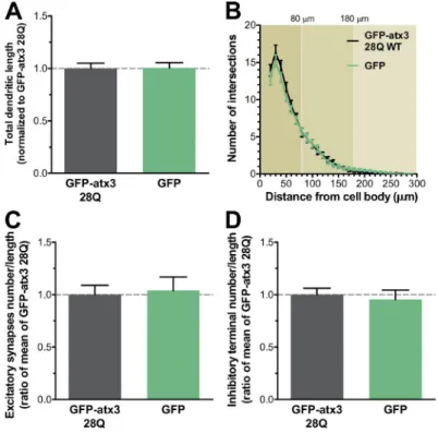
Importantly, compared with neurons expressing the empty
GFP vector, cells transfected with GFP -atx3 28Q show no
dif-ferences in any of the parameters analyzed, underscoring the
role of the polyQ expansion of GFP -atx3 in the phenotypes
ob-served (Fig. S1). Results therefore indicate that polyQ
expan-sion of atx3 in transfected cultured neurons causes morphologic
defects in neuron-specific structures.
Atx3 is phosphorylated at S12 in mammalian neurons
We moved on to explore novel phosphorylation sites of atx3
that could account for the modulation of toxicity of the
pro-tein, starting by a mass spectrometry analysis directed at
phos-phorylation site detection. GFP -atx3 28Q was purified from
transiently transfected HEK 293FT cells by
immunoprecipi-tation and SDS -PAG E (Fig. 2 A), and peptides resulting from
tryptic digestion of the GFP -atx3 bands were separated by
reverse-phase high performance liquid chromatography and
electrosprayed into the mass spectrometer. Examination of the
protein sample produced from okadaic acid–stimulated cells
re-vealed the presence of one phosphorylated peptide and mapped
S12 as the modified residue (Fig. 2 B).
We then generated a phosphospecific antibody recognizing
atx3 phosphorylated at S12 (anti-Patx3; Fig. 2 C) and used it to
probe extracts from cultured rat cortical neurons. Western blot
analysis yielded a band with the molecular weight expected of
endogenous rat atx3 (Fig. 2 D), which is absent when the protein
is knocked-down in cultures transduced with lentiviral vectors
encoding atx3-targeting shRNA s (Alves et al., 2010). Bands of
lower molecular weight yielded by the anti-Patx3 antibody
possi-bly correspond to endogenous atx3-derived fragments mentioned
in other studies (Berke et al., 2005; Pozzi et al., 2008; Koch et al.,
2011; Simões et al., 2012). Higher molecular weight endogenous
atx3 protein bands have also been observed in previous studies
(Paulson et al., 1997a; Trottier et al., 1998; Koch et al., 2011).
Neuronal extracts prepared in the absence of phosphatase
inhib-itors display decreased immunoreactivity, supporting that the
on November 2, 2017
jcb.rupress.org
anti-Patx3 antibody is labeling phosphorylated atx3 (Fig. 2 E).
The analysis of Patx3 labeling in MJD patient’s fibroblasts and in
fibroblasts of a healthy control (Fig. 2, F and G) revealed a
pro-tein band pattern similar to that observed in cultured rat neurons,
indicating that atx3 is phosphorylated in human samples.
Our observations indicate that S12 is a
phosphor-ylation site of endogenous atx3 and that phosphate
con-jugation to this residue occurs normally in cultured rat
neurons and human fibroblasts.
Mimicking S12 phosphorylation decreases atx3 DUB activity in vitro
S12 is localized in the catalytic JD (aa 8–168), the N-terminal
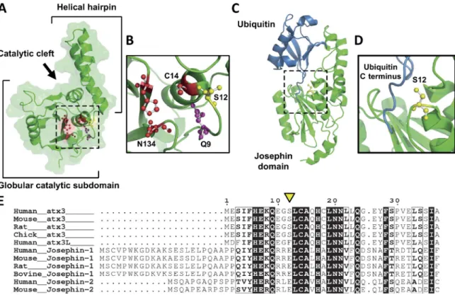
do-main of atx3 (Masino et al., 2003; Almeida et al., 2013; Fig. 3 A).
In the solution structure of atx3 JD (Nicastro et al., 2005), S12
is located at the cleft between the globular catalytic subdomain
and the flexible helical hairpin predicted to play a role in
sub-strate recognition (Komander et al., 2009; Nicastro et al., 2009).
S12 is located in the loop preceding helix
α1, where C14, the
nucleophile of the catalytic triad, is located, and nearby Q9, the
residue proposed to contribute to the stabilization of the
nega-tively charged transition state during peptide bond cleavage
(Nicastro et al., 2005; Fig. 3 B). S12 is also adjacent to the loop
centered on S72, which precedes the helical hairpin that
com-prises the docking site for the C terminus of the Ub substrate
(Nicastro et al., 2009; Fig. 3, C and D). S12 is exposed to the
solvent on the surface of the JD and is consequently accessible
for phosphate conjugation. The proximity of S12 to structural
el-ements associated to substrate binding and hydrolysis suggests
that phosphorylation at this residue affects atx3 DUB activity.
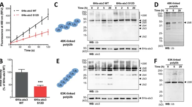
We investigated the effect of S12 phosphorylation on
atx3 enzymatic activity using in vitro DUB assays. A mutant
of hexahistidine (6His)-atx3 with S12 substituted by a
nega-tively charged aspartate, 6His-atx3 S12D, was generated to
mimic constitutive phosphorylation. Because 6His-atx3 wild
type (WT) and 6His-atx3 S12D were expressed in Escherichia
coli
, nonmutated His-atx3 WT is not phosphorylated. After
pu-rifying the proteins through a series of chromatographic steps as
Figure 1. PolyQ expansion of atx3 causes dendrite and synapse loss in cortical neurons. (A) Rat cortical neuron cultures were transfected with nonex-panded GFP -atx3 28Q and exnonex-panded GFP -atx3 84Q. The panel shows representative fluorescence microscopy images of the cell body and dendritic tree. Bars, 100 µm. (B) Neurons expressing GFP -atx3 84Q WT display a reduced total length of dendritic tracts (n = 19–21, from two independent experiments; t test: **, P < 0.01). (C) Sholl analysis reveals a contraction of dendrites caused by GFP -atx3 84Q expression, comparing with neurons expressing GFP -atx3 28Q (n = 19–21 neurons, from two independent experiments; t test: *, P < 0.05; **, P < 0.01). (D) Excitatory synapses were immunocytochemically detected as instances of PSD -95 and VGL UT1 puncta colocalization (merge); (F) inhibitory postsynaptic terminals were quantified as gephyrin-positive puncta. Panels show representative fluorescence microscopy images of dendritic tracts evidencing synaptic marker puncta. Bars, 5 µm. (E and G) Expres-sion of GFP -atx3 84Q causes a decrease in the number of excitatory synapses (n = 31–32 neurons, from three independent experiments; t test: ***, P < 0.001) and inhibitory postsynaptic terminals (n = 30–31 neurons, from three independent experiments; t test: ***, P < 0.001), compared with expression of GFP -atx3 28Q. (B, C, E, and G) Graph bars represent mean ± SEM .
on November 2, 2017
jcb.rupress.org
previously described (Gales et al., 2005), they were incubated
with the substrate Ub-C-terminal 7-amino-4-methylcoumarin
(Ub-AMC ), and the fluorescence yielded by free AMC was
re-corded as a measure of product formation. The respective
reac-tion curves reveal that the substrate cleavage rate is higher for
6His-atx3 WT comparing with the phosphomimetic 6His-atx3
S12D (Fig. 4 A). The decreased activity of the phosphomimetic
form is expressed by a 43.6 ± 4.7% (SEM ) reduction of the initial
reaction velocity, when compared with the control (Fig. 4 B).
Incubation of 6His-atx3 WT or 6His-atx3 S12D with
K48- and K63-linked chains of six Ub monomers led to the
formation of Ub species of lower molecular weight in a time-
dependent manner, as assessed by Western blot (Fig. 4, C–F).
Consistent with a reduced substrate proteolysis, 6His-atx3
Figure 2. S12 of atx3 is phosphorylated in neurons. (A) HEK 293FT cells were transfected with GFP -atx3 28Q and stimulated with phorbol-12-myristate 13-acetate (PMA ), sodium orthovanadate (SOV ), or okadaic acid (OA) to increase protein phosphorylation levels. GFP -atx3 28Q was immunoprecipitated, separated by SDS -PAG E, and the resulting gel was stained with Coomassie blue, as represented. GFP -atx3 28Q bands (marked with *) were excised and subjected to tryptic digestion and mass spectrometry analysis. (B) Online liquid chromatography followed by ion spray mass spectrometry (LC-ES-MS) de-tected one phosphorylated peptide in the sample prepared after okadaic acid stimulus and mapped the phosphorylation site to S12. Human atx3 variant MJD 1a amino acid sequence (available from GenBank under accession no. AAB33571.1) is shown with the detected phosphopeptide colored in red. (C) A phosphospecific antibody recognizing phosphorylated atx3 S12 was produced. The diagram represents the positions of the S12-containing antigen used during antibody production and the epitope recognized by the anti-atx3 antibody (clone 1H9). (D) Western blot probing of cortical neuron lysates with the anti-Patx3 antibody yielded a protein band corresponding to full-length endogenous atx3 that is absent from cells transduced with atx3-targeting shRNA (sRNA -atx3). Actin was used as a loading control. (E) The endogenous atx3 band displays no immunoreactivity when cell lysates are prepared without phosphatase (PP) inhibitors. (F) Probing of human MJD patient fibroblasts (MJD 1–3) and a matched control with the anti-Patx3 antibody reveals a similar protein band pattern to that of neurons. exp., expanded; nonexp., nonexpanded. (G) Increased resolving of patient MJD 2 fibroblast sample distinguishes two bands that match the size of expanded (exp.) and nonexpanded (nonexp.) atx3.
on November 2, 2017
jcb.rupress.org
S12D elicited a decreased formation of Ub products
compar-ing with 6His-atx3 WT. This was observed for both types of
chains; however, the difference is more prominent in the
reac-tion with the K63-linked chains.
Collectively, these in vitro assays demonstrate that the
presence of a negatively charged aspartate at position 12
de-creases atx3 DUB activity, suggesting that such is the effect of
S12 phosphorylation on atx3 DUB activity.
Mimicking S12 phosphorylation counters morphologic changes caused by expression of pathogenic atx3 in neuronal cultures
We tested whether phosphorylation of S12 interfered with the
neuromorphologic defects caused by expanded atx3 expression
in cortical neurons. Cultures were transfected with
phosphomi-metic GFP -atx3 84Q S12D or nonphosphorylatable GFP -atx3
84Q S12A (Figs. 5 A and S2), and total dendritic length was
analyzed aspreviously described (see first pragraph of Results).
The GFP -atx3 84Q S12A mutant exhibited similar effects to
those of GFP -atx3 84Q WT, but neurons expressing GFP -atx3
84Q S12D had higher total dendritic length, showing no
differ-ences relative to the total dendritic length of cells expressing
GFP -atx3 28Q WT (Fig. 5, A and B). Nonexpanded GFP -atx3
28Q S12A also led to an increased toxicity comparing with the
other nonexpanded forms. These results suggest that
phosphor-ylation of S12 protects against dendritic tract loss caused by
atx3 polyQ expansion and that compromising this modification
is enough to cause a loss of dendritic tracts.
We then investigated whether the S12 phosphomutations
affected the number of synaptic contacts in the cultures. Neurons
expressing GFP -atx3 84Q S12D show no difference in the
num-ber of excitatory glutamatergic synapses (Fig. 6, A and C) and
in-hibitory postsynaptic terminals (Fig. 6, B and D) compared with
GFP -atx3 28Q WT, suggesting that atx3 phosphorylation at S12
rescues the deleterious outcomes of expanded atx3 expression.
The differential effects caused by mutation of S12
hint to the fact that phosphorylation of this amino acid
resi-due counters the morphologic defects caused by expanded
atx3 expression in neurons.
Atx3 S12 modulates toxicity induced by pathogenic atx3 in cultured neurons and in vivo
Given the recognized role of aggregation in the toxicity
path-ways involved in polyQ expansion diseases, we tested whether
the S12 phosphomutations affected atx3 tendency to aggregate.
It has been demonstrated that atx3 JD is prone to self-assemble
in vitro, modulating aggregation of both expanded and
nonex-panded atx3 (Masino et al., 2004, 2011; Gales et al., 2005;
El-lisdon et al., 2006); changes caused by phosphorylation at S12
could therefore modulate the dynamics of atx3 aggregation.
Cortical neurons were transfected with GFP -tagged WT or
phosphomutants S12D or S12A atx3 and scanned in search for
GFP -positive accumulations. Cells were counted as having GFP
-atx3 aggregation when they presented at least one noticeable
instance of GFP -positive accumulation, regardless of size; cells
Figure 3. S12 localizes in the vicinity of atx3 catalytic site. (A) S12 (yellow) is positioned in the groove between the two subdomains of the JD, (B) in close proximity to the amino acids of the catalytic triad—C14, H119, and N134—and to the residue proposed to form the oxyanion hole—Q9 (PDB ID 1YZB; Nicastro et al., 2005). (C and D) Binding of an Ub molecule to the Ub-binding site 1 of the JD positions the C-terminal region of Ub close to S12 (PDB ID 2JRI; Nicastro et al., 2009). The structures represented in (A–D) were prepared with PyMOL (http ://www .pymol .org). (E) S12 (arrowhead) is conserved in vertebrate atx3 but is substituted by a phenylalanine residue in atx3L and by a glutamate residue in Josephin-1 and -2. Protein sequences were obtained from the UniProt database, aligned with the Crustal Omega online tool (http ://www .ebi .ac .uk /Tools /msa /clustalo /) and displayed with ESP ript 3.0 (Robert and Gouet, 2014). Strictly conserved residues are represented by white letters in a black background and highly similar residues are
represented by bold letters in framed columns.
on November 2, 2017
jcb.rupress.org
displaying only diffuse GFP signal were counted as having no
aggregates (Fig. 7 A). Expanded GFP -atx3 84Q WT displays
a profoundly increased tendency to aggregate compared with
GFP -atx3 28Q WT (Fig. 7 B), as expressed by the percentage
of cells that presented aggregates in the context of the overall
population of transfected cells (GFP -atx3 aggregates in 48.31
± 10.25% of the cells; n = 5). This is in agreement with the
well-known association between the tendency to aggregate and
the length of the polyQ sequence, serving as validation to this
aggregation assay. Mutation of S12 to aspartate or alanine
de-creases the fraction of neurons with aggregates, reaching
statis-tical significance in cells transfected with GFP -atx3 84Q S12A
(Fig. 7 B). The results suggest that S12 contributes to atx3
ag-gregation and toxicity in neurons and that modification of this
amino acid decreases these effects. The DUB inactive mutant
GFP -atx3 84Q C14A (Burnett et al., 2003) exhibits a tendency
to aggregate comparable with that of GFP -atx3 84Q WT (Fig.
S3), suggesting that the effect of atx3 phosphorylation on atx3
aggregation is not related to its effect on proteolytic activity.
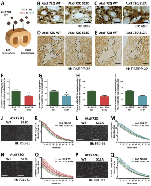
Finally, we explored the changes caused by S12
phosphor-ylation on the pathogenic events occurring on a lentiviral
ani-mal model of MJD with striatal pathology, generated by direct
delivery of viral particles encoding full-length human expanded
atx3 (72Q) to the striatum of adult animals by stereotaxic
injec-tion (Alves et al., 2008b). For each animal, particles encoding
atx3 72Q WT were injected into the striatum of the brain’s left
hemisphere, whereas the striatum of the right hemisphere was
injected with particles encoding either atx3 72Q S12D or atx3
72Q S12A (Figs. 8 A and S4). Animals were euthanized 4 wk
after injection, and coronal brain slices of the whole striatal
re-gion were immunohistochemically processed and analyzed.
Slices were labeled with an anti-atx3 antibody, and the
regions of the striatum demonstrating atx3 aggregation were
imaged and the number of inclusions was quantified (Alves et
al., 2008a; Nascimento-Ferreira et al., 2011). Transduction of
the rat striatum with atx3 72Q led to the formation of
detect-able aggregates (Fig. 8, B and C), in agreement with what has
been described for this animal model. Mutation of S12 to
aspar-tate or alanine significantly decreased the amount of inclusions
formed, comparing with atx3 72Q WT (Fig. 8, F and G).
Examination of the severity of neurodegeneration caused
by each atx3 72Q form was performed by labeling the slices
with an antibody detecting a neuronal marker: dopamine- and
cyclic AMP -regulated phosphoprotein of 32 kD (DAR PP-32).
Expression of atx3 72Q led to a localized loss of anti–DAR
PP-32 antibody immunoreactivity, compatible with the toxicity
expected of atx3 with a pathogenic polyQ expansion (Fig. 8,
D and E). Nonetheless, the volume of the DAR PP-32–depleted
region was reduced in the case of the injections with the
phos-phomutants, comparing with atx3 72Q WT (Fig. 8, H and I).
Excitatory synapses were labeled in striatal slices with
antibodies detecting the postsynaptic protein PSD -95 and the
presynaptic vesicular transporter VGL UT1, and the
immunore-activity signal of each marker was imaged in the vicinity of atx3
72Q–expressing neurons (Fig. 8, J, L, N, and P). The integrated
density of PSD -95 and VGL UT1 agglomerates was quantified
through a range of detection thresholds and found to be
in-creased in the striatal slices expressing phosphomutated (S12A
or S12D) atx3 72Q, comparing with slices expressing atx3 72Q
WT (Fig. 8, K, M, O, and Q), suggesting that phosphorylation
of S12 decreases excitatory synapse loss associated with the
ex-pression of expanded atx3 in the rat striatum.
Figure 4. Mimicking S12 phosphorylation decreases atx3 DUB activity in vitro. (A) Recombinant 6His-atx3 was incubated with Ub-AMC in vitro, and product formation was assessed by measuring AMC fluorescence. 6His-atx3 S12D shows a decreased Ub-AMC cleavage rate, comparing with 6His-atx3 WT. The panel shows the reaction curve of initial product formation of a representative experiment (n = 3 technical replicates, dots represent mean ± SEM fluorescence values, after subtracting the value of buffer and Ub-AMC only; RFU , relative fluorescence units; two-way ANO VA detected a P < 0.0001 significant difference between protein samples). (B) The initial reaction velocity of 6His-atx3 S12D is decreased to 43.6 ± 4.7% in relation to 6His-atx3 WT (n = 8 technical replicates normalized to 6His-atx3 WT, from three independent experiments; graph bars represent mean ± SEM ; t test: ***, P < 0.001). (C and E) In vitro incubation of 6His-atx3 with K48- or K63-linked hexameric polyUb chains (6Ub) leads to a time-dependent formation of lower molecular weight Ub species (Ub2–5), as assessed by Western blot labeling of Ub species. 6His-atx3 S12D elicits less polyUb cleavage than 6His-atx3 WT. (D and F) PolyUb chains showed no time-dependent degradation in the absence of 6His-atx3.
on November 2, 2017
jcb.rupress.org
The observations made in the MJD lentiviral rat model
indicate that phosphomutation of S12 decreases expanded atx3
aggregates and cytotoxicity, in vivo.
Discussion
Approaches to MJD treatment may benefit from a deepened
comprehension of the reversible pathways that interfere with
the cellular defects caused by pathogenically expanded atx3.
This study demonstrates that polyQ expansion of the protein
causes a loss of dendrites and synapses that is countered by
sim-ulating phosphorylation of the protein at S12. Importantly,
mu-tation of this novel phosphorylation site reduces the tendency
of pathogenic atx3 to aggregate and induce neuronal demise in
vivo, suggesting that phosphorylation of S12 of atx3 may have
a protective effect in the context of MJD (Fig. 9).
The phosphospecific anti-Patx3 antibody that was
gen-erated detected phosphorylated atx3 in cortical neurons and
human fibroblasts for the first time, indicating that
phosphory-lation of S12 is physiologically relevant. We investigated
neu-ron-specific changes in cellular morphology in cortical cultures,
considering the growing evidence implicating the cerebral
cor-tex in the pathogenic mechanisms (Yamada et al., 2001; Alves
et al., 2008b; Pedroso et al., 2013; Rüb et al., 2013): authors
have described reduced glucose metabolism (Soong et al.,
1997; Taniwaki et al., 1997) and atrophy in the cortex of MJD
patients’ brain (Murata et al., 1998; D’Abreu et al., 2012; Lopes
et al., 2013) and the presence of polyQ inclusions (Yamada et
al., 2001; Ishikawa et al., 2002). Although cortical neurons are
not frequently found to visibly degenerate, it is possible that
they are dysfunctional in MJD ; the presence of inclusions may
reflect this dysfunction, even if it is not causatively involved
(Schöls et al., 2004). In our experiments, the presence of an
expanded polyQ sequence in transfected GFP -atx3 led to a
mor-phologic phenotype, with contracted dendrites, fewer excitatory
synapses, and a decreased number of inhibitory postsynaptic
terminals. Previous studies using neurons from transgenic
Dro-sophila
larvae have detected dendritic abnormalities
result-ing from expanded atx3 expression (Lee et al., 2011), and a
severe impairment of dendritic arborization was also recently
described to occur in the Purkinje cells of a MJD mouse model
(Konno et al., 2014). Our results constitute the first evidence that
full-length expanded atx3 causes an observable dendritic loss/
shrinkage and synapse loss in mammalian neuron cultures,
sug-gesting that similar events may take place in MJD patient brains.
The changes we observe may also occur in brain regions other
than the cortex, representing a wider neuronal-specific
mech-anism of cellular dysfunction. The possibility of a disruption
of neuronal structure—and, consequently, function—without or
before cell death triggering supports the prevalent idea of an
in-volvement of synaptic dysfunction in the pathogenesis of MJD
(Chou et al., 2008; Boy et al., 2010; Shakkottai et al., 2011).
Mutation of S12 to negatively charged aspartate reduced
the dendritic demise and synaptic loss caused by
polyQ-ex-panded atx3 expression in cortical neurons; contrastingly, the
effects of the phospho-null mutant were similar to those of the
nonmutated expanded atx3. These results suggest that
phos-phorylation at S12 may be able to counter the cellular defects
caused by atx3 expansion. Additionally, the fact that these
defects are reversible on mutation of a single residue, which
is distant from the polyQ stretch, suggests that atx3 may be
functionally involved in the development or maintenance of
dendrites and synapse in vivo. Our results suggest that S12
phosphorylation contributes to this function, which would be
disrupted on polyQ expansion. Thus far, atx3 has never been
demonstrated to play any clear function which would be critical
for neuronal survival and activity, but it was recently reported to
be involved in neuronal differentiation of neuroblastoma cells
(Neves-Carvalho et al., 2015). Atx3 involvement with dendrite
and synapse maintenance may be mediated through its reported
participation in cell pathways, including transcriptional
regula-tion (Rodrigues et al., 2007; Chou et al., 2008), protein quality
control, and cytoskeletal organization (Rodrigues et al., 2010).
Future studies will be needed to determine if dendrite retraction
and synaptic loss on atx3 expansion result from the disturbance
of any of these functions.
Figure 5. Mimicking S12 phosphorylation reduces dendritic tract loss caused by expression of expanded atx3 in cortical neurons. (A) Rat cortical neuron cultures were transfected with phosphomutants S12D and S12A of GFP -atx3 28Q and GFP -atx3 84Q. Panels show representative fluores-cence microscopy images of the cell body and dendritic trees of trans-fected neurons. Bars, 100 μm. (B) Expression of phospho-null GFP -atx3 84Q S12A causes a reduction of total dendritic length, but transfection with GFP -atx3 84Q S12D elicits no differences, comparing with the GFP -atx3 28Q WT control (n = 16–21 neurons, from two independent experi-ments; graph bars represent mean ± SEM ; comparison with GFP -atx3 28Q WT was performed with the Mann-Whitney t test: **, P < 0.01; ***, P < 0.001; two-way analysis of variance followed by Bonferroni post hoc test
compared between other pairs of conditions: #, P < 0.05).
on November 2, 2017
jcb.rupress.org
The in vitro assays show that S12 phosphorylation
reg-ulates atx3 DUB activity, adding to the previously described
effect of ubiquitination of K117 at the JD, which enhances the
DUB activity of the protein (Todi et al., 2010). In the cell
envi-ronment, conjugation of a phosphate group to S12, localized in
the catalytic domain of atx3, might affect the neighboring amino
acid residues, as a consequence of steric constraints and/or
im-posed by the negative charge of the phosphate group. Changes
in the amino acids comprising the catalytic triad or alterations
that could affect substrate binding and/or positioning, such as
in the conformation of the closely positioned Ub-binding site
1 of the JD helical hairpin, might result in changes of
proteo-lytic activity (Nicastro et al., 2005, 2009; Hunter, 2007; Tarrant
and Cole, 2009; Huang et al., 2012; Renatus and Farady, 2012).
Notably, the crystal structure of the complex formed by Ub and
the ataxin-3–like protein (atx3L) JD revealed that the
phenylal-anine at position 12 forms a hydrophobic enclosure with other
aromatic amino acid around the active center, through which the
C terminus of the Ub molecule threads (Weeks et al., 2011). The
position appears to be suitable for interaction with the lysine
side chain forming the isopeptide bond. It has been reported
that mutation of S12 of the atx3 JD to a phenylalanine increases
proteolysis of Ub substrates (Weeks et al., 2011), consistent
with the relevance of this residue in determining atx3 activity
that our results propose. Phosphorylation of S12 may therefore
modulate important aspects of atx3 biologic activity, such as
the cleavage rate of particular atx3 substrates and/or the
molec-ular interactions established by the protein. The consequences
of these changes may have different cellular outcomes, possibly
related to the maintenance of specialized neuronal structures.
S12 is highly conserved in atx3 vertebrate homologues, but
is substituted by different residues in other JD-containing proteins:
a negatively charged glutamate in Josephin-1 and -2 and an
aro-matic phenylalanine in atx3L (Fig. 3 E). In agreement with our
results showing that S12 phosphorylation decreases the proteolytic
activity of atx3, the activity of the JDs isolated from human atx3,
atx3L, Josephin-1, and Josephin-2 has been shown to be markedly
different when tested against several different Ub model substrates
in vitro (Weeks et al., 2011). Although the variations reported may
be attributed to other sequence or structural differences between
the protein JDs, it is possible that the residue occupying the
posi-tion of S12 plays a role in modulating enzyme activity.
Figure 6. Mimicking S12 phosphorylation reduces synapse loss elicited by expanded atx3 expression in cortical neurons. (A and B) Rat cortical neuron cul-tures were transfected with phosphomutants S12D and S12A of GFP -atx3 28Q and GFP -atx3 84Q. The panels show representative fluorescence microscopy sections from dendritic tracts of GFP -atx3–express-ing neurons, evidenc-atx3–express-ing (A) glutamatergic synapse markers VGL UT1 and PSD -95–positive puncta and the respective colocalization (merge) or (B) gephyrin-posi-tive puncta. Bars, 5 μm. (C) Expression of the GFP -atx3 84Q S12A mutant causes a decrease in the number of functional glutamatergic synapses, comparing with the expression of GFP -atx3 28Q WT. Phosphomimetic GFP -atx3 84Q S12D causes no significant reduction (n = 24–32 neurons, from three independent experi-ments; graph bars represent mean ± SEM ; compari-son with GFP -atx3 28Q WT was performed with the Mann-Whitney t test: **, P < 0.01; ***, P < 0.01; two-way analysis of variance followed by Bonferroni post hoc test compared between other pairs of
condi-tions: #, P < 0.05; ##, P < 0.01). (D) GFP -atx3 84Q
S12D expression does not produce the loss of inhibi-tory postsynaptic terminals caused by GFP -atx3 84Q WT and S12A, as inferred from the number of gephy-rin-containing puncta (n = 24–30 neurons, from three independent experiments; graph bars represent mean ± SEM ; comparison with GFP -atx3 28Q WT was per-formed with the Mann-Whitney t test: ***, P < 0.001; two-way analysis of variance followed by Bonferroni post hoc test compared between other pairs of
con-ditions: ###, P < 0.001).
on November 2, 2017
jcb.rupress.org
Mutation at serine 12 to aspartate or alanine reduces
the tendency of expanded GFP -atx3 to form aggregates in
cultures neurons. It has been hypothesized that atx3
aggrega-tion is dependent on the molecular interacaggrega-tions it establishes,
and in vitro association of the JD with Ub protects against
aggregation (Song et al., 2010; Masino et al., 2011). The two
Ub-binding sites in the catalytic JD overlap with the regions
described as taking part in JD self-assembly (Nicastro et al.,
2009; Masino et al., 2011), and interestingly, S12 is in close
proximity to the Ub-binding site 1. Taking all this into
con-sideration, mutation at S12 may reduce aggregation by
in-terfering with the interactions modulated by Ub-binding site
1, possibly favoring interactions that protect against atx3
ag-gregation. The fact that the inactive C14A mutant leads to an
aggregation profile similar to that of GFP -atx3 84Q supports
the idea that this effect on aggregation is not related to the
decrease in DUB activity caused by phosphorylation.
Further-more, phosphomutation of S12 does not alter atx3 subcellular
localization (in COS -7 cells; Fig. S5, A–D), indicating that the
decrease on aggregation may not involve a decreased nuclear
distribution of the protein.
When the phosphomutants of atx3 are expressed in the
striatum of injected rats, they lead to a decreased inclusion
for-mation and neurodegeneration comparing with the nonmutated
protein, suggesting that S12 modification has a protective effect
in vivo. This similar effect of the phosphomimetic and the
phos-pho-null mutations is not unique. In a study describing the effects
of huntingtin phosphorylation at T3, although each
phosphomu-tant of T3 had opposed tendencies to aggregate, both mutations
were neuroprotective in a Drosophila model, reducing lethality
and neurodegeneration (Aiken et al., 2009). Our results indicate
that the availability of S12 is an important factor contributing to
toxicity-related events in neurons. We suggest that
nonphosphor-ylated S12 of expanded atx3 may contribute to pathways turning
the protein more toxic, precipitating events such as misfolding,
cleavage, or aggregation. Consequently, modifications targeting
S12 may reduce the toxicity of expanded atx3, with possible
pro-tective outcomes. In a biologic context, the modifications caused
by phosphorylation of S12 are likely to influence pathogenic
mechanisms mediated by this amino acid. Phosphorylation of
S12, as a biologic device that actually turns it into a different
chemical entity, could have an effect of “turning off” the toxic
mechanisms in which S12 takes part.
The ameliorating effects of the phosphomimetic
muta-tions in our cell culture and in vivo models of MJD strongly
suggest that S12 phosphorylation is protective against the
toxicity of expanded atx3. Further studies will inform on the
usability of the modulation of S12 phosphorylation state as a
target for MJD therapy.
Materials and methods
Expression plasmids and lentiviral vectors
Eukaryotic expression pEGF P-C1 plasmids encoding human atx3 vari-ant MJD 1a (available from GenBank under accession no. AAB33571.1) with 28 glutamines (GFP -atx3 28Q) or 84 glutamines (GFP -atx3 28Q) N terminally fused with GFP (Chai et al., 1999) and pFLA G-CMV -6a plasmids encoding human atx3 variant MJD 1-1 (available from Gen-Bank under accession no. NP_004984.2) with 22 glutamines (FLA G-atx3 22Q) or 80 glutamines (FLA G-atx3 80Q) N terminally fused with FLA G were a gift from H. Paulson (University of Michigan, Ann Arbor, MI). Empty vector pEGF P-C1 was obtained from BD Biosci-ences (available from GenBank under accession no. U55763). Eukary-otic expression plasmid SIN -W-PGK encoding human atx3 variant MJD 1a with 72 glutamines and an N-terminal Myc tag (atx3 72Q) and plasmid SIN -CW-PGK -nls-LacZ-LTR -TRE encoding an universal short hairpin targeting human and rat atx3 (shRNA -atx3) were previ-ously produced by our group (Alves et al., 2008b, 2010). Constructs used in the viral production (pCMV DR-8.92, pMD.G, and pRSV -Rev) have been described previously (de Almeida et al., 2002) and were a gift from N. Deglon (University Hospital of Lausanne, Lausanne, Switzerland). Bacterial expression pDES T17 plasmid encoding N-ter-minally 6His-tagged human atx3 isoform MJD 1-1 with 13 glutamines (6His-atx3) was previously generated by our group (Gales et al., 2005).
Site-directed mutagenesis of the expression constructs was performed using the QuickChange Site-Directed Mutagenesis kit (Stratagene) or the QuikChange II XL Site-Directed Mutagenesis kit (Agilent Technologies). Phosphomimetic S12D mutants, with aspartate at position 12, were generated using primers (forward) 5′-CAC GAG AAA CAA GAA GGC GAC CTT TGT GCT CAA CAT TGC CTG -3′ and (reverse) 5′-CAG GCA ATG TTG AGC ACA AAG GTC GCC TTC TTG TTT CTC GTG -3′. Phospho-null S12A mutants, with alanine at
posi-Figure 7. Mutating S12 decreases expanded atx3 aggregation in cor-tical neurons. (A) GFP -atx3 accumulates in the cell body of a fraction of transfected cortical cultures. The number of cells with GFP -atx3 aggre-gates was counted versus the number of cells presenting only diffuse GFP -atx3 signal; neurons displaying a compromised structure were excluded from the counting. The panels illustrate the diversity of the aggregates ob-tained. Bar, 20 μm. (B) Mutating S12 of GFP -atx3 84Q decreases the fraction of cells with aggregates comparing with what is caused by GFP -atx3 84Q WT. The decrease reaches statistical significance with GFP -atx3 84Q S12A (17–59 neurons were counted for each condition in n = 5 independently prepared cultures; graph bars represent mean ± SEM ; one- sample t test: **, P < 0.01).
on November 2, 2017
jcb.rupress.org
tion 12, were obtained with primers (forward) 5′-CAC GAG AAA CAA GAA GGC GCA CTT TGT GCT CAA CAT TGC CTG -3′ (reverse) 5′-CAG GCA ATG TTG AGC ACA AAG TGC GCC TTC TTG TTT CTC GTG -3′. DUB activity-deficient C14A mutants, with alanine at position 14,
were produced with primers (forward) 5′-GAG AAA CAA GAA GGC TCA CTT GCT GCT CAA CAT TGC CTG -3′ and (reverse) 5′-CAG GCA ATG TTG AGC AGC AAG TGA GCC TTC TTG TTT CTC -3′. Mutation was confirmed after automatic DNA sequencing (STA B VID A).
Figure 8. Mutating S12 reduces the neurodegen-erative phenotype of a MJD lentiviral rat model. (A) Atx3 72Q–encoding lentiviral particles were stereotaxically delivered into rat striata: the left hemisphere striatum was injected with atx3 72Q WT and the right hemisphere with one of its phos-phomutants—S12D or S12A. (B and C) Atx3 was immunohistochemically (IH) labeled and atx3 ac-cumulations were quantified. Representative im-ages of the peroxidase staining of striatal sections displaying atx3-positive aggregates are shown. Bars, 50 μm. (F and G) Mutating S12 reduces the amount of atx3 accumulations caused by atx3 72Q expression (n = 7 animals injected with atx3 72Q WT: atx3 72Q S12D; n = 6 animals injected with atx3 72Q WT: atx3 72Q S12A; graph bars represent mean ± SEM ; Mann-Whitney test: *, P < 0.05; **, P < 0.01). (D and E) Neuronal loss was assessed by determining the volume of the
DAR PP-32 immunoreactivity-depleted region in
each hemisphere. Representative images of the peroxidase staining of striatal sections evidencing immunoreactivity loss (interrupted line) are shown. Bars, 300 μm. (H and I) Mutating S12 of atx3 reduces the lesion caused by atx3 72Q injection into the rat striatum, as evaluated by DAR PP-32 immunostaining (n = 4 animals injected with atx3 72Q WT: atx3 72Q S12D; n = 5 animals injected with atx3 72Q WT: atx3 72Q S12A; graph bars represent mean ± SEM ; Mann-Whit-ney test: *, P < 0.05; **, P < 0.01). (J, L, N, and P) Excitatory synapse markers PSD -95 and
VGL UT1 were immunohistochemically labeled
and imaged in the vicinity of atx3 72Q–express-ing neurons. Panels show representative confocal images of PSD -95 (J and L) and VGL UT1 (N and P) punctuate immunostaining. Bars, 5 µm. (K, M, O, and Q) The integrated density of PSD -95 and VGL UT1 agglomerates is increased in the stria-tal slices expressing phosphomutated atx3 72Q, comparing with slices expressing atx3 72Q WT, throughout the range of detection thresholds used (n = 3 animals for each injection condition; graph bars represent mean ± SEM ; two-way analysis of variance detected a P < 0.001 significant differ-ence between hemispheres).
Figure 9. Phosphorylation of atx3 at S12 counters toxic polyQ expansion–derived changes. Atx3 is phos-phorylated at S12, an amino acid localized in the catalytic JD of the protein. Current results suggest that this modification decreases the hydrolytic activity of atx3 against polyUb chains. S12 phosphorylation in atx3 reduces expanded atx3 aggregation, decreases dendritic and synaptic loss caused by the expanded protein in neurons, and limits neurodegeneration.
on November 2, 2017
jcb.rupress.org
Cell line culture and transfection
HEK 293FT cells were grown in DME M, pH 7.2, supplemented with 44 mM NaHCO 3, 10% FBS , 0.1 mM nonessential amino acids, 6 mM
l-glutamine, 1 mM sodium pyruvate, and 500 µg/ml geneticin. COS -7 cells were cultured in DME M, pH 7.2, supplemented with 44 mM NaCO3, 10% FBS , and 1% penicillin-streptomycin. Both cell lines were
maintained at 37°C, in a humidified atmosphere containing 5% CO2.
One day before transfection, fully confluent cells were diluted (1:6) and seeded onto 100-mm culture plates, and transient trans-fection was performed after 24 h of cell growth, using the Lipofect-amine Transfection Reagent for HEK 293FT cells (Life Technologies) or the Lipofectamine LTX Transfection Reagent (Life Technologies) for COS -7 cells. Expression was left to occur for 24–48 h before extracts were prepared.
Viral production
Lentiviral vectors encoding atx3 72Q or shRNA -atx3 were produced in HEK 293T cells using the four-plasmid system described previously (de Almeida et al., 2002). In brief, HEK 293T cells (cultured as de-scribed for COS -7 cells) were plated into 100-mm dishes (4 × 10⁶ cells/
dish) and subjected the following day to transient calcium phosphate transfection. DNA complexes were formed by mixing 0.5 M CaCl2
with the DNA constructs (for each plate: 13 µg of the pCMV DR-8.92 packaging construct, 3.75 µg of pMD2G, 3 µg of pRSV -Rev, and 13 g of SIN -W-PGK -ATX 3 72Q WT, S12D or S12A, or shAtaxUNI V), to a final concentration of 0.25 M CaCl2. The solution was stirringly mixed
with an equal volume of Hepes-buffered solution (280 mM NaCl, 1.5 mM Na2HPO 4, and 100 mM Hepes, pH 7.1), and 1 ml of DNA
complexes was added to each dish. Cells were incubated at 37°C, in a humidified atmosphere containing 3% CO2, for 6 h, after which
trans-fection media was replaced with fresh culture medium. After 48 h of culture incubation at 37°C, 5% CO2, the supernatants were collected,
filtered (0.45 µm Stericup Filter Unit; Merck Millipore), concentrated by ultracentrifugation (90 m, 19.000 g, 4°C), and resuspended in PBS with 1% BSA . When the viral particles were aimed at animal striatal injections, this was followed by another ultracentrifugation step before final resuspension in the same solution. The content level of batches was assessed by p24 antigen ELI SA (Retrotek HIV -1 p24 Antigen ELI SA; Zepto Metrix), and stocks were stored at –80°C.
Immunoprecipitation and mass spectrometry analysis
HEK 293FT cells were stimulated with 20 nM okadaic acid for 16 h, 5 mM sodium orthovanadate for 30 min, or 200 nM PMA for 20 min. Cells were scraped unto immunoprecipitation buffer (20 mM Tris, pH 7.0, 100 mM NaCl, 2 mM EDT A, 2 mM EGT A, 50 mM NaF, and 1 mM Na3VO4), supplemented with 1% Triton X-100, 1 µM okadaic acid, and
protease inhibitors (1 mM DTT , 0.1 mM PMS F, 10 µg/ml chymosta-tin, pepstachymosta-tin, antipain, and leupeptin [CLA P]), sonicated, and cleared of the insoluble fraction by centrifugation. 1 mg of protein from each extract (1 mg/ml) was used for immunoprecipitation with 3 µg of an-ti-GFP antibody (clones 7.1 and 13.1; Roche Applied Science). Protein samples were eluted with 50 µl SDS -PAG E 2× loading buffer (125 mM Tris, pH 6.8, 100 mM glycine, 40% glycerol, 4% SDS , 200 mM DTT , and ∼0.01% bromophenol blue). Three samples of each extract were subjected to the i.p. procedure, but collectively eluted in sequence.
Immunoprecipitants (100 µl) were alkylated (1% acrylamide, 20 min) and separated by SDS -PAG E. Online liquid chromatography followed by ion spray mass spectrometry analysis of the protein bands was performed by the Proteomics Center at the Children’s Hospital Boston. Samples were digested in-gel with 12.5 ng/µl trypsin, over-night, at 37°C, and resulting peptides were extracted with 100 mM am-monium bicarbonate and acetonitrile and lyophilized. Samples were
resuspended in 5% acetonitrile, 5% formic acid, and directly injected into a liquid chromatography –mass spectrometry system encompass-ing a microautosampler, a Suvery high performance liquid chromatog-raphy pump, and a Proteome X (LTQ ) mass spectrometer (all acquired from Thermo Finnigan). Peptides were eluted with 0.1% formic acid in acetronitrile (30 min linear gradient 8–34% acetonitrile) and applied to the mass spectrometer by electrospray ionization. The mass spec-trometric data obtained were searched against the human international protein index database (IPI human 336) using the protein identification software Mascot (version 2.2.04; Matrix Sciences), according to the ap-propriate search criteria necessary for the detection of phosphorylation. Generation of the anti-Patx3 phosphospecific antibody
Production of the phosphospecific anti-Patx3 antibody was performed by Eurogentec. Rabbits were immunized with a phosphorylated atx3-based peptide—8KQE G-S(PO3H2)-LCA QHC LN20—coupled with the
keyhole limpet hemocyanin protein carrier. Phosphospecific antibody purification was performed on the serum of the best responding animal (as evaluated by ELI SA) through affinity chromatography procedures. Cortical neuron cultures preparation and transfection
Cortices of Wistar rat embryos with 18 d were dissected, trypsinized, and resuspended in neuronal plating medium (MEM supplemented with 10% horse serum, 0.6% glucose, and 1 mM sodium pyruvate). Cells were passed through a 0.2-µm filter and then seeded in high, medium, or low density, according to the experimental objectives: high-density cultures: 9 × 10⁵ cells/well in six-well culture plates coated with poly-d-lysine
(0.1 mg/ml); medium-density cultures: 2.5 × 10⁵ cells/well onto coated
15-mm coverslip-containing 12-well culture plates; and low-density cul-tures (Banker culcul-tures): 3.25 × 10⁵ cells/plate in 60-mm culture plates
containing coated 18-mm coverslips. For high- and medium-density cultures, 2–4 h later, platting medium was substituted by Neurobasal medium supplemented with 2% SM1, 0.5 mM glutamine, and 0.12 mg/ ml gentamycin. In the Banker cultures, after 2–3 h, coverslips were turned over an astroglial feeder layer (grown in MEM supplemented with 10% horse serum, 0.6% glucose, and 1% penicillin-streptomycin) and maintained in supplemented Neurobasal medium. Neuronal cultures were kept in an incubator at 37°C, with a humidified atmosphere con-taining 5% CO2, up to 15 days in vitro (DIV ). High- and medium-density
cultures were fed once a week, and low-density cultures were fed twice a week, by replacing 1/3 of the culture medium with fresh medium.
Expression plasmids were transfected by a calcium phosphate transfection procedure adapted from a previously described protocol (Jiang et al., 2004). In brief, DNA complexes were generated by adding 2.5 M CaCl2 in 10 mM Hepes drop-wise to the plasmid DNA
solu-tions, for a final concentration of 250 mM CaCl2. High-density cultures
were transfected at 9–10 DIV cortical neurons using 10 µg of plasmid DNA ; medium-density cultures were transfected at 4–5 DIV with 4 µg plasmid DNA ; and low-density cultures were transfected at DIV 9–10 with 3 µg DNA . Complexes were mixed with an equal volume of Hepes-buffered transfection solution (274 mM NaCl, 10 mM KCl, 1.4 mM Na2HPO 4, 11 mM dextrose, and 42 mM Hepes, pH 7.2) and
added to the cultures. Cells were incubated for 1–1.5 h, after which transfection medium was substituted by fresh culture medium. Ky-nurenic acid (2 mM) was used throughout the transfection procedure to block ionotropic glutamate receptors. Expression was left to occur for a maximum of 5 DIV under normal cell culture incubation conditions. Atx3 knock-down and neuronal lysate preparation
High-density rat cortical cultures (8 DIV ) were transduced with shRNA -atx3–encoding lentiviral particles (20 ng of p24 antigen/105 cells)
in the presence of 8 µg/ml hexadimethrine bromide. 18 h later, the
on November 2, 2017
jcb.rupress.org
infection medium was substituted by a mixture of conditioned medium and fresh medium (1:2), and expression was left to occur for 6 d under normal culture conditions.
Human fibroblast culture
Human fibroblast cells from MJD patients and a matched control were obtained from Neurology and Pathology Services, University Hospital of Coimbra. Cultures were maintained under 37°C, 5% CO2
incuba-tion, in DME M, pH 7.2, supplemented with 44 mM NaHCO 3, 10%
FBS , 0.1 mM nonessential amino acids, 2 mM l-glutamine, 1 mM so-dium pyruvate, and 1% penicillin-streptomycin.
Cell lysate preparation
Lysates were prepared by scraping cultures onto RIP A buffer (10 mM Tris-HCl, pH 7.2, 150 mM NaCl, 5 mM EDT A, 0.1% Triton X-100, 1% sodium deoxycholate, and 0.1% SDS ), supplemented with protease (1 mM DTT , 0.1 mM PMS F, and 10 µg/ml CLA P) and phosphatase inhibitors (5 mM NaF, 2 mM Na3VO4, and 1 µM okadaic acid),
son-icated, and freeze-thawed. After, protein quantification samples were denatured by adding 5× loading buffer (625 mM Tris-HCl, pH 6.8, 50% glycerol, 10% SDS , 500 mM DTT , and ∼0.01% bromophenol blue) and heating to 95°C.
In vitro activity assays
6His-atx3 produced in BL21(DE3)-SI cells was purified as described previously by our group (Gales et al., 2005). Incubation of protein sam-ples (200 µM) with 0.5 µM Ub-AMC (Boston Biochem) was performed in the presence of 20 mM Hepes, pH 7.5, 5% glycerol, and 1 mM EDT A, 0.1 mg/ml BSA , and 10 mM DTT , in 96 well plates. Product forma-tion was assessed at 30°C by fluorescence recording (excitaforma-tion: 380 nm; emission: 460 nm). For each technical replicate, initial reaction velocity was calculated as the slope of the trend line traced based on the fluorescence values (relative fluorescence units) of the first 2 min of reaction, after subtraction of the value of the negative control lacking 6His-atx3 (with an additional 0.2 M BSA ). The obtained velocity values were normalized to the mean WT velocity of the respective experiment.
Incubation of the samples (100 nM) with 250 nM K48- or K63-linked hexameric polyUb chains (Boston Biochem) was performed after mixing with 50 mM Hepes, 0.5 mM EDT A, 0.1 µg/ml ovalbumin, and 1 mM DTT and left to occur for 20 h, at 37°C. Samples (10 µl) were taken from the reaction mixture at 0, 2, 5, and 20 h and immedi-ately denatured by adding 2× concentrated sample buffer.
SDS -PAG E and Western blot
Equivalent protein amounts of each experimental condition were re-solved and probed by SDS -PAG E followed by Western blot, performed according to standard protocols. Incubation with the primary antibodies diluted in 0.5% milk TBS -Tween 20 (TBS -T) was performed for 1–2 h at RT or overnight at 4°C, using a mouse monoclonal anti-atx3 antibody (1H9, 1:1,000; Millipore), the rabbit polyclonal anti-Patx3 (1:20; pro-duction described under Generation of the anti-Patx3 phosphospecific antibody), a mouse monoclonal anti-actin (β; 1:5,000; Sigma-Aldrich), or a rabbit polyclonal anti-Ub (1:1,000; Dako). The appropriate alka-line phosphatase-conjugated secondary antibodies (goat anti–mouse IgG and mouse anti–rabbit IgG; Jackson ImmunoResearch Laborato-ries) diluted in 0.5% milk TBS -T (1:10,000) were incubated for 1 h at RT. In the case of the anti-Patx3 antibody, 5% low-fat dry milk TBS -T was used in every step instead.
Dendritic morphology analysis
Transfected cortical neurons (low density) with 14–15 DIV were fixed with 4% paraformaldehyde/4% sucrose in PBS for 15 min at RT,
and coverslips were mounted with Fluorescence Mounting Medium (Dako). GFP -atx3–expressing neurons were imaged at RT using an Axiovert 200M microscope (CCD monochromatic digital Axiocam HRm camera; Axiovision software; Carl Zeiss) and a 20× air objec-tive (LD-PlanNeofluar, 0.4 NA; Carl Zeiss), and tracing of the cell body and dendrites was drawn based on the GFP signal, using the Neurolucida software (MBF Bioscience). Sholl analysis and dendritic measurement was performed with the Neurolucida Explorer software (MBF Bioscience). Total dendritic length of the neurons was normal-ized to the mean total dendritic length of neurons transfected with GFP -atx3 28Q WT, in each independent experiment. For each indi-vidual experiment, cells were cultured and immunocytochemically processed on the same occasion. Dendritic morphology analysis was performed blindly to condition.
Synapse quantification
Low-density cultures were fixed asdescribed in the previous para-graph, permeabilized with 0.25% Triton X-100 in PBS and blocked with 10% BSA . Primary antibodies diluted in 3% BSA PBS were incubated overnight at 4°C: the mouse monoclonal anti–PSD -95 (1:200; Affinity BioReagents), the guinea pig polyclonal anti-VGL UT1 (1:100,000; Millipore), and the mouse monoclonal anti-gephy-rin (1:1,000; Synaptic Systems). Coverslips were then incubated with the secondary antibodies for 45 min at 37°C: Alexa Fluor 568 anti– mouse IgG (1:500; Invitrogen) and the Alexa Fluor 647 anti–guinea pig IgG (1:500; Molecular Probes). Coverslips were mounted with Flu-orescence Mounting Medium.
Puncta observation and imaging was performed at RT with an Axio Observer Z1 microscope (CCD digital Axiocam HRm camera; ZEN Blue software; Carl Zeiss) with a 63× oil objective (Plan-Apo-chromat, 1.4 NA), after randomly selecting dendritic tracts (min-imum 10 µm) in GFP -atx3–expressing neurons. Image analysis was performed using Fiji software (Schindelin et al., 2012) in manually thresholded images. Instances of colocalization of pre- and postsyn-aptic markers were quantified as functional synapses. Values were nor-malized per dendritic section length to the mean value of GFP -atx3 28Q WT in the respective experiment. For each individual experiment, cells were cultured and immunocytochemically processed on the same occasion, and images were acquired with the same settings. Synapse quantification was performed blindly to condition.
Quantification of aggregate-containing neurons
Transfected cortical neurons (medium-density cultures) with 8 DIV were fixed as described for the dendritic morpholoy analysis. Count-ing of GFP -atx3 aggregate-containCount-ing neurons was performed blindly to condition by manually scanning coverslips and observing the GFP fluorescence with a Zeiss Axiovert 200M microscope using a 63× oil objective (Plan-Apochromat, 1.4 NA). Aggregation was expressed as the fraction of cells with aggregates, normalized to the values obtained upon transfection with GFP -atx3 84Q WT, in each experiment. In vivo lentiviral injections and MJD rat model generation
Adult male Wistar rats (Charles River Laboratories) were anesthetized and stereotaxically injected for delivery of lentiviral vectors to the stria-tum, following the procedure previously described in Alves et al. (2008b). For each animal, particles (400,000 ng of p24 antigen, 2 µl, 0.2 µl/min) encoding atx3 72Q WT were injected in the left hemisphere and particles encoding atx3 72Q S12D or S12A were injected in the right hemisphere. Animals were housed in a temperature-controlled room and maintained on a 12-h light/dark cycle. Food and water were supplied ad libitum, and proceedings were executed in accordance with European Union Directive 2010/63/EW.