2019
UNIVERSIDADE DE LISBOA
FACULDADE DE CIÊNCIAS
DEPARTAMENTO DE BIOLOGIA ANIMAL
Immunological Characterization of Mucosal-Associated
Invariant T (MAIT) Cells in chronically Hepatitis B infected
patients
Tiago Nuno Ribeiro Alves
Mestrado em Biologia Humana e Ambiente
Dissertação orientada por:
Antony Chen
Maria Gabriela Rodrigues
The work presented in this thesis resulted from a partnership between the University of
Lisboa and Janssen Pharmaceutica NV, Beerse I. All experimental activities were
performed at Janssen Pharmaceutica NV, Beerse I, a Johnson & Johnson pharmaceutical
research and development facility in Beerse, Belgium.
i
Acknowledgements
I would like first to thank my supervisors, Antony Chen and Tine Thoné, for having accepted me as their master´s student and for all their help and supervision during the entire time of the project, as well as demonstrating enormous patience, availability and guidance along the entire process. I also would like to thank the leader of the group, Helen Horton, for accepting me in the ID&V group at Janssen Pharmaceutica and for always being present in answering every question I had and for being so kind and receptive every day during the entire time I stayed in the research group. I would like the entire Helen’s Horton research team for always being there for me every day and every step of my master’s thesis project, with a special thanks to Doreen Verstappen and Jochen Lamote for all the lab work help and all the repetitive questions I made you answer repeatedly. I would like to acknowledge my University of Lisbon supervisor, Professor Gabriela Rodrigues for having accepted me as a master thesis student and the coordinator of the Human Biology and Environment Master Program, Professora Deodália Dias, for all the support and help they have demonstrated along this entire year.
Gostava de agradecer a algumas pessoas que me fizeram o homem que sou e que me acompanharam em 8 anos de faculdade e todo o meu percurso de vida.
Aos meus pais e irmã, por todos os momentos bons e maus e por todo o apoio dado ao longo deste percurso e por terem sido 150% compreensivos durante toda a minha vida e no meu percurso académico até aos dias de hoje. Por todos os momentos em que as forças pareciam faltar e o stress e pressão pareciam tomar conta de mim, tiveram sempre uma palavra de conforto e esperança que foram recebidas como uma lufada de ar fresco e me fizeram continuar. Muito Obrigado
À minha namorada Rafaela, 5 anos de namoro contam muitas histórias mas já são 8 anos de amizade profunda, obrigado por nunca me teres deixado desistir, mesmo quando o fundo do poço parecia estar perto, por todas as conversas, discussões, por todos os risos, choros, conversas em que me chamaste à razão e me fizeste abrir os olhos para continuar a lutar em busca do meu futuro. Um sincero e cheio de amor MUITO OBRIGADO, sem ti acho que não seria a pessoa que sou hoje e cada dia que passa cresço mais e mais ao teu lado.
Às minhas amigas, Marta Brito, Inês Panão, Joana Machado e Raquel Sousa por uma eterna amizade já com mais de 20 anos, que continue por bons e frutuitos anos, muito obrigado. Ao Gonçalo Aguiar, Luis Alves, Sofia Alves e Marta Puga, camaradas de secundário e companheiros de muitas aventuras, muito obrigado. Aos meus amigos escuteiros que a cada caminhada que percorri estiveram presentes, Muito Obrigado. Aos meus amigos Turbinados, grupo que fiz na Facudade de Ciências da Universidade de Lisboa e para o qual um tremendo OBRIGADO basta pois os momentos, as festas, encontros e risadas falam por si.
Um especial OBRIGADO ao meu amigo João Santos, amigo que considero como um irmão e que independentemente do país e do tempo sem nos vermos, sei que sempre poderei contigo para enfrentar esta coisa a que se chama vida.
Por fim gostava de agradecer aos meus avós, Joana Carvalho, Omar Ribeiro, Maria Clarisse Alves, Carlos Alves, que por infortuitos da vida já não se encontram entre nós, mas que onde estiverem sei que estão orgulhosos do pequenino Tiago que conheceram e que se tornou um homem e está prestes a defender a sua tese de mestrado e a ser lançado para o mundo real do trabalho e da vida. Gostava que estivessem presentes e comigo, mas acreditem que nunca vos esqueci, Obrigado.
iii
Resumo
A Organização Mundial de Saúde considera a doença da Hepatite B como a oitava causa de mortalidade a nível mundial e estima que aproximadamente 400 milhões de pessoas estarão infetadas nos dias de hoje. O vírus da hepatite B é caracterizado por ter um tamanho reduzido, possuir uma conformação genómica baseada em 4 “open reading frames” (ORFs), ser transmitido de forma singular e com um modo ação muito efetivo. Pacientes infetados com o vírus da hepatite B, dependendo da idade em que foram infetados, podem ser diferenciados em pacientes agudos ou devido ao historial de progressão da doença ser considerados pacientes crónicas, tendo um risco elevado de virem a contrair uma de duas patologias no futuro – cirrose hepática e cancro do fígado. Actualmente existem dois regimes terapêuticos utilizados contra a hepatite B crónica, sendo estes “Nucleos(t)ide analogues” (NUCs) e tratamentos a base de interferões. Contudo, apesar de promoverem uma cura funcional, estes tratamentos são considerados ineficazes a longo prazo contra a infeção, uma vez que não conseguem erradicar o ADN viral (cccADN) dos hepatócitos infetados e o seu consumo é imprescindível a partir do momento do seu diagnóstico.
À semelhança de outras doenças virais crónicas, como HIV e Hepatite C também pacientes infectados com o virus da Hepatite B devido á exposição do mesmo por tempo indeterminado até ao final da vida, ficam sujeitos a uma continua disfunção a nivel celular. As células T, membros activos do nosso sistema imunitário, após combate contínuo perante a infeção, acabam por perder gradualmente a sua função contra o virus e acabam por entrar num estado de exaustão. Este processo está documentado em vários estudos científicos e sugere que não é necessariamente o grau e intensidade da infeção viral que aumenta, mas sim o próprio sistema imunitário que, por estar em constante esforço, acaba por ser ineficaz em erradicar o virus do corpo humano. No entanto, a comunidade científica está a direcionar esforços para a criação de novas medidas terapêuticas, que passam por fazer um targeting viral e utilizar mecanismos de imunoterapia que promovam a erradicação do vírus via a eliminação do ADN viral das células infetadas.
Como exemplo, as células “mucosal-associated invariant T” (MAIT) são vistas como uma possível nova abordagem imunoterapêutica contra o vírus da hepatite B. As células MAIT constituem um subsistema das células T, sendo caracterizadas por serem inatas, não convencionais e reconhecerem metabolitos de riboflavina microbiana (Vitamina B2). Este reconhecimento acontece no seguimento da secreção de riboflavina microbiana por parte de diversas espécies bacterianas e fungos e são apresentadas as células MAIT por intermédio da molécula não-polimórfica do complexo de histocompatibilidade principal (“major histocompatibility complex”, MHC) classe I (MR1). Estas células estão ainda evolutivamente conservadas entre várias espécies e podem ser encontradas em larga escala nos tecidos da mucosa humana, sangue periférico e fígado, existindo uma correlação direta com a acumulação por parte destas células em locais de infeção e fornecimento de proteção contra infeções. Tendo por base a sua localização celular e a sua frequência no fígado, é sugerido que em ambientes de exaustão imunitária como é o caso da hepatite B, as células MAIT podem subsistir e estar funcionalmente ativas, imitando o papel imunitário das células T contra a infeção.
Adicionalmente, as células MAIT dispõem de diversas funções effectoras quando estimuladas e, consequentemente, activas. A produção de citoquinas pro-inflamatórias, tais como IFN-ү e TNF-α, assim como degranulação, citotoxicidade, proliferação celular (marcador Ki-67) e respostas dependentes/independentes da presença do MR1, previamente referido, são algumas das funções effectoras relevantes levadas a cabo por estas células e que influenciam o seu desempenho junto do corpo humano. É importante mencionar que as células MAIT só recentemente comecaram a ser estudadas de uma forma mais abrangente estando associadas a diferentes patologias, como é o caso deste projecto, no qual nos focamos no papel destas células no contexto do virus da hepatite B. Contudo, na sua grande maioria, os marcos existentes referentes á função celular destas células
remetem para infeções bacterianas, uma vez que estas células activamente reconhecem metabolitos produzidos por espécies bacterianas, nomeadamente no caso da Eschericia coli.
O foco do nosso projecto passou pela caracterização de células MAIT, previamente isoladas de amostras de PBMCs de dadores saudáveis e doentes crónicos infetados com hepatite B, com o intuito de comparar o seu fenótipo, assim como a sua resposta funcional aquando estimulação com citoquinas recombinantes, neste caso as interleucinas IL-12 e IL-18. Além disso, estas células foram ainda avaliadas quanto à expressão de três painéis de anticorpos diferentes: 1) “Checkpoint
Inhibitors” (PD-1, TIM3 e LG3), 2) “Activation Markers” (CD25, CD38, CD69 e HLA-DR) e 3)
“Pro-Inflammatory Cytokines” (IFN-ү, TNF-α e IL-2), e analisadas através de citometria de fluxo. Ao mesmo tempo, o perfil expressivo de citoquinas e quimiocinas das células MAIT foi examinado na presença de uma linhagem celular humana (B lymphoblast - C1R) e aquando estimulação com um composto agonista do MR1 (Diclofenac). As células MAIT de 3 pacientes saudáveis e 3 pacientes crónicos infectados com o virus da hepatite B foram testadas. É sabido que os marcadores avaliados são expressos pelas células MAIT em pacientes saudáveis. Contudo, o intuito de avaliarmos os mesmos em pacientes infectados passou por perceber se, na presença de uma infeção crónica, estas células conseguiriam permanecer funcionais. Ainda assim, foi necessário perceber se o virus ou o tempo de infeção, que pode variar de paciente para paciente, promove uma alteração na frequência das células MAIT e na produção dos diversos marcadores testados.
Embora os nossos dados preliminares sejam indicativos de que as células MAIT são ativamente funcionais em pacientes crónicos com hepatite B, comparativamente com os pacientes saudáveis testados, no futuro é necessária a recriação destas experiências num maior número amostral, uma vez que o nosso valor de N neste projeto é limitado. Não obstante, foi possível concluir com este projeto que células MAIT de pacientes infectado com HBV conseguem ser estimuladas e activadas pelas duas citoquinas recombinantes anteriormente referidas, IL-12 e IL-18, produzindo diversas citoquinas necessárias para que a sua actividade citotóxica seja equiparável as células MAIT de pacientes saudáveis. No entanto, foi verificado que a frequência dos marcadores de exaustão testados aparenta ser superior em pacientes crónicos. Contudo, seria importante testar células MAIT provenientes de amostras hepáticas de pacientes crónicos, com o objetivo de comparar com os resultados obtidos neste projecto, uma vez que o fígado é um órgão de alto risco em caso de infeção por hepatite B. Não descurando, os dados obtidos neste projeto mostram que as células MAIT poderão ter um papel imunoterapêutico promissor contra o vírus da hepatite B em combinação com terapias já existentes.
O estudo cientifico realizado no âmbito de tese de mestrado demonstrou que as células MAIT não seguem o caminho clássico de exaustão celular e conseguem reter parcialmente a sua funcionalidade em ambientes desfavoráveis, como é o caso do fígado e sangue de pacientes crónicos com hepatite B. Por conseguinte, isto é indicativo de que as células MAIT são um alvo promissor quanto ao seu uso no âmbito da imunoterapia em infeções virais crónicas e no tratamento tumoral onde o local activo da infeção seja um ambiente resiliente. Para concluir, torna-se imeprativo mencionar que existem diversas questões para as quais não existem ainda respostas evidentes relativamente ao papel activo das células MAIT. Contudo, sabemos que estas células representam um facção substancial de células T especifícas circulatórias e teciduais no corpo humano. Deste modo, um maior número de estudos ciêntificos são fundamentais para compreender a contribuição das células MAIT para uma resposta imunitária contra o virus do HBV e outras infeções virais.
v
Abstract
Hepatitis B is considered by the World Health Organization (WHO) as the eight most frequent cause of mortality worldwide, with approximately 400 million infected people and without a definitive cure available. The main hallmarks of HBV are its small size, genomic conformation, transmission mechanism and mode of action. Chronic HBV patients present higher risk of disease progression to state of liver cirrhosis and hepatocellular carcinoma (HCC).
Currently, there are two treatment regimens for chronic HBV infection – Nucleos(t)ide analogues (NUCs) and IFN-based therapies – that when used can stop viral replication and induce the innate immune system to fight and control the infection, even though with less than 10% cure rate. However, these treatments are still ineffective in eradicating HBV infected hepatocytes, since its cccDNA genome formation is considered ultra-stable. Nonetheless, viral targeting and immunotherapeutic approaches are emerging, since a therapy that involves adaptive immunity (T cells) is required that can control the infection and promotes viral clearance from infected cells. Unfortunately, in chronically infected patients, HBV specific T cells are non-functional (exhausted), hence we looked into mucosal-associated invariant T (MAIT) cells because these cells are capable to sustain effector function despite having upregulated PD-1 exhaustion marker and are present in high frequency within the liver. With these qualities, MAIT cells are viewed as a possible new cell-based immunotherapeutic approach against HBV infection.
MAIT cells are unconventional innate-like T cells subset characterized for recognizing microbial riboflavin metabolites (Vitamin B2) secreted by numerous bacteria and fungi species presented via the non-polymorphic major-histocompatibility complex (MHC) class-I related molecule (MR1). Besides being evolutionary conserved among species, MAIT cells also play a protective role that can be directly related to a highly abundant frequency in human mucosal tissues, peripheral blood and liver, as well as an increased tendency to accumulate at the infection sites. Based on these cells’ localization and high frequency within the liver, it is suggested that in an HBV infected liver environment, MAIT cells can still be actively functional and can be developed into a therapeutic cure for chronic HBV infection.
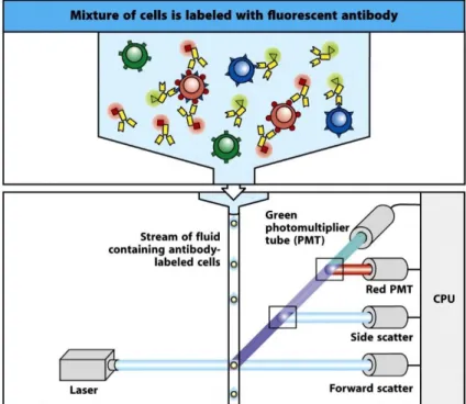
In this project, we characterized the phenotypic signature of MAIT cells isolated from healthy and chronically infected HBV patients’ PBMCs as well as their functional response upon stimulation with IL-12 and IL-18 recombinant cytokines. MAIT cells were evaluated regarding the expression of three different antibody panels – 1) checkpoint inhibitors, 2) activation markers and 3) pro-inflammatory cytokines – and analyzed using a FACS flow cytometer. Furthermore, MAIT cell cytokine and chemokine expression profile were assessed, when in contact with human B lymphoblast antigen presenting cell line and while stimulation with a MR1 agonistic compound (diclofenac).
Although our preliminary data shows that MAIT cells from chronic HBV patients are functional in ex
vivo stimulation, it will be of interest to further test the developed assays with additional human
PBMC samples, since our sample size in this project is limited. Also, it would be of high relevance to test MAIT cells retrieved from chronic HBV patients’ liver samples and compare the data with our results. Nonetheless, the data obtained in this project shows that MAIT cells are functional, even though these cells do not possess a TCR, which leads to the unrecognition of recognize infected hepatocytes. Nonetheless, MAIT cells are considered as a promising cell-based therapeutic approach that could be used against HBV in combination with the already existent therapies.
vii
Table of Contents
Chapter 1 – Introduction ... 1
1.1 Molecular Virology of Hepatitis B Virus ... 3
1.1.1 Genetic Variability of HBV ... 5
1.2 Immunopathogenesis of Chronic Hepatitis B ... 6
1.2.1 T-Cell exhaustion ... 9
1.3 Treatment Strategies ... 9
1.3.1 Nucleos(t)ide analogues and Interferon-Based therapies ... 9
1.3.2 Therapeutic Vaccination ... 10
1.4 Mucosal-associated Invariant T Cells: What are they? ... 12
1.4.1 MAIT cells Development and Tissue Distribution ... 13
1.4.2 Antigen presentation to MAIT cells ... 15
1.5 MAIT cells Effector Functions ... 19
1.5.1 – Cytokine Production ... 19
Chapter 2 – Objectives ... 24
Chapter 3 – Materials and Methods ... 28
3.1 Human Samples ... 30
3.1.1 Healthy Donors ... 30
3.1.2 CHB Patients ... 30
3.1.3 Isolation of Peripheral Blood Mononuclear Cells ... 30
3.1.4 Thawing of the PBMCs... 30
3.2 Mucosal-Associated Invariant T cells (MAIT cells) ... 31
3.2.1 Flow Cytometry ... 31
3.3 Gating Strategy of Mucosal-Associated Invariant T (MAIT) cells: ... 33
3.4 Isolation of MAIT cells ... 33
3.4.1 Isolation of MAIT cells with Microbeads Separation Kit ... 33
3.4.2 FACS Cell-Sorting ... 33
3.5 Functional Assay – C1R Cell line ... 34
3.5.1 Cell Culture ... 34
3.5.2 Stimulation of Sorted MAIT cells in combination with B Lymphoblast cell line ... 34
3.5.3 Flow cytometry ... 34
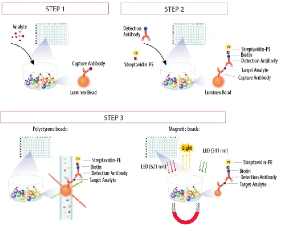
3.5.4 Luminex ... 34
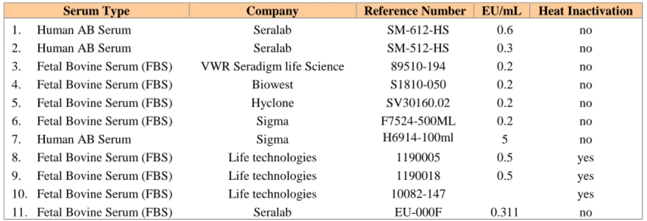
3.6 Serum Screen ... 36
3.6.2 Intracellular Staining to confirm ELISpot Results ... 37
3.6.3 Proliferation Assay ... 38
Chapter 4 – Results ... 39
4.1 Optimizing flow cytometry panel for MAIT cell analysis in human PBMC ... 41
4.2 MAIT cell phenotyping (% population in PBMC) ... 41
4.2.1 Surface markers (CD4/CD8 and CD56) within the MAIT cell population in donor PBMC ... 41
4.3 Ex vivo MAIT cell activation ... 43
4.3.1 Checkpoint Inhibitors ... 43
4.3.2 Activation Markers ... 46
4.3.3 Pro-Inflammatory Cytokines ... 49
4.4 MAIT cell isolation with beads ... 51
4.5 Serum screening for PBMC culture media ... 52
4.5.1 ELIspot ... 52
4.5.2 Intracellular Staining (ICS) ... 54
4.5.3 Proliferation Assay ... 58
Chapter 5 – Discussion & Conclusions ... 60
ix
Acronyms
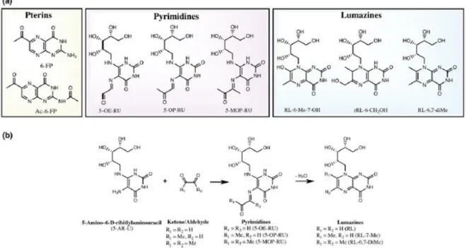
5-A-RU - 5-amino-6-D-ribitylaminouracil 5-OE-RU - 5-(2-oxoethylideneamino)-6-D-ribitylaminouracil 5-OP-RU - 5-(2-oxopropylideneamino)-6-Dribitylamonouracil 6-FP - 6-formylpterinAc-6-FP – Acetyl 6-formylpterin ALT - Alanine aminotransferase APC – Antigen presenting cell cccDNA - Closed circular DNA CTLs - Cytotoxic Lymphocytes ER – Endoplasmic reticulum CCR - C-C chemokine receptor CD – Cluster of differentiation CXCR - CXC chemokine receptors CHB – Chronic Hepatitis B DC – Dendritic cell DCF - Diclofenac
ER - Endoplasmic reticulum ETV - Entecavir
GM-CSF - Granulocyte-macrophage colony stimulating factor Gnly - Granulysin
GrzA/B – Granzyme A/B HBcAg – Core antigen HBeAg - Hepatitis B e-antigen HBsAg - Hepatitis B surface antigens HBx - Hepatitis B Virus X protein HBV – Hepatitis B virus
HCC – Hepatocellular carcinoma HCV - Hepatitis C virus
HLA-DR - Human leukocyte antigen-antigen D related IC - Immune complex
IFNα - Interferon α IFNү – Interferon ү IL– Interleukin IR - Inhibitor receptor
ISGs - Interferon-stimulated genes LAG3 - Lymphocyte activation gene-3 LAM – Lamivudine LPS - Lipopolysaccharide MAIT - Mucosal-associated invariant T MDR1 - Multidrug resistance protein 1 MHC - Major-histocompatibility complex MR1 – MHC-related protein-1
MAIT - Mucosal-associated invariant T MVA - Modified vaccinia virus Ankara
NCPT - Sodium taurocholate co-transporting polypeptide NK – Natural killers
NUCs - Nucleos(t)ide analogues ORF – Open reading frame
PBMCs - Peripheral blood mononuclear cells PD-1 - Programmed cell death protein 1 PEG-IFNα - Pegylated interferon-α pgRNA – Pre-genomic viral RNA PMA - Pphorbol myristate acetate
Pol/RT - Polymerase/reverse transcriptase
Pre-S/S, Pre-S2/M and Pre-S1/L – Small, medium and large HBV proteins Prf - Perforin
PRR – Pattern recognition receptor RcDNA – Relaxed-circular DNA SVP – Sub-viral particle TCR - T cell receptor TDF - Tenofovir Th - T helper
TIM3 - T-cell immunoglobulin domain and mucin domain 3 TLR - Toll-like receptor
TNFα - Tumor necrosis factor α WHO - World Health Organization
1
3
1.1 Molecular Virology of Hepatitis B Virus
Besides being one of the smallest virus present in nature, hepatitis B virus (HBV), is also known to have a highly compact genetic organization within its genome, where 4 open reading frames (ORFs) overlap with each other1–3. The human HBV belongs to the Hepadnaviridae viral family of enveloped and primarily hepatotropic DNA viruses4. In the presence of a suitable host this virus is capable of replication and assembly exclusively in hepatocytes where virions are discharged non-cytopathically over the cellular secretory system, as well as sub-viral particles (SVPs). Hepatitis B viral genome has a partially double stranded relaxed-circular (rc) DNA features 4 ORFs: PreS/S, preCore/Core, Pol/RT and X (Figure 1.1)1,2,5,6.
These ORFs encode for 7-8 known proteins, that are displayed during the viral life cycle of HBV7. The preS/S open reading frame encodes for three fundamental but related envelope glycoproteins. These proteins have overlapping ORFs, and synthesis of each one individually is initiated by start codons that are specific for each viral antigen present. They are termed small (S), middle (M) and large (L) proteins. There is another characterization possible, S, S2 and Pre-S1 respectively, supported by the fact that surface envelope proteins such as these may be known as Hepatitis B surface antigens (HBsAg, Figure 1.2)2,4,8. The polymerase/reverse transcriptase (Pol/RT) ORF is considered very complex due to many functions it needs to attend, meaning that this ORF encodes for the viral polymerase, which basically is a multifunctional protein with reverse transcriptase functionalities, as well as DNA-dependent DNA polymerase and RNase H activity1,2,5. Additionally, the Pol/RT ORF is also described as functioning as a terminal protein used in priming. The preCore/Core domain encodes for a structural protein of the viral nucleocapsid, the core antigen Figure 1.1. HBV genome structure, genes and mRNAs. The 3.2-kb partially double-stranded relaxed circular DNA
genome of HBV is displayed in the center. The colored arrows surrounding the HBV rcDNA are representative of the locations of the four overlapping open-reading frames (ORFs) within HBV genome – HBcAg Core (Pre-Core/Core); HBsAg Surface (Pre-S1/L, Pre-S2/M and S); HBV DNA Polymerase and HBx. All open reading frames have a clockwise direction. The outer layer arrows indicate the major HBV mRNAs, all of which end at a common polyadenylation signal located in the core open reading frame. DR1 and DR2 represent 11 base direct repeats that have an important role in viral DNA synthesis. AAA: Polyadenilated Tail; PC: preCore; DR1: Direct repeat 1; DR2: Direct repeat 2. Adapted from Seeger et al, 2016.
(HBcAg)7 and a non-structural secreted HBV protein, the hepatitis B e antigen (HBeAg)2,4. Lastly, the X region encodes a small but regulatory protein (HBx)9. This protein is of vital importance for hepatitis B viral replication and regulation of transcription upon infection modulating in concordance host and viral gene expression10.
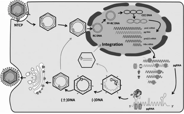
The life cycle of HBV initiates with the virus attachment to its receptor on the hepatocyte surface (Figure 1.3). Interestingly, the rationale behind this receptor function for HBV remained uncharacterized for years. However, more recently it has been noticed that sodium taurocholate co-transporting polypeptide (NCPT) performs as one possible receptor for the HBV life cycle8,11. In vitro studies confirmed that the expression of the human NCPT receptor, when placed on HBV non-permissive mouse cell lines, weakened those cells’ ability to become non-permissive. This fact may suggest the requirement of other receptors granting HBV entry into hepatocytes. Another possibility is indeed the lack of host factors that influence viral replication in these animals. Simultaneously, upon hepatocytes viral uptake, the translocation of the HBV nucleocapsid from the cytoplasm to the nucleus occurs. After viral uptake, relaxed circular DNA (rcDNA) is released to the nucleus through nuclear pores and converted into the covalently closed circular DNA (cccDNA). This phenomenon takes place within the nucleoplasm via DNA repair mechanisms of the host himself 1,3,4. The newly synthesized DNA sequence is then wrapped around histones, so that it may form an episomal chromatinized architecture as mini-chromosomes and responsible for the establishment of chronic HBV6. It is essential to point out that cccDNA displays a major function as a stable template for viral replications, as well as a key element for viral persistence. Although this entire pathway is of extreme importance for viral replication, it is used as a transcription template for the 4 poly-A-tailed viral mRNA transcripts and gives insight on some of the features behind Hepatitis B viral infection1,2,7,8 When transported to the cytoplasm, these mRNAs are translated and give origin to all the different proteins described above, within their ORFs and their place of action4,12. Also, is important to mention that this cascade of events might not take place if the nucleocapsid dimerization and self-assembly of the pre-genomic viral RNA (pgRNA), is not reversibly transcribed into rcDNA within the viral capsid. Nevertheless, DNA nucleocapsids within the cytoplasm happen to undergo one of two
Figure 1.2.HBV virion schematic representation. Structurally, the HBV virion consists of an envelope containing three
HBV secreted surface proteins (S-, M-, and L-proteins) and lipids. Also, HBV virion contains an icosahedral nucleocapsid, which is constituted by the core protein (HBcAg) enclosing the viral DNA genome in a covalent linked manner to the terminal protein of the HBV DNA polymerase. Adapted from Pollicino et al, 2014
5 resolutions: is recycled into the nucleus to preserve the cccDNA supply levels or it is wrapped and secreted via the endoplasmic reticulum due to their excess amount of proteins produced exceeding the level needed for virion assembly2. This excessive number of proteins, mainly envelope proteins, are subjected to dimerization/multimerization processes, which result is a mature growth in the endoplasmic reticulum (ER) and/or Golgi compartments as non-infectious spherical and filamentous SVPs or as virions (Figure 1.3). In a typical manner, these SVPs outnumber the virions values by 1000 to 10000-fold13–16. This characteristic may explain why SVPs are constantly found within the circulating immune complexes as well as immune tolerance induction by the mechanism of “viral apoptotic-like mimicry”13,17. Like other viruses that affect hepatocytes and the liver, HBV genome integration in the host genome may occur in a randomly manner. Despite not being mandatory for viral replication, viral genome integration is still involved in hepatocyte transformation, as well as a source for new infections13.
1.1.1 Genetic Variability of HBV
Due to the lack of reverse transcriptase proofreading activity, there is a movement towards the emergence of new viral mutations affecting HBV genome configuration. As a result, these frequent mutations can induce the appearance of genetically unique viral species, also notated as quasispecies, which will mature due to tension from the different hosts1. The relationship between the different characters of an HBV infection, the host, the virus, hepatocytes, immune responses and even antiviral treatments are believed to push towards the emergence of hepatitis B virus mutants. On their Figure 1.3. HBV life cycle. HBV enters hepatocytes through the NTCP channel, followed by the uncoating and consequent
nuclear transport of the rcDNA. The rcDNA is converted in cccDNA, which serves as the template for transcription of the preC RNA, pgRNA, preS/S mRNAs, as well as HBx mRNA. Shortly after these RNAs are transported to cytoplasm, where protein translation takes place. The pgRNA is selectively bundled inside core particles, and subjected to reverse transcription, where it is degradated and gives place to RC DNA. Such mature core particles can be enveloped for release as virions or transported to the nucleus to generate more cccDNA.Adapted from Balmasova et al, 2014.
counterpart, these HBV mutants have the competence to evade several immune stimuli or antiviral treatment responses1,5 These mutations that accumulate in the different viral genomes echo on the duration of active HBV infection and consequently the strength rate of the immune response towards the virus2. The relationship between virus/host is always present in the immunologic features of any infectious disease as a measurement for the virus itself survival or dissolution1,5,18. Presently, and throughout the analysis of genome-wide nucleotide diversity, 9 genotypes (A-I) and several sub-genotypes were identified for HBV18. Also, the existence of a 10th HBV genotype (J) is considered, but its characterization is not fully understood. HBV genotypes differ in their genomic sequences, as well as their geographic distribution around the globe acting in a diverse manner. Nonetheless, all HBV genotypes induce and affect the transmission, progression and pathologies on HBV disease progression. Despite considering that there is not a more common HBV genotype than others, it is thought that HBV genotype C can be considered as critical, since it shows an increased viral mutation rate and high replication capacity, which is correlated to the emergence of cirrhosis and hepatocellular carcinoma (HCC) in chronic HBV patients.
1.2 Immunopathogenesis of Chronic Hepatitis B
Like other infectious diseases, Hepatitis B is known for having two initial stages, an acute and a chronic stage of infection. While these two stages share some similarities, they are still distinct. The main characteristic distinguishing these two stages, besides the timeline of infection, is whether the infection occurred perinatally/early infancy or already in adulthood. The other parameter that differs between the two stages is HBV immunopathogenic mechanisms, with several immune response pathways along the course of disease progression in infected patients.
In acute resolving infections, HBV-infected adults normally acquire self-limited and short-term hepatitis, and it is estimated that in 95% of the infections indeed end with viral clearance, as well as establishment of protective antibodies19,20. It is also theorized that innate and adaptive immune responses to HBV are decisive and well timed, when encountering a case of acute resolving infection improving the chance for viral clearance1. Thus, it is essential that a strong adaptive T cell reaction takes place early for control of infection, which leads to a cytolytic dependent/independent antiviral outcome via the expression of antiviral cytokines such as interferon γ (IFNγ) and tumor necrosis factor α (TNFα), as well as B-cells stimulation 21,22. Upon stimulation, B-cells start secreting neutralizing antibodies that alongside antiviral cytokines prevent additional hepatocyte infection and virus spreading21. Furthermore, since viral spread is inhibited and hepatocyte turnover mechanisms occur there is a decline in HBV cccDNA levels1. However, when the infection progresses to a chronic state HBV specific T-cell function becomes impaired. Therefore, there is a decrease in proliferation, cytotoxic activity and production of antiviral cytokines such as IFNγ and TNFα, leading to phenomenon called T cell exhaustion 23. During its chronical state, HBV infection spread exerts its effect through various disease stages, which are strongly thought to be associated with age1. Interestingly, it was recently observed that HBV infection acquired at younger age, for example perinatally, shows a tendency for evolving to a state of chronic HBV infection. In addition, it is thought to be directly related to their immune profile, since it is less compromised when compared to older infected patients and more susceptible to disease progression21,22,24.
HBV is acknowledged for its capacity on engaging several immune elements during the natural course of infection, as it manages to adapt and progress through its pathogenesis25. Pathologically, HBV chronic patients display a greater risk to develop cirrhosis and HCC. Nonetheless, HBV is unable to have a direct cytopathic outcome on hepatocytes and currently it is well-established that hepatocytes death in chronic hepatitis B (CHB) patients results from their apoptotic state as consequence of immune responses, due to active viral replication and liver
7 injury19,26,27. HBV-specific cytotoxic lymphocytes (CTLs) are considered key players in hepatocytes killing, since they show strong reactivity responses to viral antigens in chronic HBV infection. Per contra, these lymphocytes are not strong enough to mediate the full eradication of HBV19,28,29. In concordance, CTLs try to mobilize HBV-nonspecific inflammatory cells such CD8+ T cells, natural killer (NK) cells and neutrophils19,30. All these immunological players consequently induce CHB immunopathological functions. Despite all the continuous immune responses against HBV infection, it is still not clear why viral clearance is so rare in HBV patients, since HBV-specific T cells are exhausted and incapable of killing infected hepatocytes, which means no liver injury and no control of infection19,25,31. During HBV chronic infection, there is a progressive synergistic relation between the viral replication and host immune responses to HBV1. However, not all patients exhibiting chronic HBV infection progress and develop CHB1. CHB has been divided in different stages according to its natural viral progression 25,32. Also, its nomenclature has changed over time, since new scientific breakthroughs about the disease were discovered, as well as parameters present in HBV regulation. Recently, chronic HBV infection is divided into 4/5 branches or states, depending on the report1. Chronic HBV nomenclature bears in mind distinct parameters such as the presence of HBeAg (HB e antigen), HBV DNA levels, alanine aminotransferase (ALT) values and the presence/absence of liver inflammation25,31 (Figure 1.4). Also, the new chronic states annotations are mainly based in two central attributes of chronicity: infection and hepatitis1. Nonetheless, it is wrong to establish an immediate viral infection stage classification, since a single measurement of HBV replication markers and disease activity mediators might not be enough to take solid conclusions. Thus, evaluation and monitoring of the different disease parameters is necessary to speculate about possible therapeutic approaches administered individually or in group against HBV. Curiously, despite the 4/5 existing phases for HBV chronic infection, it does not necessarily mean that all phases follow a sequential order.
CHB infection initial stage is nominated HBeAg-positive or, as previously called, “immune tolerant phase”1,4 (Figure 1.4). Also, this stage is well-characterized by the presence of HBeAg in patient’s serum, high viral load (HBV DNA) and ALT values within the documented range of approximately 40 IU/L1,4,25,31,33,34. As for the liver inflammatory level within this infection stage, there is no apparent necroinflammation or fibrosis in liver tissues1,25,34,35. However, HBV DNA integration, as well as clonal hepatocyte spread, might still occur. Furthermore, this could be an indication of initial hepatocarcinogenesis manifestations1,4,36,37. The preservation of HBV specific T-cell functionalities is more frequent and prolonged in this stage, progressing until young adulthood if patients are infected perinatally4,36–38. Importantly, loss and seroconversion of HBeAg to anti-HBeAg is normally low in this phase, which decreases the chances of disease remission4,25,39,40.
As HBV infection progresses, ALT values show a tendency to increase leading to the next chronic HBV stage - CHB HBeAg positive or “immune clearance stage”1,4 (Figure 1.4). Nonetheless, HBeAg frequency in patients’ serum and HBV DNA levels remain similar to the previous stage, whereas in the liver the signs of necroinflammation and fibrosis become moderate or even severe in singular cases1,4,38,41. This medical condition is frequently found among subjects infected during adulthood and it may arise after numerous years of the previous disease state1. CHB patients in this stage can have one of two outcomes – manage to seroconvert HBeAg and suppress HBV DNA levels or fail to control HBV infection – the latter allowing the entrance in the HBeAg negative CHB phase 1,4,38,42.
Following this, since HBeAg loss gives rise to the production of anti-HBeAg, CHB patients eventually reach the “inactive carrier stage” or currently noted as HBeAg negative CHB infection phase1,4,25,43 (Figure 1.4). At this stage, HBV patients are subjected to a change in the parameters
defining chronic HBV infection4. Patients’ serum shows traces of anti-HBeAg in constant rise, HBV DNA levels are considered minimal and ALT parameter is described as stable and in mild values. In addition, the risk for disease progression into a cirrhosis and HCC in this stage is considered low 1,38,44. Although this CHB phase is considered stable, there are cases of patients suddenly progressing deeper into the infection, which could be related to the emergence in viral load with no apparent changes in liver ALT values, but residual signs of necroinflammation45. The HBeAg negative CHB infection phase is also characterized for showing low levels of HBsAg loss, which means that seroconversion of HBV’s antigen is minimal and is extremely difficult to spot on in patient’s serum 1,45. Nonetheless, in 20%-30% of the inactive HBsAg carriers, spontaneous reactivation of hepatitis B may occur, which can lead to a disease progression and development of liver damage and decompensation 4. As HBV infection progresses, patients are likely to enter the HBeAg-negative CHB stage. Here, the lack of HBeAg in patients’ serum is characteristic, as well as fluctuations in HBV DNA levels, which might be correlated to an increase of ALT in the liver. The aforementioned phase is associated with low rates of spontaneous disease remission, perhaps since nearly all the patients within this phase demonstrate to have HBV variants accommodated within the precure/basal core promoter regions, leading to an impairment/abolishment of HBeAg manifestation.
The last official phase in chronic HBV infection is called the HBsAg-negative phase but can also be denominated by “occult HBV infection”1 (Figure 1.4). The latter denomination relies on the fact that this stage is hypothesized to occur during all the previous HBV infection phases mentioned, though its diagnostic time point is not fully understood. Furthermore, this stage is characterized for showing no detectable antibodies for HBsAg in patients’ serum, positive antibodies for HBcAg (anti-HBcAg) and normal ALT levels. Also, the majority of patients in this late HBV infection stage show undetectable traces of cccDNA) in the serum, although it is normally detected upon liver histological exams. HBsAg loss and seroconversion in this stage is correlated to disease progression into more extreme pathologies such as cirrhosis or HCC, since this HBV antigen is known to mediate HBV progression. Nonetheless, CHB patients’ survival rate is increased if HBsAg loss takes place before liver flares and cirrhosis outbreaks. However, in cases where patients show signs of cirrhosis or HCC but HBsAg loss is insignificant, it is possible that HBV reactivates, leading to a possible death. Table 1.1 Phases of chronic HBV infection.
Legend: ALT, alaninaminotransferase; IU, international units; NA, nucleos(t)ide analogues; PEG-IFN, pegylated interferon alfa; HBV, hepatitis B virus; HBsAg, hepatitis B surface antigen; LHBs, large hepatitis B surface protein; anti-HBc, antibodies to hepatitis B core antigen; HBcrAg, hepatitis B core-related antigen; RNA, ribonucleic acid; DNA, deoxyribonucleic acid.The risk for disease progression and the need for treatment is indicated by colour shading: red: high risk for disease progression / treatment required, yellow: low risk for disease progression / monitoring
9
1.2.1 T-Cell exhaustion
Innate and adaptive immunocyte activation is regulated by a set of inhibitory surface receptor-ligand pairs or checkpoint inhibitors46. It is well established that the presence of anti-viral T cells contributes to the eradication of acute infections, restrains the resurgence of latent viral infections and controls the viral loads during chronic infection, as these cells are crucial elements of the adaptive immune response47. Typically, naive T cells recognize antigen peptides presented via their T-cell receptor (TCR) and become activated, being liable to clonal expansion as well as differentiation into potent effectors48. When in a differentiated effector state, T cells show high cytotoxic levels, produce effector cytokines such as IFNγ and TNFα and express chemokine and homing receptors, which allows them to migrate to peripheral tissues and infection sites48–50. However, the host immune system fails to respond if there is a continuous TCR stimulation due to the persistence of high levels of antigens, leading T cells to enter a state nominated T cell exhaustion51,52. Fundamentally, T cell exhaustion is characterized by the endurance of high levels of antigens, that leads to a repeated T cell dysfunctional state and an increase of distinct inhibitor receptors (IR), a gradual depletion of effector cell functions translated by the loss of effector cytokine secretion levels (IL-2, IFNγ and TNFα), as well as a change of these cells metabolism and transcriptional profile 51,53–55. Also it is well-established that exhausted T cells are characterized by the upregulation of surface markers such as PD-1, TIM3 and LAG3, investigated during the course of this project.
1.3 Treatment Strategies
Hepatitis B virus (HBV) remains a major healthcare challenge, and according to the World Health Organization (WHO), hepatitis viral infection is still the eight most frequent cause of mortality worldwide56,57). HBV is estimated to affect approximately 400 million people around the globe, and despite the existence of a prophylactic vaccine, there is still no definitive curative treatment for the more than 240 million people globally that are considered chronically infected with HBV57. CHB patients show a higher risk of death due to an inherent tendency to develop other severe pathologies, including liver cirrhosis and HCC8,57 . Prophylactic vaccination against HBV contains the envelope protein S and is either produced by processing of HBsAg retrieved from HBV carriers’ plasma, or from the yeast specie Saccharomyces cerevisiae, holding a recombinant DNA plasmid that also expresses the S protein58–60. Currently, there is another type of prophylactic vaccine, which encompasses the S and M envelope proteins obtained from genetically engineered ovary cell lines58–60. Even though HBV infection can be prevented by a recombinant yeast-derived prophylactic vaccine, the treatment is mostly available in developed countries and is mainly administered to infants and newborns. It is still extremely difficult for some undeveloped countries individuals to be granted access and acquire this preventive treatment, mainly due to its limited availability and high cost. Nonetheless, it appears that 184 of the 196 countries in the world are able to get a safe and effective prophylactic vaccine, at least for children8). In compliance with the number of chronically infected HBV patients globally and high mortality rate, the formulation of new and more efficient therapies is necessary. Also, efforts are being made by the scientific community to develop different therapeutic vaccines to substitute the prophylactic treatment, which can endure the clearance of circulating HBsAg and convert these to a state of recognition by antibodies of HBsAg, also called HBsAg serocoversion28,57.
1.3.1 Nucleos(t)ide analogues and Interferon-Based therapies
Currently, there are two treatment therapies approved for CHB patients: nucleos(t)ide analogues (NUCs) and IFN-based therapies such as pegylated interferon-α (PEG-IFNα)56,57. Despite demonstrating very interesting results among hepatitis B infected patients, unfortunately these
treatment regiments are still far from a satisfactory cure and complete eradication of the virus. When used separately, these two therapies work in distinctive manners. NUCs therapy impact positively on adaptive immunity61, suppressing viral replication by directly targeting virion synthesis61,62. However, this therapeutic drug does not successfully eliminate the nuclear persistence form of the virus genome, cccDNA, from all liver infected hepatocytes56,57. This eventually leads to an HBV infection rebound and hepatic flares upon treatment withdrawal56,63,64. For chronically infected HBV patients, NUCs treatment is considered life-long, representing an immense financial liability and latent systemic drug toxicity56,57,63. Another concern about using NUCs as a therapy against HBV infection is the possible appearance of drug resistance mutants in chronic patients due to its prolonged use65. Thus, these drugs can also be nominated as antiviral inhibitors, acting on HBV reverse transcriptase activity and disturbing its viral life cycle66. There are some NUCs available for clinical treatment, such as lamivudine (LAM), adefovir, entecavir (ETV), telbivudine and tenofovir (TDF)8,67. Although the use of NUCs show some contradictory effects, this antiviral therapy has shown to be effective as a functional cure. This kind of treatment provides the normalization in ALT levels, suppression of HBV DNA in patients, histological improvements and decreased liver inflammation and fibrosis8. As for HBeAg and HBsAg, it is hypothesized that NUCs treatment leads to a possible loss and seroconversion of both HBV specific antigens, but more studies should be performed in the clinics8.
Besides using NUCs as a controlled treatment against CHB infections, it is currently very common to combine this medication with an IFN-based therapy57. IFN-based therapies, also called cytokine-mediated therapies, are new treatment strategies known for the enhancement of the immune system through the production of interferon-stimulated genes (ISGs) and their cytotoxic activity in killing HBV infected cells66,68. This therapeutic approach includes PEG-IFNα, and together with NUCs is the best line of attack against HBV infection, with particular attention for CHB patients66. In contrast to NUCs antiviral treatment, PEG-IFNα therapies currently show a higher efficacy response on the innate immune response61,69,70. Despite this, PEG-IFNα treatment and other IFN-based therapies can only grant an HBV cure to 10-15% of the treated patients57. Nonetheless, this treatment is strongly recommended for chronically infected HBV patients with no medical history of liver diseases, since PEG-IFNα is associated with severe side effects, including flu-like symptoms and the occurrence of hepatic flares57,66,71,72. Interferon α (IFNα) drugs are typically administered subcutaneously/intramuscularly on CHB patients, and lead to a decline of HBV DNA serum levels and viral replication rate. Consequently, this causes a reduction in HBV transcription, as well as enhancer activity in the host genome65,66,73. Also, IFNα therapy results in a decay of HBV virions, DNA polymerase, as well as HBcAg levels linked to HBV virions due to an inhibition of its reverse transcriptase activity65,66,74. The use of PEG-IFNα compared to standard IFNα has resulted in a significant reduction of HBV DNA and ALT levels in CHB66,75. Interestingly, IFNα was recognized as an up-regulator of APOBEC3A/C3B proteins, causing cystidine deanimation and degradation of cccDNA, without affecting the host genome3,66,76. These two drug treatment strategies are often combined, demonstrating higher reduction in HBV DNA serum levels, inhibition of HBV replication and lower ALT levels. However, even when combined, these treatments are unable to eradicate cccDNA from all infected cells, and it is very common for patients to experience a rebound of the viral load and ALT levels increase upon withdrawal of the medication. This might be due to the increase of cccDNA persistence as main cause and drug-resistance to the medication, hence it is considered a life-long treatment61,65,66.
1.3.2 Therapeutic Vaccination
Since HBV is capable of hampering adaptive and innate immune responses, it is of the utmost importance to find a new therapeutic approach that boosts the immune system and further enhances
11 the efficacy of the current antiviral therapeutic regimen58. The development of therapeutic vaccination is meant to activate the immune system in infected patients, in order to fight against, control, or ideally eliminate the established infectious pathogen57. In CHB patients, the objective is to reinforce the immune system to a point in which cccDNA clearance from HBV infected hepatocytes is achieved. Many studies have been published on the development of therapeutic vaccination against the HBV58. For the past 20 years, several clinical trials have been made regarding numerous therapeutic vaccination strategies, but the end results are far from excellent and cccDNA clearance is still impossible to accomplish57. Nonetheless, the scientific community is working to develop a therapeutic vaccine that can grant a definitive cure for CHB patients, instead of a functional one obtained with the available antiviral treatments. To the best of our knowledge, recombinant peptide-based vaccines77–85, DNA-based vaccines86–88, viral vector-based vaccines89,90 and cell-based vaccines 91,92 are being studied and are presently the most promising therapeutic vaccination approach57,58.
In a simplified manner, the primary attempts to develop a therapeutic vaccine for HBV were peptide-based vaccines, and Pol S and co-workers were the first conducting a HBV therapeutic vaccination trial to test the efficacy of this vaccine nature in CHB patients58,77. This kind of therapeutic vaccination is easily produced and induces high titers of HBV-specific antibodies. Despite the efforts, peptide-based vaccines still show weak cellular immunity responses and require adjuvant and continuous administrations58,93. However, it seems that there is a substantial decrease in serum HBV DNA levels and viral replication, but no significant diminished in HBsAg levels in most of the patients tested58,77. Still, there is evidence that peptide-based vaccines for HBV will activate T cells responses, reduce HBV viral load and quicken HBeAg seroconversion frequency in CHB patients58,94. Another peptide-based vaccine approach involves the combination of recombinant HBV core particles (HBcAg) with HBsAg58,95. The combination of recombinant HBsAg with anti-HBs antibodies to form an antigen-antibody immune complex (IC) became a motivating method for developing therapeutic HBV vaccination58. Noteworthy, it is thought that these ICs may improve the chance of HBsAg being captured by antigen presenting cells (APCs), therefore enhancing the immunogenic effects of the vaccine and inducing more potent HBs-specific T cell responses58,96. All peptide based vaccines so far have failed to induce sustained HBV-specific T cell responses in CHB patients. Though, with all the clinical trials performed so far, the medical community suggests that repetitive adjuvant vaccination might result in an inflammatory environment and stimulate pre-existing T cells in the liver58,81. This hypothesis is based on T cell responses detected upon therapeutic vaccination due to an effect registered on pre-existing T cells, indicating that patients with active liver inflammation may benefit more from therapeutic vaccination58,81,82.
On the other hand, DNA-based vaccines encoding HBV envelope proteins are also under investigation as a therapeutic vaccination against HBV chronically infected patients57,97,98. This vaccination type induces humoral and cellular immune responses, including CD8+ and CD4+ T cell responses58,99. It seems that patients’ immune response after DNA-based vaccines administration is similar to that of individuals whose HBV infection was cleared. Furthermore, since HBV-specific IFN-γ-secreting T cells show upregulated levels upon treatment with this vaccination regimen, this strategy is promising, since it induces effective but transient T cell responses58,88,99,100. However, DNA-based vaccines fail to achieve HBeAg and HBsAg seroconversion, and they are not able to induce a consistent T cell response with reduction of viremia levels in chronic HBV patients57,97,98.
Viral vector-based vaccines are another therapeutic vaccination approach to cure HBV, as these vaccines foundation is based on live attenuated viruses capable of broad stimulation and sustained immune responses58. In particular, adenoviral and modified vaccinia virus Ankara (MVA)-based vectors have been tested in clinical trials, since these vaccines have an extensive stimulation
Entry Inhibitors Interaction with sodium (Na+) - Taurocholate Cotransporting Polypeptide (NTCP) and decreases viral infection rate i.e Myrcludex
Silencing & Eliminating cccDNA
cccDNA targeting through use of antiviral cytokines, blockade of RcDNA or via epigenetic regulation possibly leads to viral DNA degradation and elimination from infected hepatocytes i.e CRISPR/Cas9 and histone deacetylase (HDAC) inhibitors
Secretion Inhibitors Inhibition of HBsAg secretion i.e nucleic acid polymers
HBV Polymerase Inhibitors Promote the inhibition of the reverse transcriptase mechanism by heapatitis B polymerase and help in suppressing viral expansion
Core Allosteric Modulators (CpAM) These agents stimulate the inhibition of nucleocapsid assembly, during hepatitis B life cycle, which incapacitates pgRNA encapsidation and results in the capture of viral RcDNA
Silencing RNA Prevent HBV replication by using HBV-specific small molecules that interfere and silence RNA resulting in reduced cccDNA levels in infected hepatocytes i.e RNA interference (RNAi)
Immune Stimulation
Induce a stimulatory effect in the immune system. Agents for immune stimulation include pattern recognition receptor (PRR) agonists, Toll-like receptor 7 (TLR7, GS-9620), as well as TLR1/2, retinoic acid inducible gene I (RIG-I), and stimulator of interferon genes (STING)
Immune Modulation & Cytokines
Innate cells have been identified for their role in immune tolerance. For example, NK cells demonstrate to have a regulatory role upon HBV-specific T cells via upregulation of a death receptor and myeloid derived suppressor cells have been shown to influence the adaptive immune response. On the other hand, the modulation of innate-adaptive interactions could also hold therapeutic promise, as cytokines such as TNFα, IL-2 and IL-12 have been implicated in HBV replication inhibition in vitro.
Checkpoint Inhibitors
HBV-specific T cell are exhausted and overexpress inhibitory molecules such as programmed death-ligand 1 (PD-1), TIM3 and LAG3. However, in case of these molecules blockade as shown potential in vitro, with special attention in hepatocellular carcinoma medical conditions.
T cell Therapies
Increasing the number of HBV-specific T cells by autologous infusion of T cells expressing chimeric antigen receptors (CARs) or by engineering T cells to overexpress human leukocyte antigen (HLA)-restricted HBV-specific T cell receptors (TCRs).
Viral Targets
Immune Targets
range in immune responses like T cell-mediated immunity57. Nevertheless, vaccination strategies should combine different viral vector-based vaccines with other vaccination therapies like DNA-based vaccines, and recombinant antigens57,90. Finally, a vaccine strategy called cell-based vaccination was developed based on the role of innate immunity in HBV infection, as it aims to stimulate pattern recognition receptors (PRR). The objective is to induce a stimulatory response on different immune regulators like toll-like receptors (TLRs) and APC receptors (e.g. dendritic cell, or DC, receptors), which in turn can stimulate specific CD4+ and CD8+ T cells and direct them at HBV-specific infected cells58,101.
New and more compelling therapeutic vaccination strategies have been developed and entering clinical trials recently (Table 1.2). However, these likely require new potent vaccine components and schemes that could induce more effective humoral and cellular immune responses. Besides this, it is necessary to include checkpoint inhibitors or other similar strategies that could overcome T cell exhaustion in chronic HBV infected patients, as well as lowering HBV specific antigen levels as a preventive action for T cell attrition in patients with low antigen loads57.
1.4 Mucosal-associated Invariant T Cells: What are they?
From an immunological point of view, the liver is considered one of the most important organs in the human body102. This aspect is supported by the fact that, even after being continuously exposed to non-self-food and microbial-derived products from the gut, the liver remains sterile and tolerogenic in homeostasis103. Nonetheless, in situations where the intestinal mucosal defenses are Table 1.2. New therapeutic strategies against chronic HBV infection. These strategies aim to target either a viral target,
or modulate immune system components to overcome the viral infection. Adapted from Gill, Upkar S and Kennedy, Patrick T.F, 201.
13 Figure 1.4 MAIT cells developmental stages, phenotype and frequency in the thymus, cord and adult peripheral
blood. Adapted from Dias Joana, 2017.
ruptured, or in presence of a systemic infection, this organ acts as a second ‘firewall’, through its enrichment with innate effector cells, which rapidly respond to infections or tissue damage102,103.
Mucosal-associated invariant T (MAIT) cells are described as the most abundant subset of unconventional and innate-like T cells in humans104. MAIT cells are evolutionary conserved among different species and seem to be highly abundant in human mucosal tissues, peripheral blood and liver104. Also, there are evidence suggesting that MAIT cell can recognize microbial riboflavin metabolites (Vitamin B2) secreted by numerous bacteria and fungi species presented via the non-polymorphic major-histocompatibility complex (MHC) Class-I related molecule (MR1)105,106. Due to the strong evolutionary conservation of this molecule (MR1) and the lack of riboflavin metabolite antigens variety, it is proposed that these cells are homogenous in their responses against several microbes that hold the biosynthesis pathway for riboflavin metabolites104–106. These T cells subset are defined by the expression of a semi-invariant αβ TCR) combined with a specific segment (Vα7.2) and high expression levels of the C-type lectin receptor CD161. These two receptors are well-established for describing, define and isolate MAIT cells in healthy humans. However, it seems that recent studies showed that the IL-18 receptor α subunit (IL-18Rα), in combination with the TCRVα7.2 marker, can be used to define MAIT cells in humans107–109. It is known that MAIT cells demonstrate higher tendency to accumulate at the infection sites, providing a protective role in some experimental infection models104.
1.4.1 MAIT cells Development and Tissue Distribution
In resemblance to other T cells subset like NK T cells, MAIT cells development starts in the thymus110,111, where these cells undergo a pre-selection by MR1-expressing CD8+CD4+ thymocytes112. Using transgenic mouse models and thymic organ cultures, Seach and colleagues were able to determine that MR1-expressing double positive thymocytes have a vital role in MAIT cells selection mechanisms during their maturation development113,114. Also, other cells types such as thymic B cells and DCs are apparently redundant in MAIT cells selection in the thymus112. MAIT cells phenotype shows differences, depending on these cells’ location in the human body, as well as their developmental stage. When in the thymus and cord blood, MAIT cells exhibit a naive phenotype, characterized by being CD45RA+CD45RO- 111,115,116. On the other hand, peripheral blood MAIT cells are CD45RA-CD45RO+CD28+CCR7-CD62L-, which marks these cells as effector memory cells110,111,115. Nevertheless, Koay and colleagues proposed that MAIT cells maturation and development consists on a three-stage pathway, and is based on the expression of two markers in the thymus, CD161 and CD27 (Figure 1.5) 117.
In stage 1, in thymus, CD161-CD27- MAIT cells would be predominantly CD8+CD4+ or CD4+ only, whereas in stage 2, besides the latter described, MAIT cells could also be CD8+ only. Nonetheless, in stage 2 MAIT cells could be defined for being CD161-CD27+, characterizing this development stage in the thymus. Lastly, during stage 3, MAIT cells are CD161+CD27+, in the thymus as well as mostly CD8+ and CD8-CD4- in the thymus. However, MAIT cells in the latter stage are rare in the thymus, while abundant of cord and peripheral blood, which suggests that MAIT cell functional maturation occurs after these cell are translocated out of the thymus117. Besides this, MAIT cells are known for expressing the CD8αβ co-receptor exclusively in the thymus and cord blood116,117, while adult peripheral blood MAIT cells show evidence of CD8αβ or CD8αα expression (Figure 1.5)111,116,117. Due to their effector memory phenotype, human peripheral blood MAIT cells are referenced as expressing different transcriptional factors such as PLZF, RORγt115,118,119, as well as T-bet, Helios, and Eomes at low, intermediate and high levels, respectively118. These transcription factors are known for having a role in effector function, proliferation and activation in CD8+ T cells120–122. Furthermore, adult peripheral blood MAIT cells also express receptors for IL-12, IL-23, and IL-7 115,123,124, adding up to IL-18R, a well-established MAIT cell receptor. The NK cell receptor NKG2D, the T cell activation antigen CD26, and the multidrug resistance protein 1 (MDR1) are also described as being expressed by MAIT cells at different degrees115,125. It is strongly suggested that peripheral blood MAIT cell population tends to gradually expand with age126–128 and reaches a frequency ten times larger than initially seen in the thymus and cord blood117. MAIT cell populations range between 1 and 10% of the total circulating T cells111,115, although their frequency is associated to a high variability among individuals. Also, the frequencies of CD8+ and CD4+ MAIT cells show a tendency to inversely decrease and increase with age, respectively127,128.
Adult peripheral blood MAIT cells are known to express a very distinct mixture of chemokine receptors which moderate their cellular trafficking to the peripheral tissues102. Indeed, MAIT cells prefer to home to the peripheral tissues, mainly due to the expression of CCR6 and CXCR6, liver-homing receptors, as well as gut-liver-homing integrin αβ47 and intermediate levels of CCR9, which is also expressed by T cells102,115,129. These receptors have an interesting function in lymphocyte migration to the gut. Human MAIT cells percentage is enhanced in the liver, constituting approximately 15% to 50% of the hepatic T cells, staging as the most predominant T cell subset population expressing the markers CD161 and CD56115,123,130–132. Despite being considered the largest T cell subset and preferably allocated within the gut, MAIT cells present highly variable frequencies, according to which peripheral tissue they are encountered in102,104,132. MAIT cells have been found in several compartments of the small intestine, ileum, duodenum and jejunum, as well as within the large intestine, colon and rectum102. MAIT cells are also abundant in peripheral blood (≈1-10% of T cells) in which they are representative of a homogenous subset of CD4- T cells known to express the TRAV1-2 chain together with the CD161, CD26 or IL-18Rα markers 104,111,115,116. The presence and expression of chemokine receptors, such as CXCR6 and CCR5, illustrates these cells ability to transit to the lungs (≈2-4% of T cells)115,132. There is also evidence of the presence of MAIT cells in the stomach (≈2.5% of T cells). More recently it was discovered that MAIT cells have a small frequency in the endometrium and cervix (≈1-2% of T cells), and partially in the human skin. Transcripts for MAIT cells T cell receptor were also detected in other human organs, including kidneys, the ovaries and prostate 133. Although MAIT cells are widely distributed among the human tissues, studies show traces of these cells in the lymphoid tissues, but in a low frequency115. This aspect may be related to the lack of expression of CCR7 and CD62L, two receptors known to have an important function in lymph node homing132,134,135. Since MAIT cells are widely distributed within the human body and are highly frequent in the liver, these cells might be a new approach for HBV, as well as other liver