Prevalence of obstructive sleep apnea in children
and adolescents with sickle cell anemia*
Prevalência da apneia obstrutiva do sono em crianças e adolescentes portadores da anemia falciforme
Cristina Salles, Regina Terse Trindade Ramos, Carla Daltro, Andréa Barral, Jamocyr Moura Marinho, Marcos Almeida Matos
Abstract
Objective: To estimate the prevalence of obstructive sleep apnea syndrome (OSAS) in children and adolescents with sickle cell anemia (SCA); to investigate the possible correlation between mean annual hemoglobin level and total sleep time with SpO2 < 90%, as well as between mean annual hemoglobin level and total sleep time with
SpO2 < 80%; and to investigate the possible correlation between apnea-hypopnea index (AHI) and painful crisis. Methods: The study involved 85 patients with SCA. The patients completed a questionnaire, were submitted to polysomnography and underwent clinical evaluation (by a pediatrician and an otolaryngologist). An AHI > 1 was considered indicative of a diagnosis of OSAS. Results: The prevalence of OSAS was 10.6%. We found a negative correlation between mean annual hemoglobin level and total sleep time with SpO2 < 90% (r = −0.343; p = 0.002), as well as between mean annual hemoglobin level and total sleep time with SpO2 < 80% (r = −0.270; p = 0.016).
There was no association between AHI and painful crisis. Conclusions: The prevalence of OSAS in this population was high (10.6%). Therefore, it is important to identify signs of OSAS as soon as possible and to determine the mean annual hemoglobin level because of the inverse correlation between that level and the total sleep time with SpO2 < 90% or < 80%.
Keywords: Prevalence; Sleep apnea, obstructive; Anemia, sickle cell; Polysomnography; Sleep apnea syndromes.
Resumo
Objetivo: Estimar a prevalência da síndrome da apneia obstrutiva do sono (SAOS) em crianças e adolescentes com anemia falciforme (AF) e investigar a possível correlação entre hemoglobina anual média e tempo total de sono com SpO2 < 90% e tempo total de sono com SpO2 < 80%, assim como investigar a possível correlação entre o índice de apneia-hipopneia (IAH) e episódios de crise álgica. Métodos: Participaram 85 pacientes com AF, que responderam a um questionário, foram avaliados por um pediatra e um otorrinolaringologista, e submetidos a estudo polissonográfico. O diagnóstico de SAOS foi definido como IAH > 1. Resultados: A prevalência da SAOS foi 10,6%. Observou-se uma correlação negativa entre hemoglobina anual média e tempo total de sono com SpO2 < 90% (r = −0,343; p = 0,002) e tempo total de sono com SpO2 < 80% (r = −0,270; p = 0,016). Não foi observada associação entre IAH e episódios de crise álgica. Conclusões: A prevalência da SAOS nesta população foi alta (10,6%). Portanto, é importante identificar precocemente os sinais de SAOS e avaliar hemoglobina anual média, devido à correlação inversa entre essa e o tempo total de sono com SpO2 < 90% ou < 80%.
Descritores: Prevalência; Apneia do sono tipo obstrutiva; Anemia falciforme; Polissonografia; Síndromes da apneia do sono.
* Study carried out at the Bahia School of Medicine and Public Health, Oswaldo Cruz Foundation, Hospital Especializado Octávio Mangabeira – HEOM, Octávio Mangabeira Specialized Hospital – and Centro de Hematologia e Hemoterapia da Bahia – HEMOBA, Bahia State Center for Hematology and Transfusion Medicine – Salvador, Brazil.
Correspondence to: Cristina Salles. Centro Médico Hospital da Bahia, Av. Professor Magalhães Neto, 1541, sala 2010, CEP 41.810‑011,
Salvador, BA, Brasil.
Tel/Fax 55 71 2109-2210. E-mail: [email protected]
Financial support: This study received financial support from the Fundação de Amparo à Pesquisa do Estado da Bahia (FAPESB, Foundation for the Support of Research in the state of Bahia).
mean annual hemoglobin level and total sleep time with SpO2 < 80% (observed during poly-somnographic recording), and to evaluate the possible correlation between the apnea-hypopnea index (AHI) and painful crisis.
Methods
This observational, cross-sectional study involved non-probabilistic sampling of consecu-tive patients with SCA enrolling for treatment at a referral center for hematology and transfu-sion medicine between May of 2007 and May of 2008. The study sample comprised 85 patients. The inclusion criteria were as follows: having been diagnosed with SCA through the quan-titative analysis of hemoglobin by hemoglobin electrophoresis or HPLC, using the Variant II equipment (Bio-Rad Laboratories, Bossier City, LA, USA); being aged 2 to 19 years; being clinically stable; completing the questionnaire; allowing a pediatric and otorhinolaryngological examination; and undergoing nocturnal poly-somnography. The exclusion criteria were as follows: presenting other genetic syndromes, debilitating diseases, acute hepatitis, a history of OSAS treatment or a history of recent trauma; using hypnotics; having been treated with corti-costeroids; being pregnant; and presenting an infection during the evaluation. To calculate sample size, the software PEPI-Sample (Sagebush Press, Salt Lake City, UT, USA) was used, and the following parameters were adopted: confi-dence level of 95% and prevalence of OSAS in children and adolescents of 5% (a prevalence of
4.9% was an acceptable difference). The popu -lation from which the sample was selected was composed of approximately 1,000 children and adolescents with SCA, registered at a referral center for hematology and transfusion medicine. Therefore, to meet the objectives, the calculated sample size was 71 patients. Considering the possibility that the losses would be 10%, the calculated sample was 78 patients.
Age was calculated in whole years from the date of birth. Race was self reported, in accordance with to the official nomenclature of demographic censuses, skin color (white, brown or black—corresponding to White, Mulatto and Black, respectively) being adopted as a refer-ence. Data regarding mean hemoglobin level in the last 12 months (g/dL) were also collected from medical charts.
Introduction
There have been few studies describing the overlap between obstructive sleep apnea syndrome (OSAS) and sickle cell anemia (SCA). Therefore, the exact prevalence, etiology and natural history of OSAS in patients with SCA has yet to be defined.(1) It is known that OSAS is associated with hypoxemia, hypercapnia and acidosis, which might induce the polymerization of hemoglobin S (Hb S), the potentiation of the sickling process and the onset of vaso-occlusive crises.(2) In addition, approximately 80% of chil-dren with SCA present nocturnal desaturation, which might be the result of OSAS.(3)
Few studies have attempted to estimate the prevalence of OSAS in children and adoles-cents with SCA. In addition, those studies had methodological limitations with regard to the populations studied, the number of patients in the samples and the diagnosis of OSAS, which is made through polysomnographic study. The studies described in the literature have not evaluated the problem of OSAS in the general population but rather under specific conditions. For example, the authors of a study conducted in 1992 evaluated a sample composed of chil-dren with SCA and chilchil-dren with thalassemia, applying a questionnaire and performing clinical evaluations.(4) However, since those authors did
not perform polysomnography, they were able to determine only the prevalence of sleep-disor-dered breathing (SDB) in the children with SCA. In 2008, another group of authors conducted a similar study, in which polysomnographic studies were performed.(1) However, the popula-tion investigated again comprised children with SCA and children with thalassemia, and only those presenting a daytime SpO2 ≤ 94% were
ized Sonolab 620 device (Medtron, São Paulo, Brazil). All results were issued by the same observer.
Through polysomnography the following
were recorded: electroencephalogram (C4‑A1,
C3-A2, O2-A1 and O1-A2), electro-oculogram, electromyogram (anterior tibial and mentalis nerves) and electrocardiogram. Respiratory movements were observed using a chest band and an abdominal band, and SpO2 was observed through pulse oximetry. An oronasal cannula and a thermistor were also used to measure oronasal flow. A microphone was placed near the neck to record snoring.
We used the criteria proposed by Rechtschaffen & Kales for staging of sleep: sleep efficiency was calculated as TST divided by the time in bed. Sleep latency was defined as the interval between the turning off of the lights and the first minute of stage 1 sleep. Rapid eye movement (REM) sleep latency was defined as the interval between sleep onset and the first period of REM sleep. Arousals were defined as an abrupt change in the frequency of the electroen-cephalogram lasting 3 s or longer, preceded by at least 10 s of sleep. The total index of arousals corresponded to the number of arousals divided by the total number of hours of sleep.
The following definitions were adopted:
• Obstructive apnea—interruption in airflow,
with a duration ≥ 2 respiratory cycles, despite the persistence of chest effort, abdominal effort or a combination of chest and abdominal effort
• Index of obstructive sleep apnea—number
of events/h of sleep
• Hypopnea—reduction ≥ 50% in airflow amplitude associated with one arousal or reduction > 3% in relation to the basal SpO2
The patients were weighed using a mechan-ical scale (model 131; Filizola, São Paulo, Brazil). Height was measured using an anthro-pometer or stadiometer. These measures were compared with the growth charts of the United
States National Center for Health Statistics and
converted to Z scores of body mass index and height/age, based on age and gender, using the
software Epi Info, version 3.4.1.
All oral examinations were performed by the same otolaryngologist. Pharyngeal and palatine tonsils were classified in accordance with the criteria proposed by Brodsky(5); those classified
as grade 3 or 4 were considered obstructive.
Pharyngeal tonsils were observed through nasopharyngoscopy with a flexible optical fiber (Machida, Tokyo, Japan) attached to a light source after the use of three drops of nasal vasoconstrictor in each nostril. These tonsils were scored from 0% to 100%, according to the occupation of the cavum by lymphoid tissue. The size of the pharyngeal tonsil was estimated according to the percentage of the posterior choanae covered by adenoid tissue; the pharyn-geal tonsil was considered obstructive when over 70% of this area was occupied. Patients were diagnosed as having obstructive adenotonsillar
hypertrophy (ATH) when a grade 3 or 4 pala -tine tonsil was observed, or when a pharyngeal tonsil occupied more than 70% of the posterior choanae.
The patients were accompanied by their legal guardians for the polysomnographic study, which lasted at least 10 h and was conducted in a quiet environment, with appropriate light and temperature. Polysomnography was conducted during spontaneous sleep; no sedation or sleep deprivation was used, and stimulating foods and beverages (coffee, chocolate, soda and black tea) were avoided. Polysomnography was carried out in a hospital environment, using the
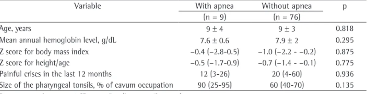
computer-Table 1 - Clinical profile of the sample of children and adolescents with sickle cell anemia.
Variable With apnea Without apnea p
(n = 9) (n = 76)
Age, years 9 ± 4 9 ± 3 0.818
Mean annual hemoglobin level, g/dL 7.6 ± 0.6 7.9 ± 2 0.295
Z score for body mass index −0.4 (−2.8‑0.5) −1.0 (−2.2 ‑ −0.2) 0.875
Z score for height/age −0.5 (−1.7‑0.9) −0.7 (−1.4 ‑ −0.1) 0.775
Painful crises in the last 12 months 12 (3-26) 20 (4‑60) 0.936
ians of the participating patients gave written informed consent.
For data tabulation and analysis, the soft-ware Statistical Package for the Social Sciences, version 12.0 (SPSS Inc., Chicago, IL, USA) was used. The quantitative variables were expressed as mean ± SD or as median (Md) and interquartile range, being compared using the Mann-Whitney test. The qualitative variables were expressed as simple and relative frequencies. To test the correlation between the variables, Spearman’s test was used. The level of statistical significance was set at p < 0.05.
Results
We evaluated 85 patients, 58.8% of whom were male. With regard to race, the patients identified themselves as Mulatto (71.8%), Black (20.0%) or White (8.2%). Table 1 shows the clinical profile of the children and adolescents with SCA.
• AHI—number of obstructive apnea or
obstructive hypopnea events/h of sleep
• Index of oxygen desaturation—all oxygen
desaturation events > 3% based on the basal SpO2/h of sleep
Patients with an AHI > 1 event/h of sleep were classified as having apnea.
In the present study, the AHI was adopted for the diagnosis and classification of OSAS for the following reasons: first, because it is relatively uncommon to observe complete obstruction of the upper airways in children; second, because in a study involving OSAS children (diagnosed on the basis of the AHI), the authors reported that the children in which hypopnea was not accompanied by desaturation events had low cognitive scores(6); third, because in this same study, the children in which hypopnea was accompanied by desaturation events presented arterial hypertension.
The project was approved by the research ethics committee of the institution (Protocol 197; ruling no. 98/2006). The parents or legal
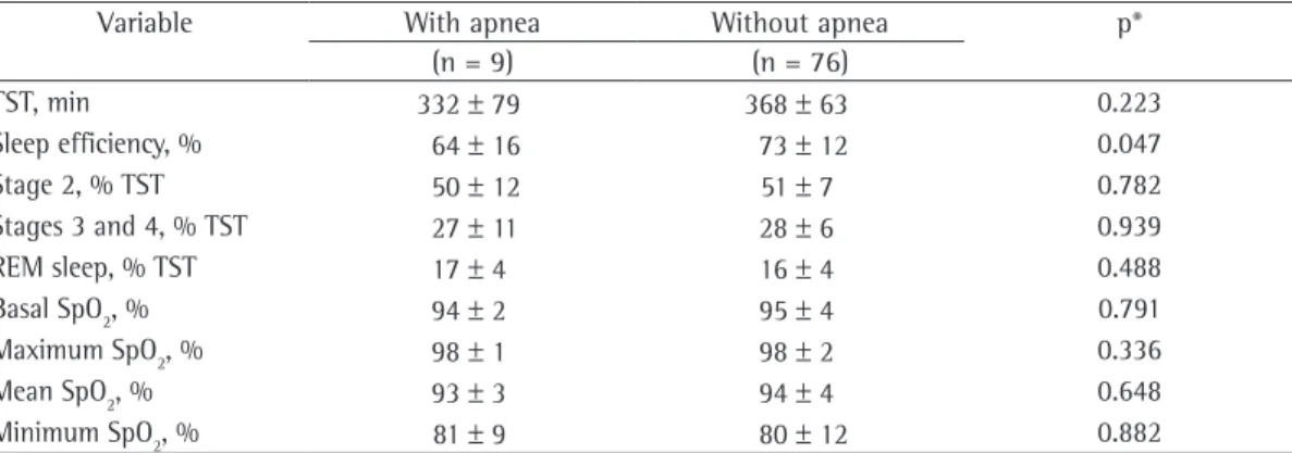
guard-Table 2 - Comparison of polysomnographic data between the patients with apnea and those without.
Variable With apnea Without apnea p*
(n = 9) (n = 76)
TST, min 332 ± 79 368 ± 63 0.223
Sleep efficiency, % 64 ± 16 73 ± 12 0.047
Stage 2, % TST 50 ± 12 51 ± 7 0.782
Stages 3 and 4, % TST 27 ± 11 28 ± 6 0.939
REM sleep, % TST 17 ± 4 16 ± 4 0.488
Basal SpO2, % 94 ± 2 95 ± 4 0.791
Maximum SpO2, % 98 ± 1 98 ± 2 0.336
Mean SpO2, % 93 ± 3 94 ± 4 0.648
Minimum SpO2, % 81 ± 9 80 ± 12 0.882
TST: total sleep time and REM: Rapid eye movement. Data presented in mean ± SD. *Student’s t-test.
Table 3 - Comparison of the polysomnographic data between the patients with apnea and those without.
Variable With apnea Without apnea p*
(n = 9) (n = 76)
Sleep latency, min 31 (18-50) 22 (8‑45) 0.227
Apnea-hypopnea index, events/h of sleep 1.3 (1.9-5.1) 0 (0-0) 0.000
REM sleep latency, min 205 (98-258) 139 (107-197) 1.000
Stage 0, % TST 154 (107‑302) 115 (80-172) 0141
Stage 1, % TST 3.5 (2.6-5.8) 3.7 (2.5-5.2) 0.732
Arousals, events/h of sleep 57 (30‑147) 43 (29‑67) 0.145
Desaturations, events/h of sleep 13 (1.5-29) 5 (1-11) 0.083
SpO2 < 90%a 10 (1-29) 0.6 (0.1‑4.9) 0.105
SpO2 < 80%b 0.1 (0-2) 0 (0-0) 0.021
TST: total sleep time; REM: Rapid eye movement. Data presented as median (interquartile range). apercentage of TST
during which SpO2 was < 90%. bpercentage of TST during which SpO
TST with SpO2 < 80% than did those without (p = 0.021), as shown in Figure 2.
Nocturnal desaturation was observed in
69 patients (81.2%); however, this parameter was not associated with OSAS, obstructive events or obstructive ATH. Of the patients studied, 66 (77.6%) presented a basal SpO2 ≤ 94%. Among these patients, 6 (9.1%) presented an AHI ≥ 1 event/h of sleep.
Obstructive ATH was observed in 55.3% of the patients (previously published finding).(7) Patients with obstructive ATH, in comparison with those without, presented more episodes of obstructive events (Md: 1 vs. 0; p = 0.010), as well as obstructive events that lasted longer (Md: 8.1 vs. 0; p = 0.015). However, obstructive ATH was not associated with the number of desatu-ration events, with the AHI or with painful crises in the last 12 months.
Painful crises in the last 12 months occurred
in 47 patients (55.3%). The correlations between
polysomnographic data, the size of pharyngeal tonsils and the characteristics of SCA were as
follows: sleep efficiency with the AHI = −0.214 (p = 0.049); size of pharyngeal tonsils with the
AHI = 0.256 (p = 0.018); size of pharyngeal
tonsils with desaturation = 0.064 (p = 0.571); the AHI with painful crises = −0.067 (p = 0.545);
desaturation with painful crises in the last
12 months = −0.150 (p = 0.181); painful crises
in the last 12 months with TST at an SpO2 < 80% = 0.062 (p = 0.589).
Discussion
In the present study, the prevalence of OSAS in children and adolescents with SCA was 10.6%. This result reflects the prevalence of OSAS in the general population of children and adolescents with SCA at a referral center. This result differs from that of the study conducted by Kaleyias et al.,(3) who applied a question-naire to 100 children, selected only the 19 most severe cases of suspected SDB to undergo poly-somnography and concluded that 53% of the selected children had OSAS. In another study,(1) only 35% of the 20 patients studied were found to have OSAS. However, the study population was not exclusively composed of patients with SCA, since patients with thalassemia were also included. In addition, only patients presenting a daytime SpO2 ≤ 94% and having undergone
polysomnography were included. Therefore, the The prevalence of OSAS in the sample was
10.6%; the prevalence of snoring was 44.7%. The
distribution of polysomnographic data related to patients with apnea and those without is shown in Tables 2 and 3. We did not find OSAS to be associated with race, gender, age, Z score for body mass index or Z score for height/age.
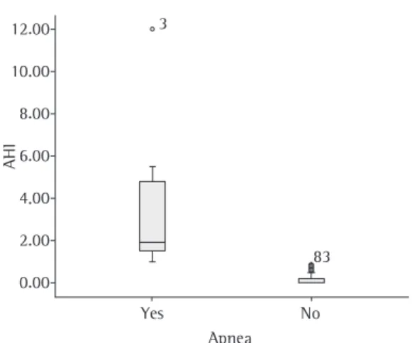
A box plot (Figure 1) shows that the group of patients that had obstructive sleep apnea presented significantly higher AHIs than the group of patients that did not have obstructive sleep apnea (p < 0.001). The number of arousals was not statistically different between patients
with apnea and those without (p = 0.145).
Children and adolescents with sleep apnea presented a significantly higher percentage of
.
.
.
.
.
.
.
Yes No
AHI
Figure 1 - Box plot graph comparing the apnea-hypopnea index (AHI) in children and adolescents with sickle cell anemia with and without obstructive sleep apnea.
Figure 2 - Box plot graph comparing the total sleep time in which SpO2 was lower than 80% in children and adolescents with sickle cell anemia, with or without obstructive sleep apnea.
Apnea No
Total sleep time with SpO2 < 80%, %
.
.
.
.
.
arousals during sleep.(3) In the present study, the frequency of nocturnal desaturation was increased (81.2%). A similar result was obtained by another group of authors,(3) who carried out a study using polysomnography and capnography. Those authors observed that 83% of the patients with SCA presented nocturnal desaturation. Oxygen desaturation is common in patients with SCA and is related to the process of intracellular sickling.(13) However, when oxygen desatura-tion occurs during sleep, it can be accompanied by hypoventilation and can be exacerbated by obstruction of the upper airways.(14) The
obstruction of the upper airways by ATH is one of the principal causes of OSAS in children,(15) as shown in the present study—patients with ATH presented more episodes of obstructive events (p = 0.010), as well as obstructive events that lasted longer (p = 0.015); in addition, we found a positive correlation between the size of the pharyngeal tonsils in these patients and the AHI (p = 0.018). One group of authors(7) observed a high prevalence of obstructive ATH in chil-dren and adolescents with SCA, reporting a prevalence of obstructive palatine tonsil hyper-trophy of 18.8% and a prevalence of obstructive pharyngeal tonsil hypertrophy of 53.3%. The authors attributed this elevated prevalence to the fact that individuals with SCA present a greater susceptibility to severe infections due to asplenia, to the reduced capacity for opsonization and to alterations in the reticuloendothelial system and phagocytic function. Another group of authors(4)
reported that 36% of the patients with SCA presented obstruction of the upper airways. In children, these episodes are frequently associated with ATH, so that the partial occlusion and the complete occlusion of the upper airways during sleep can both be present from the first years of life.(15) In another study,(16) multiple linear
regres-sion analysis showed that 74.3% of the upper
airway obstructions in individuals with SCA were caused by palatine tonsils, pharyngeal tonsils or the hard palate.
In the present study, there was a negative correlation between mean annual hemoglobin level and TST with SpO2 < 90%, as well as between mean annual hemoglobin level and TST with SpO2 < 80%. A similar result was obtained in a study involving 390 patients with SCA, in which basal SpO2 at routine medical visits ranged from 86% to 99%; however, only 2.3% of the patients present study is the first study in the literature
that investigates the prevalence of OSAS exclu-sively in children and adolescents with SCA. The present study is relevant because little is known regarding the consequences of the clinical manifestations of OSAS in patients with SCA; it is known, however, that when OSAS is not adequately treated it can lead to serious compli-cations, among which is a delay in the growth curve.(8) According to one study,(9) the delay in the growth curve of children with OSAS is related to increased respiratory effort during sleep, which generates increased caloric expenditure; in addi-tion, obstructive events might cause a reduction in growth hormone release.(10) According to one review,(11) individuals with SCA present a reduc-tion in serum concentrareduc-tion of growth hormone, as well as a reduced response to growth hormone stimulation, probably secondary to the hypoxic-ischemic injury in the hypothalamic-pituitary axis after one or more episodes of vaso-occlusive crisis, which contributes to a delay in growth.
In the present study, the patients with SCA presented reduced TST. A similar result was observed by one group of authors,(12) who studied 50 patients with a mean age of 13.9 ± 2.5 years; the authors associated that result with the “effect of the first night” at the sleep laboratory, since the night spent at the laboratory can be different from that spent at home and is characterized by a reduction in TST. In the present study, we observed that sleep architecture was altered,
since the values for stages 3 and 4, as well as
the percentage of REM sleep, were higher than expected, although an increase in the number of brief arousals was also observed. In addi-tion, sleep latency and REM sleep latency were increased. It was observed that sleep efficiency was reduced and was correlated with the AHI
(p = 0.049). It is noteworthy that patients with
apnea presented lower values for sleep efficiency
than did those without apnea (p = 0.047). This
result coincides with that observed by one group of authors,(12) who characterized the quality of sleep of patients with apnea as fragmented, since the number of arousals, movements during sleep and changes in sleep stages were increased for their ages. This same group of authors noted impairment of the slow-wave sleep, which was reduced and showed increased latency.
as the volume of the oropharynx, were strongly correlated with the AHI.(25)
Of the patients studied, 77.6% presented basal SpO2≤ 94%; among these, 9.1% presented an AHI ≥ 1 event/h of sleep. The percentage of TST with SpO2 < 80% was higher for individuals with OSAS than for those without (p = 0.021); however, we did not find a statistically significant correlation between these variables and painful crises. Similarly, a group of authors(26) did not observe a correlation between the frequency of painful crises and OSAS; however, they described the association between painful crises and recur-rent infections (p = 0.02). Studies have suggested that OSAS can induce the polymerization of HbS, potentiating the sickling process and the development of vaso-occlusive crises.(2) Based on what has been reported, we should consider that conditioning factors can enhance or impair the sickling process. For the aggregation of HbS molecules, a high concentration of deoxygen-ated molecules is necessary, which facilitates the association between the molecules. The sickling process is not instantaneous, occurring after an interval. Therefore, if hemoglobin is oxygenated during this interval, cell sickling does not occur. As a consequence, cell sickling does not occur in most red blood cells at each cycle through the capillaries. It occurs in a small percentage of cells, since the cells that become oxygenated resume their normal aspect. Therefore, the sick-ling process, for a large number of red blood cells in a blood vessel, is principally caused by the lack of time for red blood cells to pick up oxygen, leading to vaso-occlusion, and not only by deoxygenation itself.(27)
The principal limitation of the present study is related to the wide age bracket of the patients, which ranged from 2 to 18 years. According to one study,(28) a great increment in growth tends to occur in the first years of life; at birth, the craniofacial skeleton of a white American corre-sponds to 60% of the cephalic size of an adult; at 6 months, it corresponds to 80%; at 3 years, it corresponds to 90% and, at 9 years, the cranio-facial skeleton has developed almost entirely, corresponding to 95% of the cephalic size of an adult. Therefore, further studies should focus on more specific age brackets to investigate the possible correlation between craniofacial char-acteristics and OSAS in individuals with SCA. presented SpO2 < 90%, and when a multivariate
analysis was performed, the authors observed that SpO2 was inversely associated with hemo-globin level.(17) Hypoxemia has been described as a precipitating factor for painful crises, for vaso-occlusive events at the microcirculatory level(18) and for “silent” ischemic cerebrovascular accident, which causes a number of neurocog-nitive deficits, such as learning problems and reduced intelligence quotient, affecting the frontal lobes and causing attention deficit and lack of executive functions, as well as short-term and long-term memory loss.(19) In addition, it is believed that patients with OSAS present higher fibrinogen plasma levels, exacerbated platelet activity and reduced fibrinolytic activity than do individuals without apnea, characterizing a state of hypercoagulability. It is likely that the state of hypercoagulability is correlated with OSAS due to the elevated levels of oxidative stress and inflammation. Therefore, some studies have reported that SDB is intimately associated with an increased risk for cerebrovascular accident.(20)
Oxygen desaturation events were not corre-lated with the size of pharyngeal tonsils or the AHI. A group of authors(21) investigated the mech-anisms of nocturnal desaturation in 20 children and adolescents with SCA and concluded that, although nocturnal hypoxemia was common in those children, OSAS did not appear to play a central role; the authors also reported the need to consider desaturation as the result of chronic pulmonary involvement due to the repeti-tive episodes of acute chest syndrome, leading to pulmonary fibrosis, chronic hypoxemia and consequently to the development of pulmonary hypertension,(22) or due to the reduced affinity of HbS for oxygen.(23)
In the present study, the size of the pharyn-geal tonsils correlated with the AHI. It is know that the air space of the pharynx tends to be smaller in children with OSAS than in individ-uals without OSAS.(24) Sedated children (mean
age of 4.8 years) were studied using magnetic
management of childhood obstructive sleep apnea
syndrome. Pediatrics. 2002;109(4):704‑12.
9. Marcus CL, McColley SA, Carroll JL, Loughlin GM, Smith PL, Schwartz AR. Upper airway collapsibility in children with obstructive sleep apnea syndrome. J Appl Physiol.
1994;77(2):918‑24.
10. Standards and indications for cardiopulmonary sleep studies in children. American Thoracic Society. Am J Respir Crit Care Med. 1996;153(2):866-78.
11. Veríssimo MP. Growth and development in sickle cell
disease. Rev Bras Hematol Hemoter. 2007;29(3):271‑4.
12. Souza LC, Viegas CA. Quality of sleep and pulmonary function in clinically stable adolescents with sickle cell anemia. J Bras Pneumol. 2007;33(3):275-81.
13. Samuels MP, Stebbens VA, Davies SC, Picton-Jones E, Southall DP. Sleep related upper airway obstruction and hypoxaemia in sickle cell disease. Arch Dis Child. 1992;67(7):925-9.
14. Block AJ, Boysen PG, Wynne JW, Hunt LA. Sleep
apnea, hypopnea and oxygen desaturation in normal
subjects. A strong male predominance. N Engl J Med.
1979;300(10):513-7.
15. Guilleminault C. Obstructive sleep apnea syndrome and its treatment in children: areas of agreement and
controversy. Pediatr Pulmonol. 1987;3(6):429‑36.
16. Fregosi RF, Quan SF, Morgan WL, Goodwin JL, Cabrera R, Shareif I, et al. Pharyngeal critical pressure in children with mild sleep-disordered breathing. J Appl Physiol.
2006;101(3):734‑9.
17. Quinn CT, Ahmad N. Clinical correlates of steady‑state
oxyhaemoglobin desaturation in children who have
sickle cell disease. Br J Haematol. 2005;131(1):129‑34.
18. Gualandro SF, Fonseca GH, Gualandro DM. Cardiopulmonary complications of sickle cell disease [Article in Portuguese]. Rev Bras Hematol Hemoter. 2007;(29)3:291-8.
19. Angulo IL. Stroke and other vascular complications of the central nervous system in sickle cell disease [Article in Portuguese]. Rev Bras Hematol Hemoter. 2007;29(3):262-67.
20. Mohsenin V. Sleep-related breathing disorders and risk of stroke. Stroke. 2001;32(6):1271-8.
21. Needleman JP, Franco ME, Varlotta L, Reber‑ Brodecki D, Bauer N, Dampier C, et al. Mechanisms of
nocturnal oxyhemoglobin desaturation in children and adolescents with sickle cell disease. Pediatr Pulmonol.
1999;28(6):418‑22.
22. Machado RF. Sickle cell anemia-associated pulmonary arterial hypertension. J Bras Pneumol. 2007;33(5):583-91.
23. Figueiredo MS. Modifiers factors of clinical severity in sickle cell anemia [Article in Portuguese]. Rev Bras Hematol Hemoter. 2007;29(3):215-7.
24. Arens R, Marcus CL. Pathophysiology of upper airway
obstruction: a developmental perspective. Sleep.
2004;27(5):997‑1019.
25. Fregosi RF, Quan SF, Kaemingk KL, Morgan WJ, Goodwin JL, Cabrera R, et al. Sleep-disordered breathing, pharyngeal size and soft tissue anatomy in children. J Appl Physiol. 2003;95(5):2030-8.
26. Hargrave DR, Wade A, Evans JP, Hewes DK, Kirkham
FJ. Nocturnal oxygen saturation and painful sickle cell crises in children. Blood. 2003;101(3):846‑8.
27. Zago MA, Pinto AC. The pathophysiology of sickle cell disease: from the genetic mutation to multiorgan dysfunction [Article in Portuguese]. Rev Bras Hematol
Hemoter. 2007;29(3):207‑14.
28. Meredith HV. Growth in head width during the first
twelve years of life. Pediatrics. 1953;12(4):411‑29.
The present study is relevant because it is the first investigation of the prevalence of OSAS in children and adolescents with SCA, which allowed the understanding of the high prevalence of OSAS (10.6%) in this population. The data draw attention to the need for the early identification of the signs of OSAS and for the evaluation of certain factors, such as mean annual hemoglobin level, since we observed a negative correlation between mean annual hemoglobin level and TST with SpO2 < 80%. Although there are reports in the literature indicating that hypoxia favors the sickling process, no association between the AHI and painful crises was observed in the present study. These data can contribute to minimize the clinical manifestations of SCA, a pathology that does not have a specific treatment yet but a treatment based on prevention and control of symptoms.
Acknowledgments
The present article is part of the disserta-tion of Cristina Salles for the Graduate Course in Medicine and Human Health, Bahia School of Medicine and Public Health, Salvador, Brazil.
References
1. Spivey JF, Uong EC, Strunk R, Boslaugh SE, DeBaun MR. Low daytime pulse oximetry reading is associated with nocturnal desaturation and obstructive sleep apnea in children with sickle cell anemia. Pediatr Blood Cancer. 2008;50(2):359-62.
2. Brooks LJ, Koziol SM, Chiarucci KM, Berman BW. Does sleep-disordered breathing contribute to the clinical severity of sickle cell anemia? J Pediatr Hematol Oncol. 1996;18(2):135-9.
3. Kaleyias J, Mostofi N, Grant M, Coleman C, Luck L,
Dampier C, et al. Severity of obstructive sleep apnea in children with sickle cell disease. J Pediatr Hematol Oncol. 2008;30(9):659-65.
4. D’Aloya N. Sleep related upper airway obstruction
and hypoxaemia in sickle cell disease. Arch Dis Child. 1993;68(5):715.
5. Brodsky L. Modern assessment of tonsils and adenoids.
Pediatr Clin North Am. 1989;36(6):1551‑69.
6. Kaemingk KL, Pasvogel AE, Goodwin JL, Mulvaney SA, Martinez F, Enright PL, et al. Learning in children and sleep disordered breathing: findings of the Tucson Children’s Assessment of Sleep Apnea (tuCASA)
prospective cohort study. J Int Neuropsychol Soc.
2003;9(7):1016-26.
7. Salles C, Ramos RT, Daltro C, Nascimento VM, Matos
MA. Association between adenotonsillar hypertrophy, tonsillitis and painful crises in sickle cell disease. J
Pediatr (Rio J). 2009;85(3):249‑53.
About the authors
Cristina Salles
Physician specializing in Otolaryngology and Sleep Medicine. Bahia Hospital Medical Center, Salvador, Brazil.
Regina Terse Trindade Ramos
Adjunct Professor. Department of Pediatrics, Federal University of Bahia School of Medicine, Salvador, Brazil.
Carla Daltro
Assistant Professor. Bahia School of Medicine and Public Health, Salvador, Brazil.
Andréa Barral
Physician. Octávio Mangabeira Specialized Hospital, Salvador, Brazil.
Jamocyr Moura Marinho
Adjunct Professor. Foundation for Scientific Development, Salvador, Brazil.
Marcos Almeida Matos