ABSTRACT
Objective: To evaluate lung function and inspiratory muscle strength, correlating them with exercise tolerance, in obese individuals with obstructive sleep apnea syndrome (OSAS). Methods: The sample comprised 31 adult subjects with moderate-to-severe OSAS diagnosed by polysomnography. We used spirometry to measure FVC, FEV1, and FVC/FEV1 ratio, using pressure manometry to measure MIP and MEP. The incremental shuttle walk test (ISWT) and the six-minute walk test (6MWT) were used in order to determine functional exercise capacity. Results: In this sample, the mean values for FVC (% of predicted), FEV1 (% of predicted): MIP, and MEP were 76.4 ± 12.3%, 80.1 ± 6.3%, 60.0 ± 21.9 cmH2O, and 81.3 ± 22.2 cmH2O, respectively. The mean distances covered on the ISWT and 6MWT were 221 ± 97 m and 480.8 ± 67.3 m, respectively. The ISWT distance showed moderate positive correlations with FVC (r = 0.658; p = 0.001) and FEV1 (r = 0.522; p = 0.003). Conclusions: In this sample of obese subjects with untreated OSAS, lung function, inspiratory muscle strength, and exercise tolerance were all below normal. In addition, we found that a decline in lung function, but not in respiratory muscle strength, was associated with exercise tolerance in these patients.
Keywords: Sleep apnea syndromes; Exercise tolerance; Respiratory function tests; Respiratory muscles.
Correlation of lung function and respiratory
muscle strength with functional exercise
capacity in obese individuals with
obstructive sleep apnea syndrome
Thays Maria da Conceição Silva Carvalho1,a, Anísio Francisco Soares2,b,
Danielle Cristina Silva Climaco3,c, Isaac Vieira Secundo3,d,
Anna Myrna Jaguaribe de Lima2,e
Correspondence to:
Thays Maria da Conceição Silva Carvalho. Rua Dom Manoel de Medeiros. s/n, Dois Irmãos, CEP 52171-900, Recife, PE, Brasil. Tel.: 55 81 9899-0222. E-mail: [email protected]
Financial support: This study received fi nancial support from Coordenação de Aperfeiçoamento de Pessoal de Nível Superior (CAPES, Offi ce for the
Advancement of Higher Education). INTRODUCTION
Obstructive sleep apnea syndrome (OSAS) is characterized by partial or total obstruction of the upper airways during sleep.(1-3) Obstructive events are associated with oxyhemoglobin desaturation, sleep fragmentation or deprivation, hypoxemia, hypercapnia, dyspnea, as well as diurnal symptoms such as excessive daytime sleepiness.(3,4) The etiology of OSAS is multifactorial, including craniofacial anatomical changes and obesity. Obese individuals present a higher risk of pharyngeal occlusion and altered respiratory mechanics.(5)
An increase in the volume of adipose tissue in the thoracic and abdominal regions impairs diaphragmatic function and reduces chest cavity compliance and lung volumes, leading to an increase in inspiratory muscle work. (6,7) Episodes of recurrent hypoxia in OSAS are usually accompanied by microarousals to reestablish normal ventilation after occlusion of the upper airways. (8) In addition, recurrent episodes of hypoxia and reoxygenation related to upper airway obstruction in OSAS are associated with abnormal partial pressure of oxygen and of carbon dioxide, reducing respiratory muscle activity and lung volumes. These factors generate new episodes of apnea-hypopnea throughout the night, limiting the ability to perform activities of daily living.(9-11)
Obesity and OSAS are factors that potentially alter aerobic capacity and exercise tolerance in different ways. The initial impairment of lung function and respiratory muscle function seen in obese individuals is related to reductions in functional exercise capacity and quality of life.(12,13) There is also evidence that the lower-than-normal exercise tolerance in OSAS patients is due to OSAS-related episodes of dyspnea, intermittent hypoxemia, respiratory muscle dysfunction, and pulmonary hypertension.(14,15) In obese patients with OSAS, ca rdiovascular diseases such as arterial hypertension, cardiac arrhythmia, and systolic dysfunction can also limit exercise tolerance, as can increased work of breathing, hypoventilation, and a sedentary lifestyle.(16,17)
The present study aimed to evaluate lung function and inspiratory muscle strength, correlating them with exercise tolerance, in obese individuals with OSAS.
METHODS
This was a cross-sectional, descriptive, observational study with quantitative analysis. We recruited individuals of either gender with a diagnosis of OSAS, confi rmed through polysomnography, who were evaluated and treated at the Pulmonology Outpatient Clinic of the 1. Programa de Pós-Graduação em Ciência
Animal Tropical, Universidade Federal Rural de Pernambuco – UFRPE – Recife (PE) Brasil.
2. Departamento de Morfologia e Fisiologia Animal, Universidade Federal Rural de Pernambuco – UFRPE – Recife (PE) Brasil.
3. Hospital Geral Otávio de Freitas – HGOF – Recife (PE) Brasil.
a. http://orcid.org/0000-0001-8686-0834
b. http://orcid.org/0000-0003-1493-7964
c. http://orcid.org/0000-0003-1935-1540
d. http://orcid.org/0000-0003-0794-1228
e. http://orcid.org/0000-0002-4224-4009
Submitted: 7 February 2017.
Accepted: 7 December 2017.
Hospital Otávio de Freitas, located in the city of Recife (PE), Brazil. We applied the following inclusion criteria: having been diagnosed with moderate or severe OSAS; being between 50 and 70 years of age; being able to perform stress tests for functional capacity assessment; and having a body mass index (BMI) between 18 kg/m2 and 40 kg/m2. Patients with mild apnea—defi ned as a apnea-hypopnea index (AHI) between 5-15 events/h—were excluded, as were those with cardiopulmonary, neuromuscular, or orthopedic diseases that could infl uence or limit their ability to perform the tests, those with a BMI > 40 kg/m2, and those being treated with continuous positive airway pressure. We initially evaluated 150 patients for eligibility, and 81 of those patients did not meet the inclusion criteria. Therefore, 69 patients were selected to perform the tests. However, 38 were excluded during the evaluation. Consequently, the fi nal sample comprised 31 patients (Figure 1).
The sample size calculation was performed with MedCalc software, version 17.9.5 (MedCalc Software, Mariakerke, Belgium), considering as parameters a probabilistic error of 0.05 (5% alpha) and a statistical power of 80%. Thus, the minimum number of subjects required was calculated to be 24.
The research was approved by the human research ethics committee of the institution. All participants gave written informed consent.
All of the patients completed the Pittsburgh Sleep Quality Index questionnaire, which evaluates
sleep quality in the last month.(18) The Epworth Sleepiness Scale was used for the evaluation of excessive daytime sleepiness.(19) The heart rate was measured with a heart monitor (model FT1; Polar, Kempele, Finland). Blood pressure was measured with an aneroid sphygmomanometer with an arm cuff (Premium model; Missouri Mikatos, Embu, Brazil) and a Rappaport premium stethoscope (Accumed, Rio de Janeiro, Brazil). The heart rate was measured at rest and immediately after the test. Systolic blood pressure (SBP) and diastolic blood pressure (DBP) were measured at rest, 1 min after the test, 3 min after the test, 5 min after the test, and 10 min after the test. The measurement of MIP and MEP followed the methodological recommendations of the American Thoracic Society/European Respiratory Society(20) and the Brazilian Thoracic Association,(21) with the use of a manometer (model MVD300; Globalmed, Porto Alegre, Brazil). We selected three (not necessarily sequential) tests that were considered acceptable (i.e., that met the reproducibility criteria). We used the measurement with the highest value, as long as it did not vary more than 10% in relation to the other values.
Through spirometry, also following the methodological recommendations of the Brazilian Thoracic Association,(21) the following variables were obtained: FVC, FEV1, and the FEV1/FVC ratio.
We applied the incremental shuttle walk test (ISWT), in which the patient has to walk a 20-m path (10 m forward and 10 m back), according to the protocol
Figure 1. Flowchart of the study selection, allocation, follow-up, and analysis. ISWT: incremental shuttle walk test; 6MWT: six-minute walk test; SBP: systolic blood pressure; and DBP: diastolic blood pressure
Excluded (n = 81) • Did not meet the inclusion criteria (n = 13) • Declined to participate (n = 13) • Other reasons (n = 28)
All patients allocated to perform spirometry, manometry, the ISWT, and the 6MWT (n = 69)
Losses to follow-up (n = 38)
18 patients signed up for study participation but did not attend, 16 were unable to participate due to acute or chronic non-stabilized disease, and 4 were not able to perform the test because they presented
an SBP ≥ 160 mmHg or a DBP ≥ 100 mmHg
Analyzed (n = 31) Inclusion
Allocation
Follow-up
Analysis
Evaluated for
developed by Singh et al.(22) The path was delineated with two traffi c cones. We also applied the six-minute walk test (6MWT), which was performed in accordance with the American Thoracic Society specifi cations.(23)
Descriptive analyses were presented as a mean ± standard deviation or as median and interquartile range, as appropriate, according to the results of the Kolmogorov-Smirnov test. The correlations between variables were analyzed by Spearman’s test, and the differences between the means were evaluated with the Student’s t-test for independent samples. Values of p < 0.05 were considered signifi cant. The data were processed with the Statistical Package for the Social Sciences, version 16.0 (SPSS Inc., Chicago, IL, USA).
RESULTS
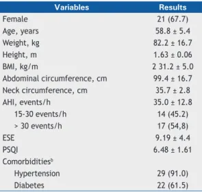
Table 1 presents demographic and polysomnographic characteristics of the population studied. We evaluated 31 patients, of whom 10 (32.2%) were male and 21 (67.8%) were female. The mean age was 58.8 ± 5.4 years, and the mean BMI was 31.2 ± 5.0 kg/m2. The mean AHI was 35.0 ± 12.8 events/h. Of the 31 patients evaluated, 14 (45.2%) had moderate OSAS and 17 (54.8%) had severe OSAS. Although all of the patients had poor sleep quality according to the Pittsburgh Sleep Quality Index, none showed excessive daytime sleepiness according to the Epworth Sleepiness Scale.
Table 2 presents data on lung function and respiratory muscle strength.
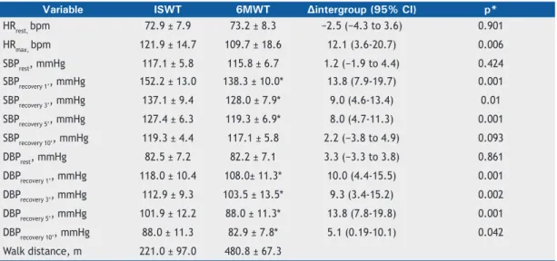
Table 3 presents comparisons of heart rates and blood pressure levels during the ISWT and the 6MWT. The following variables were signifi cantly higher during the ISWT than during the 6MWT: SBP 1 min after the test (p = 0.004), SBP 3 min after the test (p = 0.01), SBP 5 min after the test (p = 0.03), DBP 3 min after the test (p = 0.07), and DBP 5 min after the test (p =
0.001). The other variables did not present signifi cant differences in the comparison between the two tests.
Table 4 shows Spearman’s correlation coeffi cients for the anthropometric, spirometric, and inspiratory muscle strength variables related to the distances covered on the ISWT and the 6MWT. There was a strong, positive correlation between FVC and the distance covered on the ISWT (r = 0.658, p = 0.001), as well as a moderate positive correlation between the FEV1 and the distance covered on the ISWT (r = 0.522; p = 0.003). However, we found no correlations in relation to any of the other parameters evaluated.
DISCUSSION
The main fi ndings of the present study show that, in our obese patients with OSAS, lung function and inspiratory muscle strength were impaired. In addition, the functional exercise capacity was reduced and there was a signifi cant positive correlation between the distance covered on the ISWT and the variables related to pulmonary function.
In the present study, the values obtained for spirometric parameters and respiratory muscle strength were lower than the reference values for the healthy Brazilian population.(24) In the study conducted by Tassinari et al.,(25) no impairment of lung function or the respiratory musculature was observed in patients with OSAS. However, the OSAS patients evaluated in that study were of normal weight, unlike those in our sample, all of whom were obese. Likewise, Gontijo et al.,(26) studying obese individuals who did not present OSAS, obtained spirometric values within the limits of normality. The authors concluded that obesity would not be a factor associated with impairment of lung function. Studies indicate that obese individuals, due to the deposition of fat in the chest wall and abdomen, present a reduction in thoracic compliance, thus increasing the total work of breathing.(3,27,28) The upper airway collapse and the apnea-hypopnea events resulting from OSAS have respiratory consequences, such as hypoxemia, alveolar hypoventilation, and hypercapnia. Hypoxia-reoxygenation events lead to the activation of peripheral chemoreceptors leading to increased ventilation to correct alterations in blood gases.(10) Changes in O
2 and CO2 concentrations result in a decrease in respiratory muscle activity and a reduction in lung volumes.(8,9)
Table 1. Characteristics of the sample (N = 31).a
Variables Results
Female 21 (67.7)
Age, years 58.8 ± 5.4
Weight, kg 82.2 ± 16.7
Height, m 1.63 ± 0.06
BMI, kg/m 2 31.2 ± 5.0
Abdominal circumference, cm 99.4 ± 16.7
Neck circumference, cm 35.7 ± 2.8
AHI, events/h 35.0 ± 12.8
15-30 events/h 14 (45.2)
> 30 events/h 17 (54,8)
ESE 9.19 ± 4.4
PSQI 6.48 ± 1.61
Comorbiditiesb
Hypertension 29 (91.0)
Diabetes 22 (61.5)
BMI: body mass index; AHI: apnea-hypopnea index; ESS: Epworth Sleepiness Scale; and PSQI: Pittsburgh Sleep Quality Index.aValues expressed in n (%) or mean ± SD.bPatients with these comorbidities were receiving antihypertensive or hypoglycemic medications.
Table 2. Data on pulmonary function and respiratory muscle strength.a
Variable Result
FVC, L 2.4 ± 0.6
FVC, % predicted 76.4 ± 12.3
FEV1,% predicted 80.1 ± 6.32
FEV1, L 2.0 ± 0.4
FEV1/FVC ratio 79.6 ± 5.8
MIP, cmH2O 60.0 ± 21.9
MEP, cmH2O 81.3 ± 22.2
Regarding the association between pulmonary function and exercise tolerance, we found that FVC and FEV1 showed signifi cant positive correlations with the distance covered on the ISWT. This fi nding demonstrates that smaller lung volumes translate to lower exercise tolerance, underscoring the idea that pulmonary function affects exercise capacity in individuals with OSAS. Impairment of exercise capacity in obese individuals was also shown in another study, in which the authors concluded that being overweight is associated with a loss of self-esteem and lower psychic well-being.(29) Factors such as dyspnea, abnormalities in respiratory mechanics, respiratory muscle dysfunction, and arterial hypoxemia contribute to limiting exercise tolerance in OSAS patients.(14)
In our study, the distance covered on both walk tests was lower than the reference distance for healthy individuals in the Brazilian population,(30-32) indicating that exercise tolerance was below normal in our obese patients with OSAS. The patients in our sample also had comorbidities such as hypertension and type 2 diabetes mellitus. Exercise tolerance is directly related to the good performance of the cardiopulmonary system. Therefore, the impairment of functional exercise capacity in OSAS patients is multifactorial,
being associated with obesity, a sedentary lifestyle, cardiovascular diseases, dyspnea, and respiratory abnormalities.(14,15) However, there are confl icting data regarding the impaired exercise capacity resulting from the combination of OSAS with obesity in relation to exercise capacity. A study conducted by Beitler et al.(11) showed that peak oxygen consumption (VO
2peak) was signifi cantly lower in obese OSAS patients than in controls, also reporting an association between VO2peak and the AHI. In contrast, Rizzi et al.,(15) in a study of normal-weight OSAS patients, observed no impairment of the functional exercise capacity. However, in a subsequent study—a randomized clinical trial involving obese and normal-weight individuals, with or without OSAS—Rizzi et al.(33) observed a signifi cant difference in exercise tolerance between the obese and non-obese groups regardless of the presence ofOSAS, in terms of CO2 production and the maximum VO2. The authors concluded that obesity would be the main condition of low functional capacity, given that the obese patients presented low exercise tolerance, regardless of the presence or absence of OSAS.
Regarding the cardiovascular responses to the 6MWT in the present study, the cardiac parameters measured immediately after the test demonstrated marked Table 3. Variables collected during the incremental shuttle walk test and the six-minute walk test.a
Variable ISWT 6MWT ∆intergroup (95% CI) p*
HRrest, bpm 72.9 ± 7.9 73.2 ± 8.3 −2.5 (−4.3 to 3.6) 0.901
HRmax, bpm 121.9 ± 14.7 109.7 ± 18.6 12.1 (3.6-20.7) 0.006
SBPrest, mmHg 117.1 ± 5.8 115.8 ± 6.7 1.2 (−1.9 to 4.4) 0.424
SBPrecovery 1’, mmHg 152.2 ± 13.0 138.3 ± 10.0* 13.8 (7.9-19.7) 0.001
SBPrecovery 3’, mmHg 137.1 ± 9.4 128.0 ± 7.9* 9.0 (4.6-13.4) 0.01
SBPrecovery 5’, mmHg 127.4 ± 6.3 119.3 ± 6.9* 8.0 (4.7-11.3) 0.001
SBPrecovery 10’, mmHg 119.3 ± 4.4 117.1 ± 5.8 2.2 (−3.8 to 4.9) 0.093
DBPrest, mmHg 82.5 ± 7.2 82.2 ± 7.1 3.3 (−3.3 to 3.8) 0.861
DBPrecovery 1’, mmHg 118.0 ± 10.4 108.0± 11.3* 10.0 (4.4-15.5) 0.001
DBPrecovery 3’, mmHg 112.9 ± 9.3 103.5 ± 13.5* 9.3 (3.4-15.2) 0.002
DBPrecovery 5’, mmHg 101.9 ± 12.2 88.0 ± 11.3* 13.8 (7.8-19.8) 0.001
DBPrecovery 10’, mmHg 88.0 ± 11.3 82.9 ± 7.8* 5.1 (0.19-10.1) 0.042
Walk distance, m 221.0 ± 97.0 480.8 ± 67.3
ISWT: incremental shuttle walk test; 6MWT: six-minute walk test; SBP: systolic blood pressure; and DBP: diastolic blood pressure. aValues expressed as mean ± SD. *t-test for independent samples.
Table 4. Correlation between the selected variables and the distances covered on the incremental shuttle walk test and six-minute walk test.
Variables ISWT 6MWT
r p r p
BMI −0.320 0.07 −0.062 0.741
FEV1 0.522 0.003 0.117 0.532
FVC 0.658 0.001 0.189 0.308
MIP 0.075 0.069 −0.105 0.575
Abdominal circumference 0.056 0.996 −0.110 0.858
Neck circumference −0.032 0.862 −0.121 0.574
AHI 0.070 0.710 −0.111 0.551
increases in relation to the data collected during the test, results that are in agreement with those obtained by Rizzi et al.(33) in their study of obese patients with OSAS. However, the cardiovascular responses were even more pronounced during and after the ISWT, as was also reported by Gonçalves et al.(34) in a study of healthy individuals. In another study involving obese patients with OSAS, in which cardiopulmonary exercise testing (CPET) with the Bruce protocol was employed,(34) patients with severe apnea were found to show an increase in blood pressure during peak exercise with a return to basal blood pressure levels after exercise. Green et al.(35) stated that the ISWT provokes a physiological response to exercise similar to that observed during CPET.
Although the 6MWT is a standard test in the clinical assessment of effort in patients with OSAS(25) and obese patients,(36) the ISWT has also proven to be feasible and reliable in the assessment of effort tolerance in individuals with OSAS. Green et al.(35) studied patients with heart failure, comparing the responses obtained with CPET, the ISWT, and the 6MWT. They concluded that the ISWT provided a valid index to determine functional capacity in individuals with heart failure and that the predictive power of the ISWT could exceed that of the 6MWT. In a study evaluating patients with moderate or severe OSAS, Billings et al.(37) used the ISWT to compare physical fi tness before and after treatment with continuous positive airway pressure. The authors concluded that the ISWT is safe, well tolerated, and easily reproducible in OSAS patients,
supporting the idea that it can be used safely in the evaluation of exercise tolerance in such patients.
Our study has limitations, such as the small sample size. However, the number of patients evaluated was within the limits set in the sample calculation. In addition, we did not perform CPET, which is the gold standard test in the analysis of functional capacity and of cardiopulmonary impairment and would have allowed a more reliable evaluation of the cardiovascular parameters, as well as their subsequent correlation with those found on the two other tests. Another limitation was the fact that our study evaluated only obese patients with OSAS and there was no control group of obese patients without OSAS. Furthermore, data regarding cardiovascular comorbidities were not available for the patients in the sample studied. Therefore, studies with greater methodological rigor, such as randomized clinical trials, are needed in order to increase knowledge about the subject in the future.
The results found in the present study show that obese subjects with untreated OSAS presented below-normal lung function, inspiratory muscle strength, and physical capacity. In addition, it was observed that the decline in lung function, but not inspiratory muscle strength, is associated with physical effort tolerance in these patients, which makes it necessary to use therapeutic interventions to improve variables such as physical exercise. We can also emphasize that the ISWT was able to evaluate the exercise tolerance in OSAS, making it quite useful in the clinical investigation of the disease, due to its low cost, reproducibility, and ease of application.
REFERENCES
1. Huang JF, Chen LD, Lin QC, Chen GP, Yu YH, Huang JC, et al. The relationship between excessive daytime sleepiness and metabolic syndrome in severe obstructive sleep apnea. Clin Respir J. 2016;10(6):714-721. https://doi.org/10.1111/crj.12276
2. Hsia JC. Anatomy and physiology of the upper airway in obstructive sleep apnea. Oper Tech Otolayngol Head Neck Surg. 2015;26(2):74-77. https://doi.org/10.1016/j.otot.2015.03.005
3. Wimms A, Woehrle H, Ketheeswaran S, Ramanan D, Armitstead
J. Obstructive sleep apnea in women: specifi c issues and
interventions. Biomed Res Intern. 2016;2016:1764837. https://doi. org/10.1155/2016/1764837
4. Cholidou KG, Manali ED, Kapsimalis F, Kostakis ID, Vougas K, Simoes D, et al. Heart rate recovery post 6 minute walking test in obstrtuctive sleep apnea: cycle ergometry versus 6-minute walking test in OSA patients. Clin Res Cardiol. 2014;103(10):805-15. https:// doi.org/10.1007/s00392-014-0721-3
5. Martins AB, Tufi k S, Moura SM. Physiopathology of obstructive
sleep apnea-hypopnea syndrome. J Bras Pneumol. 2007;33(1):93-100. https://doi.org/10.1590/S1806-37132007000100017 6. Salome CM, King GG, Berend N. Physiology of obesity and effects
on lung function. J Appl Physiol (1985). 2010;108(1):206-11. https:// doi.org/10.1152/japplphysiol.00694.2009
7. Melo LC, Silva MA, Calles AC. Obesity and lung function: a systematic review. Einstein (Sao Paulo). 2014;12(1):120-5. https:// doi.org/10.1590/S1679-45082014RW2691
8. Haggstram FM, Zettler EW, Fam CF. Apnéia obstrutiva do sono e alterações cardiovasculares. Scientia Med (Porto Alegre). 2009;19:122-8.
9. Yokhana SS, Gerst DG 3rd, Lee DS, Badr MS, Qureshi T, Mateika JH. Impact of repeated daily exposure to intermittent hypoxia and mild sustained hypercapnia on apnea severity. J Appl Physiol (1985). 2012;112(3):367-77. https://doi.org/10.1152/japplphysiol.00702.2011
10. Mateika JH, Syed Z. Intermittent hypoxia, respiratory plasticity and sleep apnea in humans: present knowledge and future investigations. Respir Physiol Neurobiol. 2013;188(3):289-300. https://doi.org/10.1016/j.resp.2013.04.010
11. Beitler JR, Awad KM, Bakker JP, Edwards BA, DeYoung P, Djonlagic I, et al. Obstructive sleep apnea is associated with impaired exercise capacity: a cross-sectional study. J Clin Sleep Med. 2014;10(11):1199-204. https://doi.org/10.5664/jcsm.4200
12. Çiçek D, Lakadamyali H, Gökay S, Sapmaz I, Muderrisoglu H. Effect of obstructive sleep apnea on heart rate, heart rate recovery and QTc and P-wave dispersion in newly diagnosed untreated patients. Am J Med Sci. 2012;344(3):180-5. https://doi.org/10.1097/ MAJ.0b013e318239a67f
13. Rasslan Z, Saad R Jr, Stirbulov R, Fabbri RM, Lima CA. Evaluation of pulmonary function in class I and II obesity. J Bras Pneumol. 2004;30(6):508-14. https://doi.org/10.1590/S1806-37132004000600004
14. Lin CC, Hsieh WY, Chou CS, Liaw SF. Cardiopulmonary exercise testing in obstructive sleep apnea syndrome. Respir Physiol Neurobiol. 2006;150(1):27-34. https://doi.org/10.1016/j. resp.2005.01.008
15. Rizzi CF, Cintra F, Risso T, Pulz C, Tufi k S, de Paola A, et al. Exercise
capacity and obstructive sleep apnea in lean subjects. Chest. 2010;137(1):109-14. https://doi.org/10.1378/chest.09-1201 16. Ryan S. Adipose tissue infl ammation by intermittent hypoxia:
mechanistic link between obstructive sleep apnea and metabolic dysfunction. J Physiol. 2017;595(8):2423-2430. https://doi. org/10.1113/JP273312
17. Brum PC, Forjaz CL, Tinucci T, Negrão CE. Adaptações agudas e crônicas do exercício físico no sistema cardiovascular. Rev Paul Educ Fis. 2004;18:21-31.
Barba ME, et al. Validation of the Brazilian Portuguese version of the Pittsburgh Sleep Quality index. Sleep Med. 2011;12(1):70-5. https:// doi.org/10.1016/j.sleep.2010.04.020
19. Bertolazi AN, Fagondes SC, Hoff LS, Pedro VD, Menna Barreto SS, Johns MW. Portuguese-language version of the Epworth sleepiness scale: validation for use in Brazil. J Bras Pneumol. 2009;35(9):877-83. https://doi.org/10.1590/S1806-37132009000900009
20. American Thoracic Society/European Respiratory Society. ATS/ERS Statement on respiratory muscle testing. Am J Respir Crit Care Med. 2002;166(4):518-624. https://doi.org/10.1164/rccm.166.4.518 21. Sociedade Brasileira de Pneumologia e Tisiologia. Diretrizes para
testes de função pulmonar. J Pneumol. 2002;28(Suppl 3):S1-S238. 22. Singh SJ, Morgan MD, Scott S, Walters D, Hardman AE.
Development of a shuttle walking test of disability in patients with chronic airways obstruction. Thorax. 1992;47(12):1019-24. https:// doi.org/10.1136/thx.47.12.1019
23. ATS Committee on Profi ciency Standards for Clinical Pulmonary
Function Laboratories. ATS statement: guidelines for the six-minute walk test. Am J Respir Crit Care Med. 2002;166(1):111-7. https://doi. org/10.1164/ajrccm.166.1.at1102
24. Pereira CA, Sato T, Rodrigues SC. New reference values for forced spirometry in white adults in Brazil. J Bras Pneumol. 2007;33(4):397-406. https://doi.org/10.1590/S1806-37132007000400008
25. Tassinari CC, Piccin CF, Beck MC, Scapini F, Oliveira LC, Signori LU, et al. Capacidade funcional e qualidade de vida entre sujeitos saudáveis e pacientes com apneia obstrutiva do sono. Medicina (Rib Preto). 2016;49(2):152-9. https://doi.org/10.11606/issn.2176-7262. v49i2p152-159
26. Gontijo PL, Lima TP, Costa TR, Reis EP, Cardoso FP, Cavalcanti Neto FF. Correlation of spirometry with the six-minute walk test in eutrophic and obese individuals. Rev Assoc Med Bras (1992). 2011;57(4):380-6. https://doi.org/10.1016/S0104-4230(11)70081-1 27. Zieliński J. Effects of intermittent hypoxia on pulmonary
haemodynamics: animal models versus studies in humans. Eur Respir J. 2005;25(1):173-80. https://doi.org/10.1183/09031936.04.0 0037204
28. Cordeiro AL, de Melo TA, Neves D, Luna J, Esquivel MS, Guimarães AR, et al. Inspiratory Muscle Training and Functional Capacity in Patients Undergoing Cardiac Surgery. Braz J Cardiovasc Surg. 2016;31(2):140-4.
29. Tavares TB, Nunes SM, Santos MO. Obesidade e qualidade de vida: revisão de literatura. Rev Med Minas Gerais. 2010;20:359-66. 30. Soares KK, Gomes EL, Junior AB, Oliveira LV, Sampaio LM, Costa D.
Avaliação do desempenho físico e funcional respiratório em obesos. Fisioter Mov. 2011;24(4):697-704. https://doi.org/10.1590/S0103-51502011000400014
31. Fernandes PM, Pereira NH, Santos AC, Soares ME. Teste de caminhada de seis minutos: avaliação da capacidade funcional de indivíduos sedentários. Rev Bras Cardiol. 2012;25(3):185-91. 32. Dourado VZ, Guerra RL, Tanni SE, Antunes LC, Godoy I.
Reference values for the incremental shuttle walk test in healthy subjects: from the walk distance to physiological responses. J Bras Pneumol. 2013;39(2):190-7. https://doi.org/10.1590/S1806-37132013000200010
33. Rizzi CF, Cintra F, Mello-Fujita L, Rios LF, Mendonca ET, Feres MC, et al. Does obstructive sleep apnea impair the cardiopulmonar response to exercise? Sleep. 2013;36(4):547-53. https://doi. org/10.5665/sleep.2542
34. Gonçalves CG, Hayashu D, Mesquita R, Pitta F, Fernandes KB, Probst VS. Teste de campo “incremental shuttlle walking test” impõe esforço máximo a indivíduos saudáveis de diferentes faixas etárias? Rev Bras Fisioter. 2012;16:364-74.
35. Green DJ, Watts K, Rankin S, Wong P, O’Driscoll JG. A Comparison of the shuttle and 6 walking tests with measured peak oxygen consumption in patients with heart failure. J Sci Med Sport. 2001;4(3):292-300. https://doi.org/10.1016/S1440-2440(01)80038-4 36. Perecin JC, Domingos-Benício NC, Gastaldi AC, Sousa TC, Cravo
SL, Sologuren MJ. Teste de caminhada de seis minutos em adultos
eutrófi cos e obesos. Rev Bras Fisioter. 2003;7(3):245-51.