Anna Vadymivna Kladova
Metals in proteins from sulphate-reducing bacteria: adenylate kinase
and ATP sulfurylase
Proteins containing cobalt, zinc, iron (II) ions
Ph.D. Thesis
A Thesis submitted at the Faculty Science and Technology of the New University of Lisbon for a degree in Doctor of Philosophy in Biochemistry with specialization in Physical Biochemistry
- nº de arquivo
Acknowledgments
I have been fortunate enough to have been taught and allowed to work in the Laboratory of Biochemistry and Biophysics of Proteins at the Department of Chemistry at the New University of Lisbon under the supervision of Professor Dr. Sergey Bursakov. These years in my life represent a unique experience at the professional and personal levels.
I would like to thank Professor Dr. Isabel Moura and Professor Dr. José João Galhardas de Moura for receiving me at the Department and at the laboratory.
I would like to thank Professor Dr. Maria João Romão for her steadfast interest and attention to the work I was developing with cobalt/zinc-containing proteins. Over the years, I have had several colleagues who created an enjoyable and pleasant working environment at the Laboratory. Many thanks are due to Professor Dr. Cristina Costa, Professor Dr. Carla Carneiro, Professor Dr. Maria dos Anjos Macedo, Professor Dr. Jorge Caldeira, and Professor Dr. Stephane Besson.
My Thesis at the New University of Lisbon has had the invaluable help of many people that provided essential contribution to my work. I would like to thank the members of crystallography group of Professor Dr. M.J. Romão: Dr. Ana Luísa Carvalho, Cecília Bonifácio, Dr. Teresa Santos-Silva, Dr. Jóse Trincão, Dr. Abhik Mukhopadhyay and Dr. Shabir Najmudin.
Acknowledgements are also due to Ana Teresa Lopes, Celia Silveira, Alexandra Serra, Rui Almeida, Dr. Marta Santos, Dr. Marta Carepo, Dr. Raquel Grasina, Dr. Gabriela Almeida, Dr. Rui Duarte, Dr. Sofia Pauleta.
My gratitude goes also to several collaborators on this project from outside the New University of Lisbon for having made substantial contributions each in their own very personal way. I received excellent advice and assistance in the area of structural stability from Professor Valery Shnyrov from the University of Salamanca, Spain. I thank him for his friendship and for influencing much of my internal thought processes. I would like to thank also Dr. Juan J. Calvete from the Instituto de Investigaciones Biomédicas, CSIC, Valencia, Spain for his help and advice
in the work with mass spectrometry measurements.
But my fondest thanks go to my parents. Only with your love, genuine interest in what I do and infinite care could I to start and complete this work.
Finally, thanks to everyone for lots of great memories!
Summary
This work is devoted to the biochemical, biophysical and structural characterization of four proteins: cobalt-, zinc- and iron (II) - forms of adenylate kinase and Co2+/Zn2+ form of native ATP sulfurylase from two anaerobic Gram-negative strains of the sulphate-reducing bacteria
Desulfovibrio (D.) gigas NCIB 9332 and D. desulfuricans ATCC 27774, respectively. Both
enzymes were found to be involved in the bioenergetic metabolism of SRB: AK contributes to the maintenance of constant levels of cellular adenine nucleotides and nucleic acid synthesis, ATP sulfurylase catalyses the first step of sulphate activation to accomplish the reduction of sulphate to sulphide that is used to drive oxidative phosphorylation.
The methods for the production of fully cobalt- zinc- and iron (II) -substituted forms of adenylate kinase of D. gigas overexpressed into E. coli were developed. As a result,
homogeneous (with the ratio metal/protein 1/1) Co2+-, Zn2+- and Fe2+- forms of the protein were produced.
The presence of metal ions in the LID domain affects the kinetic properties of AK. Holo-AKgig shows the highest affinity of Fe2+-AKgig to the substrates of the backward reaction (MgADP/ADP), while Co2+- and Zn2+-AKgig have the highest affinity to the substrates of the forward reaction AMP and MgATP, respectively. Thus, the magnitude of the Km(AMP) and Km(MgATP) could be depicted in the following order: Fe2+-AKgig>Co2+-AKgig≈Zn2+-AKgig,
while for Km(ADP,MgADP) this order is Co2+-AKgig> Zn2+-AKgig> Fe2+-AKgig.
According to the disparities in the apparent optimal temperatures of Co2+-, Fe2+- and Zn2+ -AKgig for their AK activity (32 °C for Fe2+-AKgig, and 36 °C for Co2+- and Zn2+-AKgig) the type of the metal ion in the LID mobile domain to some extent controls the temperature dependence of the catalytic activity.
Far UV-CD spectra analysis indicates no differences between secondary structural elements of holo-AKgig, while tertiary structures of them, observed by near UV CD, are different. The major difference observed in tyrosine region for Fe2+- form of AKgig as compared to Co2+- and Zn2+- forms of AKgig.
The thermal denaturation of Co2+- and Zn2+-forms of AKgig (Tm values of 43.7 and 45.3 °C,
respectively) was determined as a cooperative two-state process, sufficiently reversible at pH 10, that can be correctly interpreted in terms of a simple two-state thermodynamic model. In contrast, the thermally induced denaturation of Fe2+-AKgig is irreversible and strongly dependent upon the scan rate, suggesting that this process is under kinetic control. The changes between Ts
Well ordered diffracted crystals of Co2+-, Zn2+- and Fe2+-AKgig were obtained, and the structures were resolved with a resolution of 2.0, 2.1 and 3 Å, respectively. The carbon skeleton superposition of Zn2+- AKgig with Co2+- and Fe2+-AKgig gives the rmsd values of around 0.1 and 0.37 Å, respectively, indicating very similar crystal structures for all of them. The NMA analysis indicates that metal ion nature does not influence the directionality of the substrate-binding domains (LID and AMPbd).
The thermal denaturation process of ATP sulfurylase from D. desulfurycans was found to be
strongly dependent upon the scan rate, suggesting that it is under kinetic control. The Tm and ΔHcal values of the ATPS thermal denaturation process were found to be independent within the
protein concentration range of 0.4 – 2.1 mg mL-1, suggesting, that the dimer does not dissociate into monomers when unfolds. Thus, the mechanism of ATPS denaturation process can be presented on the basis of the simple kinetic scheme: N2k D2 with the value of the free
stabilization energy of around 105 kcal per mol at 25 °C.
Phylogenetic analysis shows that the sequence of ATP sulfurylase from D. desulfuricans
is posted together with other ATPS from mesophiles, whereas ATP sulfurylase from thermophiles or psychrophiles are located in different clades. At that point clade of psychrophiles ATPS is situated more close in comparison to that of thermophiles.
Well diffracted crystals of native ATP sulfurylase from D. desulfuricans were obtained. The
Sumário
Este trabalho tem como o objectivo a caracterização do ponto de vista bioquímico, biofísico e estrutural de duas enzimas, a cinase do adenilato (AKgig) nas suas formas recombinantes contendo cobalto(II), zinco(II) e ferro(II), expressas em E. coli e Co/Zn ATP
sulfurilase na sua forma nativa (ATPS), que está presente nas bactérias redutoras de sulfato,
Desulfovibrio (D.) gigas NCIB 9332 and D. desulfuricans ATCC 27774 respectivamente.
Ambas as enzimas estão envolvidas no metabolismo bioenergético das bactérias redutoras de sulfato (BRS), sendo essencias para o seu crescimento e desenvolvimento. A cinase do adenilato contribui para a manutenção de níveis constantes da adenina e da síntese de ácidos nucleicos dentro da célula. A ATPS cataliza o primeiro passo da activação do sulfato para a sua posterior redução a sulfito, usado na redução de oito electrões envolvidos na fosforilação oxidativa.
A enzima AK recombinante foi sobre expressa em E. coli (na razão metal/proteína igual à
1/1) com obtenção das formas protéicas homogéneas contendo Co2+-, Zn2+- e Fe2+ .
A presença de iões metálicos no domínio LID da AKgig afecta as suas propriedades cinéticas. Entre as holo-formas da AKgig analisadas, o complexo Fe2+-AKgig demonstra a afinidade mais elevada para os substratos da reacção no sentido inverso (Mg2+ADP/ADP), enquanto que os complexos Co2+- e Zn2+-AKgig tem a maior afinidade para os substratos da reacção no sentido directo, que são AMP e Mg2+ATP. Assím, a magnitude dos parâmetros Km(AMP) e
Km(Mg2+ATP) da reacção no sentido directo, presenta a seguinte ordem: Fe2+-AKgig > Co2+
-AKgig ≈ Zn2+-AKgig, com os valores de Vm muito semelhantes. Os valores de
Km(ADP,Mg2+ADP), para a reacção no sentido inverso seguem a ordem de Co2+-AKgig > Zn2+
-AKgig > Fe2+-AKgig.
Foi testada a diferença nos valores da temperatura óptima para os complexos Co2+-, Fe2+- e Zn2+-AKgig na actividade da enzima. Os três perfis para a temperatura apresentam as formas semelhantes, com o valor máximo de 32 ºC para Fe2+-AKgig e 36 ºC para Co2+-e Zn2+-AKgig. Apesar de as diferenças na eficiência catalítica entre as formas enzimáticas contendo iões metálicos distintos serem relativamente pequenas, este resultado demonstra claramente que o tipo do metal presente no domínio móvel LID condiciona a dependência da actividade catalítica com a temperatura.
A maior diferença é observada nas regiões ricas em resíduos da tirosina para o complexo Fe2+ -AKgig , ao comparar com os complexos contendo cobalto e zinco.
Foi detectado o estado da desnaturação térmica reversível no valor de pH igual a 10, para os complexos enzimáticos com Co(II) e Zn(II), o que pode ser descrito pelo modelo simples de equilibrio de dois estados. Foram registados os valores de Tmde 43.7 ºC e 45.3 ºC para os iões de cobalto e zinco, respectivamente. No entanto, o processo da desnaturação térmica irreversível para Fe2+-AKgig apresentou o valor de T* de aproximadamente 45 ºC. Uma diferença acentuada
entre os valores Ts para Fe2+-AKgig , ao comparar com Co2+- e Zn2+-AKgig demonstra que as
propriedades hidrofóbicas de Fe2+-AKgig alteram como sendo o resultado da mudança na estrutura terciária.
Co2+-, Zn2+- e Fe2+-AK foram cristalizados. Através do método de cristalografia de raio X, foram detectadas as estruturas cristalinas para todas as formas enzimaticas, livres do substrato, na resolução de 2.0, 2.1 e 3.0 Å para Co2+-, Zn2+- e Fe2+- AKgig, respectivamente. A sobreposição do esqueleto carbónico do complexo Zn2+-AKgig com Co2+- e Fe2+-AKgig deu os valores de EQM (erro quadrático médio) de cerca de 0.1 e 0.37 Å, respectivamente, o facto que indica que todas as holoformas da enzima apresentam a estrutura cristalina semelhante.
O NMA (Normal Mode Analysis) identifica que as três formas enzimáticas exibem a mesma direccionalidade de dominios de ligação com os substratos (LID e AMPbd), independentemente da natureza do ião presente no complexo.
Demonstrou-se que a pH 9.5 o processo da desnaturação térmica do ATPS de D. desulfuricans ATCC 27774 depende fortamente “upon scan rate” o que sugere que está
submetido ao controlo cinético. Os valores de Tm e ΔHcal para a desnaturação térmica de ATPS, à
pH 9.5 não dependem da concentração proteica entre 0.4 à 2.1 mg mL-1 ao sugerir que o dímero no estado não dobrado não se dissocia para as formas monoméricas. O mecanismo da desnaturação da ATPS pode ser apresentado com base na esquema cinética simples: N2k D2,
com o valor de energia livre de estabilização cerca de 105 kcal/mol à 25 ºC. A análise filogenética demonstrou que a sequência da ATPS de D. desulfuricans é semelhante às
sequências para a mesma enzima em outros mesófilos, enquanto que ATPS de termófilos ou psicrófilos pertencem aos ramos filogeneticamente distintos. Conclui-se ainda que ATPS dos psicrófilos, é filogeneticamente mais próxima comparando com os termófilos.
Table of contents
Chapter Title Page
Summary 5
Sumario 7
Table of contents 9
Abbreviations and symbols 10
Microorganisms 12
1 General introduction 15
2 Zinc-, cobalt- and iron-chelated forms of adenylate kinase from the Gram-negative bacterium Desulfovibriogigas.
35
3 Cobalt, zinc and iron forms of adenylate kinase from the sulphate-reducing bacteria Desulfovibrio gigas: purification, crystallization and preliminary
X-ray diffraction analysis.
53
4 Metal containing adenylate kinase from Desulfovibrio gigas: Crystal structure
of a novel metal containing adenylate kinase from Gram negative bacteria.
61
5 Purification, crystallization and preliminary X-ray diffraction analysis of adenosine triphosphate sulfurylase from the sulfate-reducing bacterium
Desulfovibrio desulfuricans ATCC 27774.
77
6 Structural stability of ATP sulfurylase from sulphate-reducing bacteria
Desulfovibrio desulfuricans ATCC 27774
83
Discussion 104
Conclusions 111
Abbreviations and symbols
ε molar extinction coefficient,
Abs absorption
AK Adenylate kinase
ADP adenosine-5´- diphosphate
AMP adenosine-5´-monophosphate
ATP adenosine-5`-triphosphate
BSA bovine serum albumin
CD circular dichroism
DSC differential scanning calorimetry
Cp partial specific heat capacity
IPTG isopropyl β-D-thiogalactoside
Km Michaelis-Menten constant
LB Luria-Bertani Broth, Miller
LMCT ligand-to-metal charge transfer
MALDI-TOF matrix-assisted laser desorption-ionization - time of flight
SDS-PAGE sodium dodecyl sulfate-polyacrylamide gel electrophoresis
Tm temperature at the midpoint of the unfolding transition
UV ultraviolet
rmsd root mean square deviation
SRB Sulphate-reducing bacteria
PPi Inorganic pyrophosphate
ATPS Adenosine triphosphate sulfurylase
Ts the temperature of maximum stability
∆G free energy changes
∆H calorimetric enthalpy changes
∆HvH van’t Hoff enthalpy changes
Km Michaelis constant
Vmax maximum initial velocity
AKcol adenylate kinase from Escherichia coli
AKglo adenylate kinase from Bacillus globisporus
AKsub adenylate kinase from Bacillus subtillis
AKaeol adenylate kinase from Aquaflex aeolicus
AKvol adenylate kinase from Methanococcus voltae
AKac adenylate kinase from Sulfolobus acidocaldarius
ATPS Adenosine-triphosphate sulfurylase
Da Dalton
FPLC Fast Performance Liquid Chromatography
PCR Polymerase chain reaction
Microorganisms
A. vinelandii Azotobacter vinelandii
B. stearothermophilus Bacillus stearothermophilus
B. subtilis Bacillus subtilis
D. desulfuricans Desulfovibrio desulfuricans
D. gigas Desulfovibrio gigas
D. vulgaris Desulfovibrio vulgaris
E. coli Escherichia coli
P. denitrificans Paracoccus denitrificans
T. brucei Tripanosoma brucei
M. maripaludis Methanococcusmaripaludis
M. tuberculosis Mycobacterium tuberculosis
Pn. chrysogenum Penicillium chrysogenum
A. aeolicus Aquifex aeolicus
Sc. cervisiae Saccharomyces cervisiae
R. sphaeroides Rhodobacter sphaeroides
T. thermophilus Thermus thermophilus HB8
A. fulgidus Archaeoglobus fulgidus
Chapter 1
Sulphate-reducing bacteria
Sulphate-reducing bacteria (SRB) are those prokaryotic microorganisms, both bacteria and archaea, that can use sulphate as the terminal electron acceptor in their energy metabolism, i.e.
that are capable of dissimilatory sulphate reduction. SRB are widespread in nature and play
important roles in sulphur cycles in soil, sediments from freshwater and marine environments, and deep-sea hydrothermal vents [1-3]. Generally they require a complete absence of oxygen and a highly reduced environment to function efficiently. Hydrogen sulphide- the final product of their respiratory metabolism process is chemically reactive and quite toxic to animals and plants. SRB are usually classified by their oxygen sensitivity and their ability to use sulphate as a terminal electron acceptor [4].
Members of this genus Desulfovibrio are the most readily cultured and best studied of the
SRB. The bioenergetics of sulphate reduction in the genus Desulfovibrio is summarized in
Scheme 1 when the bacteria grow in presence of lactate and sulphate. The bacteria generate two ATP molecules by phosphorylation at substrate level coupled to the oxidation of two molecules of pyruvate. On the other hand, one ATP is consumed to activate sulphate to adenosine -5´-phosphosulphate (APS) and another ATP is used to regenerate two ADP molecules by adenylate kinase. The net production of ATP cannot be attained by substrate-level phosphorylation alone. Therefore, for growth on lactate and sulphate, Desulfovibrio has an obligatory requirement for
electron transfer-coupled phosphorylation for the net production of ATP. The reduction of bisulphite to sulphide must compensate the energy investment of sulphate activation and yield additional ATP for growth. Three molecules of ATP are formed in an electrontransfer-coupled manner during the reduction of one molecule of sulphite to sulphide.
Reaction Enzyme
2lactate ↔ 2pyruvate + 4e- + 4H+ lactate dehydrogenase 2pyruvate + 2CoA ↔ 2acetyl-CoA+ 2CO2 + 2e- + 2H+ pyruvate: ferredoxin
oxidoreductase 2acetyl-CoA+2Pi ↔ 2acetyl phosphate + 2CoA phosphotransacetylase 2acetyl phosphate + 2ADP ↔ 2acetate + 2ATP acetate kinase
SO42-+ ATP ↔ APS + PPi ATP sulphurylase
MgPPi + H2O ↔ 2Pi + Mg2+ inorganic pyrophosphatase
APS + 2e- ↔ SO32- + AMP APS reductase
AMP + ATP ↔ 2ADP adenylate kinase
SO32- + 6e- + 8H+ ↔ H2S + 3H2O sulfite reductase 2lactate + SO42-+ 2H+↔2acetate + 2CO2 + H2S + 2H2O net reaction
Scheme 1. The bioenergetics of sulphate reduction [5, 6].
Adenylate kinase
Adenylate kinase (EC 2.7.4.3. ATP:AMP phosphotransferase) catalyzes the reversible transfer of the γ- phosphate group from Mg2+ATP to the phosphate moiety of AMP, producing Mg2+ADP and ADP with high-energy turnover [7]:
AMP + Mg2+ATP ADP + Mg2+ADP AK also catalyses, to a lesser extent, the following reaction [8, 9]:
ADP + Mg2+ATP AMP + Mg2+Ap4
This reaction has an absolute requirement for Mg2+ and occurs at a lower rate by way of an associative type in-line displacement without an enzyme bound intermediate [10].
The role of AK is to facilitate the storage and use of the high energy of adenine nucleotides in cell. AK is an essential catalyst for bacterial growth and multiplication and belongs to a family of essential enzymes since its inactivation is not compatible with cell survival [11].
There are two oligomeric classes of AK: monomeric as found in Eubacteria and trimeric as found in Archaebacteria [12]. The sequence identity and structure of AK from Eubacteria and Archaea are related only distantly, suggesting that archaeal AK belong to a different AK class.
The structure of the trimeric AK from the mesophile Archaebacteria Methanococcus maripaludis (AKmar), which has Tm (temperature at the midpoint of the unfolding transition)
value close to 74 ° C, has been solved to the resolution of 2.5 Å in Ap5A inhibitor presence. Consistent with other methanococcal AK, the trimeric interface is comprised of a three-helix bundle formed by the long helix of each subunit (Fig. 1). The potentially important intrasubunit ion-pair was identified at the trimeric interface of AKmar between Glu150 and Arg156 with the
average distance of 3.35 Å.
Fig. 1 Trimeric structure of AKmar, the trimeric interface consists of a three helix bundle.
The bound Ap5A molecule (green) spans the active site of the enzyme [12].
All AK share a highly related tertiary structure with the five-stranded parallel β-sheet with helices on both sides and could be divided into three main domains: the CORE, the LID and AMP binding domain (AMPbd) or also called NMPbd (Fig. 2):
The CORE (1Met-29Ile, 60Thr-121Val, 160Gln-214Gly for AK from E. coli (AKcol)) is the largest
domain that includes the central parallel β- sheet and the immediately packed helices (1-4 and
Fig. 2 Structure of AKcol on the open conformation [17].
The AMPbd domain (30Ser-59Val for AKcol) is helical (α2 and α3 for AKcol) and contributes to the AMP binding, closing over bound AMP.
The LID domain (122Gly-159Asp for AKcol) includes a four stranded antiparallel β-sheet (
5-8 for AKcol). In long-type AK this domain consists of around ~37 amino acid residues and only a short loop in the other AK. The LID domain is well solvent exposed domain that undergoes large movement during catalysis and upon substrate binding, closing over the active site [18] and protecting the MgATP/AMP-ternary complex from bulk water, facilitating phosphoryl transfer and preventing hydrolysis [19]. The amino acid sequences of the LID domains are highly conserved among the long type AK. The LID domain of AK containing Zn2+ has its chain fold the same as those of other LID domains [20]. LID has been suggested to interact with other cellular components, fulfilling further functions [21].
Generally, AK from Gram-positive bacteria contain a Cys-X2-Cys-X16-Cys-X2-Cys/Asp structural motif in its LID domain that is responsible for the binding of zinc ion [13, 20, 22], whereas the AK from Gram-negative bacteria are usually devoid of metal ions, since their Cys residues are substituted by another four highly conserved amino acids - His, Ser, Asp and Thr, respectively [23]. Nevertheless, previously published exceptions are AK from Desulfovibrio gigas and Desulfovibrio desulfuricans ATCC 27774, that contain either cobalt or zinc [24], the
AK from Paracoccus denitrificans, overproduced in E. coli [15, 16], that binds either zinc or
iron, and the AK from Chlamidia pneumoniae and Thermotoga neapolitana, that contain zinc
[25, 26]. Metal ion in AK was proposed to play rather structural than catalytic role [23, 27], enhancing thermal stability of the enzyme. Thus, mutant of AK from E. coli with genetically
with comparable kinetic parameters [23]. Removal of the metal ion in AKgig was accompanied with decreasing of Tm up to 5.3 °C with invariable heat capacity, indicating that removal of metal
ion does not result in any appreciable decomposition of the structure of the enzyme [28]. Previous studies have shown also that zinc depletion reduces Tm value by 7.5 °C and 6.3 °C in
AK from Bacillus stearothermophilus and T. neapolitana, correspondingly [26, 29].
The study of series of chimeric AK based on mesophile Bacillus subtilis and the
thermophile Bacillus stearothermophilus, that share high sequence homology (74 %), indicates
that CORE domain governs overall stability of the enzyme, while two other mobile domains themselves (the AMP and LID domains) control the temperature dependence of the catalytic activity [30].
The catalytic mechanisms of bacterial and eucaryotic AK have been studied extensively, mainly due to their biological importance and ubiquitous distribution of the enzyme [7, 17, 31-35]. The presence of two substrate binding sites per AK molecule has been shown: the one site for metal-free substrates (AMP and ADP) and another for metal-bound substrates (Mg2+ATP and Mg2+ADP). The basic kinetic mechanism was proposed to be random Bi Bi, where binding of any of two substrates (Mg2+ATP, for instance) favours bound of the second one (AMP), that causes the products production (Mg2+ADP and ADP) [31]. Later on, an iso- random Bi Bi mechanism was proposed [36]. The major difference of the iso- Bi Bi mechanism is the presence in equilibrium at least two forms of enzyme, where the first form can bind substrates of the forward reaction (Mg2+ATP and AMP), whereas the other can bind substrates of the reversed reaction (Mg2+ADP and ADP). The conformation of the ternary complex of AK in a complex with AMP and Mg2+ATP is different from that of enzyme binding with ADP and Mg2+ADP. Thus, AK undergoes at least two steps of conformational changes. One step is the change of a ternary complex with bound substrates to one with bound products during the reaction, and the other is the change of free AK into different forms. In a catalytic cycle, the conformational changes of the free enzyme and the ternary complexes are the rate-limiting steps. This mechanism is based on the fact that AK undergoes large domain movements upon substrate binding [37].
AK are in general very specific with respect to phosphate acceptors [7, 38]. However, in several enzymes some activity was observed when AMP was substituted by 5´-AMP and dAMP [7, 39]. The short form of AK from Tripanosoma brucei can use AMP, CMP and UMP as an
[42]. AK are not very specific with respect to phosphate donors. All AK except SK3 and AK6 prefer Mg2+ATP at the ATP site and can be substituted with 2`-dATP, GTP, CTP, UTP, or ITP to some extent [7, 38]. AK3 can use both GTP and ITP as effective phosphate donors, but GTP is most probably the physiological substrate [7]. AK6 can use all NTPs and dNTPs with CTP as the best phosphate donor [41].
A bivalent metal ion is required for AK activity. Thus, for rabbit and yeast AK the order of reactivity is Mg2+ > Mn2+ > Ba2+ and Mg2+> Ca2+ > Mn2+ > Ba2+, respectively, while for bovine liver AK the order is different: Mg2+ >Mn2+ >Ca2+, Co2+. In spite of the individual differences observed, the Mg2+ ion shows the highest activity [7]. The ATP, ADP and AMP species bind magnesium ion with very different stability constants. Moreover, Mg2+-bound and Mg2+-free species have quite diverse, sometimes opposite, effects on enzymes. Nucleoside triphosphates bind magnesium very tightly, and their actions as cofactors and energy-rich compounds are carried out in complexes with Mg2+. Nucleoside diphosphates bind Mg2+ with a less extent (the stability constant is 2500 M-1) [7], and the enzymes that use Mg2+ADP are usually strongly inhibited by free form of ADP. The uncomplexed forms of both ADP and ATP may cause appreciable inhibition of the kinase-type phosphotranferase enzyme systems [43, 44]. Nucleoside monophosphates, particularly AMP, that allosterically regulates many key enzymes, bind Mg2+ very weakly [45] and participate in metabolic processes in the magnesium-free form [43].
AK require a bivalent metal ion for its efficient catalytic activity and, as was mentioned before, Mg2+ shows the highest activity through binding with ATP forming Mg2+ATP. Magnesium ion serves to neutralize the highly negative charge of the ATP, thereby reducing electronic repulsion of the transferred phosphate and increasing the efficiency of nucleophilic attack. In AK, conserved serine, threonine, or aspartate residues participate in binding the Mg2+, coordinating it either directly in the first coordination sphere or indirectly through water molecules. In the structure of AK from B. stearothermomophilus (AKbst), magnesium ion is
coordinated to β- and γ-phosphates of ATP and four water molecules (300-303) [20]. Binding of the Mg2+ activates catalysis by orienting phosphate donor and acceptor for further phosphoryl transfer. Two of the four water molecules (300 and 303), are within hydrogen bound distances from the α-phosphate of AMP. Water molecule 300 is hydrogen bounded to 33Asp and 36Arg. The last residue interacts with the phosphate of AMP and is highly conserved. Water molecules 301 and 303 are hydrogen-bonded to 84Asp, while 301 to 14Gly. Water molecule 302 reinforces the linkage between phosphate and Mg2+, interacting with the oxygen atoms of the α-, β-, and γ-phosphates of ATP.
chains at the active site [20]. This assembles a functional ternary complex in which the phosphate oxygen atoms of AMP is free to attack the Mg2+polarized -phosphate of ATP. Once phosphate transfer has occurred, the interactions between two water molecules (300 and 303) with the new ADP -phosphate and residues 33Asp and 36Arg connected with these molecules provides a new scaffolding for the Mg2+ coordination complex and a starting point for the back reaction. Water molecule 302 may then act to facilitate the back reaction by hydrogenbonding back to the terminal phosphate of the ADP in the Mg2+ADP/Mg2+ATP binding site.
The partially open position of the LID domain has increased access of the bulk water to the active site compared to the fully closed position. This serves to hydrate both the phosphates chains of the substrates/products and the polar residues of the LID. Bulk water, which is excluded from the active site during the reaction, can then interrupt the binding of the phosphate chains by replacing hydrogen bonds to the protein with hydrogen bonds to water molecules [20].
Two water molecules 218 and 220 in the crystal structure of AKcol (Fig. 3), that correspond to water molecules 300 and 302 from AKbst have additional H-bonds interactions with 156Arg [46]. 156Arg has been implicated in the stabilization of the transition state of the phosphoryl transfer reaction of the enzyme. The 156Arg side chain mobility between binding states suggests the stabilizing role of the top (the LID domain side) of this amino acid, while 13Lys and the Mg2+ stabilize the bottom and the front [47].
Fig. 3 Structure of AK from E. coli on the closed conformation; ATP, and AMP are in stick representation; the Mg2+ atom and its 4 coordinating water molecules are in a ball-and-stick representation [46].
chain. 84Asp also forms a salt bridge with 13Lys in P-loop. 13Lys stabilizes the active site by forming H-bonds and salt bridge-like interactions with both ATP (-PO4) and AMP (-PO4) using its flexibility and multifunctionality. In addition to Lys, Mg2+, its coordination waters, and some surrounding charged residues maintain the geometry and distances of the AMP -phosphate and ATP - and -phosphates in a configuration that suggests that phosphoryl transfer occurs by associative mechanism in AK [46]. The results of oxygen-18 kinetic isotope effect experiments support the notion that phosphoryl transfer occurs through an associative transition state [48].
Recent analysis shows, that the Lys171, in human AK4, which is located in a hinge that binds LID and CORE domains and is strickly conserved in all AK family is essential for orientation of the LID domain (Fig. 4). Thus, the provided mutation of Lys171Pro dramatically changes the orientation of the LID domain, which could be described as a novel twistedand- closed conformation in contrast to the open and closed conformations in other AKs [49].
Fig. 4 Overal structural comparison of human AK4 Lys171Pro mutant with the native AK4 in open and closed form (left is native AK4 open form, middle is native AK4 bound with GP5 in close form and right is the AK4 Lys171Pro [49].
Adenylate kinase from D. gigas is monomeric enzyme with the molecular mass of 24.7
kDa that bind either cobalt (II) or zinc (II) ions [27]. The UV-Vis spectrum of AKgig contains the
zinc-binding motif in another AK. Both metal ions were reported to play rather structural than catalytic role.
ATP sulfurylase and sulphate activation
The sulphate reduction plays a very important role in the sulphur cycle. Most plants and microorganisms can use inorganic sulphate as their sole source of sulphur. Because sulphate is nonreactive at cellular temperatures and pH, the anion must first be ″activated″ in order to enter the mainstream of metabolism [50]. In biological systems, sulphate is chemically activated via adenylation. Activated substrate is ready to be used by essential biological processes such as the biosynthesis of reduced sulphur metabolites, driving oxidative phosphorylation; sulphuryl group donation, modification of neuropeptides and peptide hormones, cell-cell adhesion, detoxification, homeostasis processes that may be critical for life.
N N N N NH2 O OH OH H H H H O P O OH O S O O OH O OH O H H H H O P O OH O S O O OH P O OH HO APS PAPS N N N N NH2
Fig. 5 Structural formulas of APS and PAPS.
ATP sulphurylases (EC 2.7.7.4. MgATP:SO42- adenylyltransferase) are ubiquitous enzymes that catalyze the transfer of the adenylyl group from ATP to inorganic sulphate, yielding adenosine-5´-phosphosulphate (APS) (Fig. 5) and pyrophosphate (PPi):
MgATP + SO42- APS + MgPPi
The phosphoric-sulfuric acid anhydride bond of APS has a free energy of hydrolysis that is approximately twice that of the replaced pyrophosphate bond of ATP [51], making the synthesis of APS and the inorganic pyrophosphate a highly unfavourable reaction. This energy dilemma is resolved differently in different organisms.
coupling APS synthesis to GTP hydrolysis [52, 53]. However, the APS-synthesizing enzyme in these bacteria (known as CysD) is unrelated to the APS-synthesizing enzyme found in most other organisms [54]. The GTP-hydrolyzing enzyme (known as CysN) is also specific to these bacteria [55].
In the reductive branch of the pathway, APS-reductase reduces APS to sulfite, with thioredoxin, providing the reducing potential. Additional enzymatic steps subsequently reduce sulfite to sulfide and incorporate the latter into thiol- and sulfur-containing molecules as previously mentioned. Alternatively, APS may undergo further activation into 3'-phosphoadenosine 5'-phosphosulfate (PAPS), the activated sulfate donor needed for sulfation. APS is activated into PAPS by the phosphorylation activity of APS-kinase (EC 2.7.1.25. ATP:adenylylsulfate-3-phosphotransferase):
MgATP + APS MgADP + PAPS
PAPS is used by all sulfotransferase enzymes to covalently attach sulfate to carbohydrates, proteins, and small organic molecules. PAPS can also be reduced to sulfite via a PAPS-reductase [56].
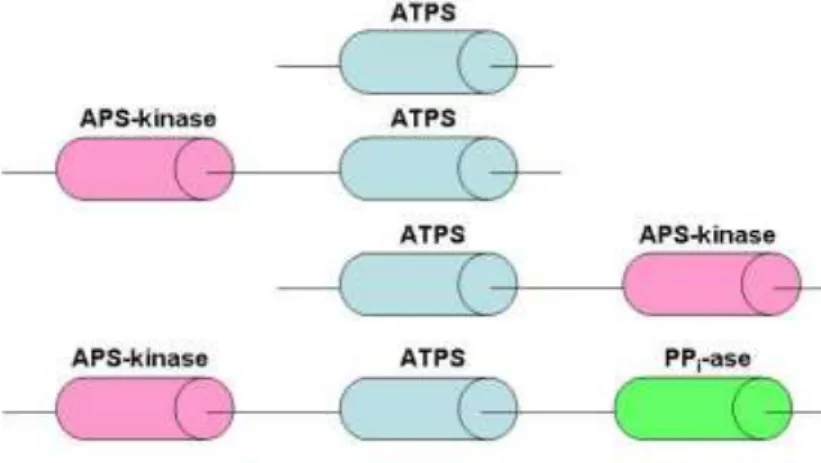
Fig. 6 Structural architecture of the ATPS family of proteins.
In most plant and microbial cells, ATP sulfurylase, APS kinase and PPi-ase are separate proteins. However, animals and some bacteria possess a ″PAPS synthetase″ in which the two sulphate activating enzymes are co-located on a single polypeptide chain in some cases [57]. Besides, ATP sulfurylase can fused also with APS-kinase and pyrophosphatase (PPi-ase) are known (Fig. 6) [56].
argued that the kinase-sulfurylase fusion protein enables the labile APS intermediate to be rapidly ‘channeled’ from the sulfurylase active site into the neighboring kinase active site [61-66]. In some fungal and bacterial species, a fusion of the ATP sulfurylase and APS kinase domains is found with a reversed domain order (Fig. 6) relative to the metazoan gene have been described from the filamentous fungi Penicillium chrysogenum [67], the chemolithotrophic
bacterium Aquifex aeolicus [68, 69] the budding yeast Saccharomyces cerevisiae [70], and the
proteobacterium Rhodobacter sphaeroides [66]. The kinase domain of the sulphurylase-kinase
fusion protein is not well conserved, and a range of functions has been attributed to the kinase domain in different organisms. In S. cerevisiae the APS kinase domain of sulfurylase-kinase
fusion is important for the quaternary structure of the enzyme but is highly degenerate at the sequence level [71]. In Penicillium chrysogenum the kinase domain of sulfurylase-kinase protein
serves as a Pbinding allosteric regulator of the sulfurylase domain and has lost all APS-kinase activity [72, 73]. In Aquifex aeolicus and Rhodobacter sphaeroides the kinase domain of
sulfurylase-kinase fusion protein still functions as an APS-kinase [66, 69].
In several protozoan genomes within the Stramenopile lineage was discovered a novel triple fusion protein of ATP sulfurylase, APS and pyrophosphatase (PPi) also located on a single polypeptide chain (Fig. 6) [56].
It appears, that isolated from different sources, ATP sulfurylase family is heterogeneous in terms of amino acid sequences, molecular masses, subunit composition and behaviour. Two completely different, unrelated types of ATP sulfurylase can be distinguished:
1. The heterodimeric type, which occurs exclusively in sulphate-assimilating prokaryotes, e.g. two subunits with masses of 23 and 53 kDa in Escherichia coli K-12 [51], 35 kDa and 68
kDa in M. tuberculosis [74], nodulating, and some other bacteria [51].
2. All other ATP sulfurylases characterized in sufficient detail are monomers, dimers and hexamers with a range of molecular masses from 38 to 69 kDa per subunit [75, 76]. Correspondingly, size variations are due to APS kinase, PAPS or PPi-binding allosteric domains residing on the same polypeptide [56, 57, 64, 67, 70, 75, 76].
sulfurylase binds and stabilizes the PPi group of ATP before and during hydrolysis of the α, β-bond, and assist the dissociation process [70].
ATP sulfurylase are in general very specific to ATP. Thus, only ATP and to a lesser extent dATP serve as effective substrates, although the latter was not tested for ATP contamination [79, 80]. The preference of adenine nucleotides can be explained by a hydrogen bond in which the N1 nitrogen of the adenine ring is the hydrogen acceptor and the main chain amide of Val333 of ATP sulfurylase from Penicillium chrysogenum is the hydrogen donor [81]. Pyrimidine
nucleotides would be too far from the Val333 main chain amide to make this hydrogen bond and N1 nitrogen of guanine nucleotides is protonated and thus cannot be a hydrogen bond acceptor.
The real substrate for ATP sulfurylase catalysis is magnesium complex of ATP, while free ATP is the competitive inhibitor. Magnesium ions required for catalysis and not for nucleotide binding [82]. In the reverse direction, only MgPPi is ATPS´s substrate, while divalent oxyanions [X = molybdate (MoO42–), selenate (SeO42–), tungstate (WO42–), chromate (CrO42–), and also
arsenate (AsHO42–)] can inhibit ATP sulfurylase [79]. They render adenosine phosphate
complexes (APX) unstable and spontaneously hydrolyze to form AMP. The monovalent oxyanions (perchlorate (ClO4–), nitrate (NO3–), chlorite (ClO3–), and fluorosulphonate (FSO3–) also compete with sulphate for ATP sulfurylase, thereby inhibiting APS formation. However, they differ from the divalent oxyanions in that APX complexes are not formed [79, 80].
The stereochemical course of the reaction catalyzed by the ATP sulfurylase from yeast has been determined. It proceeds with an inversion of the stereochemistry at the α-phosphoryl moiety, suggesting that the reaction follows an ″in-line″ nucleophilic attack by SO42–at the
α-phosphoryl group of ATP [83]. The structural data confirm this substitution mechanism (see below) that leads to direct APS formation with PPi as the leaving group and without covalent adenylyl-enzyme intermediates formation [70]. The enzyme provides suitable binding sites for ATP and sulphate, brings the reactive groups in close contact, and promotes stabilization of the nucleotide conformation that is most favourable for ″in line″ attack of the sulphate. Thus, the β-phosphate group of ATP is hydrogen-binding to the one His residue of HXXH and Arg of GRD motifs, while other His coordinates the γ-phosphate of ATP, SO42-binds to QXRN and GRD motifs. The mechanism for ATP sulfurylase from Penicillium crysogenum, Aquifex aeolicus and
rat liver was proposed to be random for the forward reaction substrates (ATP and SO42-) and preferred order for the backward reaction substrates (for the first APS ligation and only then PPi) [69, 80, 84]. The mechanism operates in a similar way for both spinach leaf and, probably, Riftia
enzymes [85, 86].
The crystal structure of ATP sulfurylase from Thermus thermophilus HB8 [87] has special
be much more similar to that of D. desulfuricans. The ATP sulfurylase of Thermus thermophilus
is composed of a N-terminal domain (residues 1-34), a catalytic domain (residues 135-290) and domain III (residues 291-347), but it lacks a C-terminal domain. APS is located in the active site of ATP sulfurylase, which contains several conserved motifs (QXRN, HXXH and GRD). The active site pocket of the Thermus thermophilus ATP sulfurylase-APS complex is more compact
than the active site pocket of Saccharomyces cerevisiae and Penicillium chrysogenum
ATPS-APS complex, indicating adaptation to a hot environment (Fig. 7).
The zinc ion is located in the N-terminal half of linker domain, immediately after catalytic domain and is tetrahedrally coordinated by Cys294, Cys297, Cys306, and His310, and could not be removed from the protein by treatment with EDTA. The zinc-binding region appears to be a hinge between catalytic and linker domains and also has been involved in protein-protein interactions.
The linker domain covers the active site of Thermus thermophilus ATP sulfurylase and the
adenine moiety of bound APS. The distances from the zinc ion to the adenine ring of the bound APS and to His from HXXH motif are 14 and 17 Å, respectively, and zinc is distant from the active site. It has been suggested that Zn2+binding and/or the dimer interaction may contribute to tight active site and to holding the conformation of this region in the correct orientation [87].
Fig. 7 Comparison of the active sites of ATP sulfurylase from Thermus thermophilus HB8
and Saccharomyces cerevisiae. The ATP sulfurylase of Sc. cerevisiae (grey) was superimposed
over that of the ATP sulfurylase from Thermus thermophilus [domain III (green) and other
(purple)]. APS (purple) and zinc (green) are shown as ball-and-stick, respectively [87]
ATP sulfurylase from Saccharomyces cervisiae and Penicillium chrysogenum are
363 equivalent α-carbons. Hovewer, the yeast enzyme is not allosterically inhibited by PAPS because of the lacks of many C-terminal residues responsible for sulfonucleotide binding [50].
The crystal structure of ATP sulfurylase from Saccharomyces cerevisiae as substrate-free
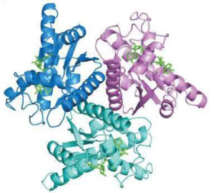
and product complex (with APS and PPi bound) have been determined to the resolution of 1.95 Å and 2.6 Å, respectively [70]. The protein crystallizes reproducibly within 2 days from solution containing Cd2+ as an additive. The enzyme is a homohexamer with the D3 symmetric structure,
where two trimer rings assemble in a staggered configuration with the twist of 60 ° (Fig. 8). Interactions within the hexamer are mediated through hydrogen bonding and salt bridges including a broad system of solvent molecules. A number of cadmium ions also contribute to the formation of the monomer-monomer interfaces.
Fig. 8 Hexameric structure of ATPS from Saccharomyces cerevisiae[70].
The monomer of Sc. cerevisiae is characterized by a high ratio of regular secondary
structure elements and can be divided into four domains: the N-terminal, catalytic, linker and C-terminal domains (Fig. 9). The N-terminal part of the peptide chain (residues 2-167), comprises a main β-barrel motif formed from five anti-parallel β-strands with several helical insertions, and an additional short β-hairpin motif. The N-terminal and catalytic domains interact mainly through hydrogen bonds and salt bridges.
Fig. 9 Ribbon plot of the monomer of yeast ATP sulfurylase. The colour-coding represents the four domains, N-terminal (green), catalytic (gold), linker (red) and C-terminal (blue). The location of the active site is indicated by the binding position of a phosphate ion [70].
The linker domain (residues 328-393) is relatively short and links the C-terminal and catalytic domains. This domain can assist in the formation of the upper section of the ATP sulfurylase substrate binding pocket. The highly conserved PFR motif allows some flexibility when ATP binds to the enzyme. Nucleotide binding causes significant conformational changes, which lead to a rigid-body structural displacement of C-terminal and linker domains towards N-terminal and catalytic domains of the ATP sulfurylase monomer and may serve for substrate recognition.
The C-terminal domain (residues 394-511) displays an α, β-fold with five-twisted parallel β-strands. The C-terminal domain of yeast ATP sulfurylase is structurally quite independent from the rest of the molecule, and not essential for catalytic activity. However, C-terminal domain is intimately involved in the association of three monomers in a trimer ring and also in the association of two of those rings in the mature hexamer. Truncation of this domain results in a monomeric enzyme with slightly enhanced catalytic efficiency [71]. Structural alignment of the C-terminal domain indicates that it is extremely similar in its fold to fungal APS kinase, although not catalytically competent, indicating a potential evolutionary relationship with a bifunctional PAPS synthetase. The ATP sulfurylase structure supports the hypothesis that the primary function of the APS kinase-like domain is to stabilize the oligomeric state of the enzyme.
Yeast ATP sulfurylase (Fig. 9) exhibits a sufficiently deep and wide, as well as properly positioned, surface groove that connects the active site of the adenylyltransferase and defunct active site of the APS kinase-like domain. It is tempting to speculate that this groove represents a substrate channel between the ATP sulfurylase and the APS kinase-like domains of the enzyme.
and β- phosphates of a nucleotide in many nucleotidyl transferases, are located in the ATP sulfurylase product complex far away from the α-phosphorous of the APS, with a distance of 5.7 Å. This suggests that histidines of ATP sulfurylase are not directly involved in the cleavage of α-β-phosphodiester bond, but play a role in pyrophosphate binding and stabilization of a productive ATP conformation.
The structure of ATP sulfurylase from Saccharomyces cerevisiae was also solved in a
complex with its second substrate ATP. Due to the horseshoe-like conformation of ATP in the pocket, this model allows an ″in-line″ nucleophilic attack of the adjacent sulphate (Fig. 10). In support of kinetic data and stereochemical investigations [89] the structural data [70] confirm a substitution mechanism with stereochemical inversion at the α-phosphorus leading directly to the formation of APS, with PPi as leaving group and without covalent adenylyl-enzyme intermediates. The enzyme provides suitable binding sites for its substrates, ATP and sulphate, that brings the reactive groups in close contact and promotes the stabilization of the nucleotide conformation most favourable for the ″in-line″ attack at the sulphate. During the reaction cycle, it stabilizes the pentavalent transition state of the reactants. As typical for ATP-binding enzymes, a Mg2+ ion could coordinate to the β and γ-phosphate groups but has not been identified in the crystal structure.
Fig. 10 Stereoview of the active site of ATP sulfurylase with sulfate and a modelled ATP molecule in the typical horseshoe conformation. Hydrogen bonds are shown in cyan; the direction of the nucleophilic attack of the sulfate is marked in green [70].
ATP sulfurylase from Penicillium chrysogenum is a homohexameric enzyme that is subject
of allosteric inhibition by PAPS (the product of APS kinase). The crystal structures of ATP sulfurylase from Penicillium chrysogenum were solved in the presence of either APS or PAPS
not induce a totally new subunit conformation but rather may exploit the existing flexibility of the enzyme [50]. The major difference between ATP sulfurylase from yeast and Penicillium chrysogenum is the C-terminal domain, while the catalytic domains have the same topology.
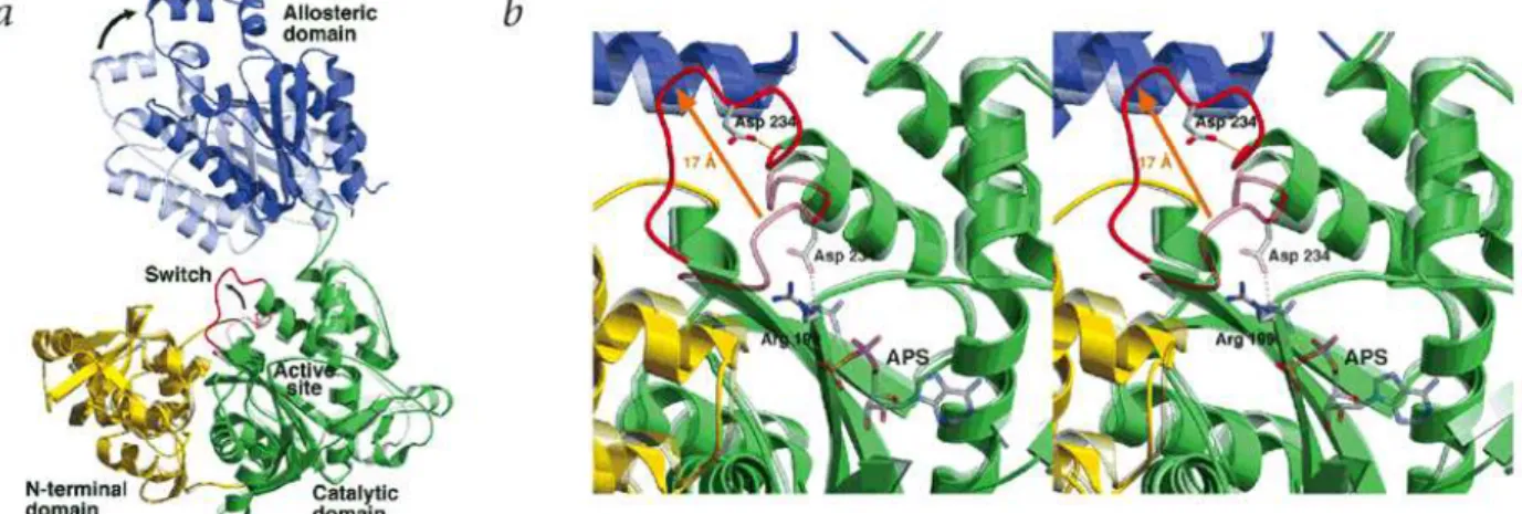
Fig. 10. Conformational changes observed in the R-to-T allosteric transition.
a, R- and T-state subunit superimposition. Domains of a single subunit are colored as follows: N-terminal domain (yellow), catalytic domain (green) and allosteric domain (blue). The R-state subunit is shown partially transparent to distinguish it from the T-state. The active site switch (residues 228−238) is red b, Active site switch. The active site region of the T-state structure is shown superimposed on the R-state active site. The R-state structure and bound APS are shown partially transparent to distinguish them from the T-state structure. The active site switch is red [90].
Penicillium chrysogenum ATP sulfurylase missing the C-terminal allosteric domain is
catalytically active but it is monomeric and much less stable than the hexameric wild type enzyme. In contrast to the wild type, all kcat values were decreased. Additionally, the Michaelis
constants for Mg2+ATP and sulphate (molybdate), the dissociation constant of E-APS, and the monovalent oxyanion dissociation constants of dead end E-Mg2+ATP- oxyanion complexes were
all increased. The cumulating results indicate that besides serving as a receptor for the allosteric inhibitor, the C-terminal domain stabilizing the hexameric structure and indirectly, individual subunits [50].
ATP sulphurylase from D. desulfuricans ATCC 27774 (ATPSdes) was described as
Co2+/Zn2+- containing homotrimer with molecular mass of 141 (3 × 47) kDa
. The UV-Vis
The objectives of this work were:
1. To develop methods for the production in vivo of fully cobalt- zinc- and iron-substituted
forms of recombinant adenylate kinase from D. gigas overexpressed in E. coli.
2. To develop methods for crystallization of Co2+, Zn2+ and Fe2+-AKgig and Co2+/Zn2+ -ATPSdes enzymes and resolve their structures.
3. To determine the role of the three metal ions (Co2+, Zn2+ and Fe2+) in the LID domain of adenylate kinase from D. gigas by different biochemical and biophysical tecniques. To study the
mechanism of thermal denaturation of holo-AKgig and role of these metals to the stability and activity of these proteins.
Chapter 2
Zinc-, cobalt- and iron-chelated forms of adenylate kinase from the Gram-negative bacterium Desulfovibriogigas.
This chapter was published as:
Anna V. Kladova, Olga Yu. Gavel, Galina G. Zhadan, Valery L. Shnyrov, Sergey A. Bursakov. 2009 Zinc-, cobalt- and iron-chelated forms of adenylate kinase from the Gram-negative bacterium Desulfovibrio gigas. International Journal of Biological Macromolecules
Abstract
Adenylate kinase from the sulphate-reducing bacterium Desulfovibrio gigas (AKgig) has been
characterized earlier as a Co2+/Zn2+-containing enzyme that is an unusual characteristic for adenylate kinases from Gram-negative bacteria, in which these enzymes are normally devoid of metal ions.AKgig was overexpressed in E. coli and homogeneous Co2+-, Zn2+- and Fe2+-forms of
the enzyme were obtained under in vivo conditions. Their structural stability, spectroscopic and
Introduction
Adenylate kinase (AK, ATP:AMP phosphotransferase, EC 2.7.4.3), a member of the nucleoside monophosphate kinase family, is a small monomeric protein that mediates the reversible transfer of phosphate groups between adenine nucleotides, which are the main substrates, co-factors, or allosteric effectors in a series of key metabolic reactions [7].
Like many nucleotide-binding proteins, AK belongs to the α/β class, with a five-stranded β -sheet surrounded by several α-helices [98]. The formation of the ternary complex stabilizes the enzyme in a form where the mobile small LID and AMP-binding sub-domains close over the remaining CORE region. This rearrangement of the two mobile sub-domains is necessary for the accommodation of the nucleotides in an optimal catalytic geometry, and the resulting closed enzyme conformation provides a solvent-free environment for phosphoryl transfer [18].
Generally, the AK from Gram-positive bacteria contain a Cys-X2-Cys-X16-Cys-X2-Cys/Asp structural motif in the LID domain that is responsible for the binding of zinc ion [13, 20, 22], whereas the AK from Gram-negative bacteria are usually devoid of metal ions, since their Cys residues are substituted by another four highly conserved amino acids - His, Ser, Asp and Thr, respectively [23]. Nevertheless, exceptions are the AK from Desulfovibrio gigas and Desulfovibrio desulfuricans ATCC 24774, which contain either cobalt or zinc [24], the AK from Paracoccus denitrificans, overproduced in E. coli [15], which binds either zinc or iron, and the
AK from Chlamidia pneumoniae and Thermotoga neapolitana, which contain zinc [25, 26].
Thus, to date three different metal ions -zinc, cobalt and iron- have been found to be present in the AK from a few Gram-negative bacteria.
Besides the somewhat rare presence of metal ions in the AK of several Gram-negative bacteria, attention has to be focused on the varieties of the metal-binding centres (Cys-X25-Cys/His-X14–19-Cys-X2-Cys/Asp) involved in different organisms [24] whose specific properties
may be involved in metal selection.
Metal ions play a variety of roles in natural proteins, including nucleophilic catalysis, electron transfer, and the stabilization of protein structure. They also play a structural role in AK, as confirmed in several crystallographic studies [19, 20, 37]. Mutations in the LID domain lead to considerable differences in the overall stability of AK [23, 99, 100]. Zinc binding at the genetically engineered zinc-chelating site of E. coli AK affords the microorganism considerably
higher thermal stability, with a Tm value of 61.6 °C versus 52.5 °C for the AK from the
wild-type, with hydrogen bonding in the LID domain [23, 100]. Previous studies have also shown that zinc depletion reduces the Tm value of D. gigas by 5.3 °C [28]; that of T. neapolitana by 6.3 °C,
A method for the production chimeras by exchange of certain parts of thermophilic and mesophilic organisms has been applied to confirm their main responsibilities as regards the activity and stability of AK [30]. This, together with mutagenesis studies, has shown that the CORE of AK is responsible for the structural stability, while dynamics of the LID and AMPbind domains have been suggested to be related to catalysis [18, 30, 37, 101]. DSC results show that the stabilities of the mesophilic and thermophilic AMPbind and LID domains are similar, and hence their overall stabilities are limited by the stabilities of their CORE domains. Additionally, the results of activity assays clearly show that the two mobile domains themselves (the AMPbind and LID domains) may control their own functional dynamics and may cause differences in activity. Bae and Phillips confirmed their suggestion that AK catalysis is regulated by the intrinsic properties of the moving domains [30].
Knowledge of structural stability and functional activity is important for understanding the binding mode and the putative role of different metal ions in AK. Thus, in the present work we describe a detailed investigation of the thermal stability of homogeneous Co2+-, Fe2+- and Zn2+ -forms of AKgig, using different independent methods such as differential scanning calorimetry (DSC), UV-Vis-spectroscopy, circular dichroism (CD) and kinetic assays.
Materials and methods
Materials.
The reagents hexokinase, pyruvate kinase, lactate dehydrogenase, phosphoenolpyruvate, NADP+, NADH, AMP, ADP, MgATP were from Sigma. Glucose-6-phosphate dehydrogenase was from Merck.
Gene Cloning and Expression Screening.
The gene coding for the AK of D. gigas (672-bp DNA fragment) (EMBL Nucleotide
Sequence Database accession number FN424087) was amplified (annealing temperature 64 ºC) from D. gigas genomic DNA by polymerase chain reaction (PCR) with the appropriate upstream
primer 5’-GGGGCTCGAGCA/TATGAACATCCTGATCTTCGGTCCGAACGGC-3’ and the downstream primer 5’-CCCCGGATCCA/AGCTTTTAGGCAAGCTGGGCCAG-3’. The PCR fragments containing upstream NdeI and downstream HindIII restriction sites were cloned into
the pMOSBlue vector (GE Healthcare) and the nucleotide sequence of the PCR product was
verifiedby DNA sequencing. The NdeI and HindIII digestion product of the DNA fragment was
then cloned into the expression vector 22b(+) (Novagen). The resulting plasmid pET-22b(+)/AK with the gene inserted was used for protein expression in E. coli strain BL-21(DE3)
(Stratagene).
Translation of the insert gene induced in mid-log phase (A660 nm 0.6 – 0.7) by the addition of 1 mM isopropyl 1-thio-β-D-galactopyranoside (IPTG) to minimal M63B1 medium (0.1 M KH2PO4, 15 mM (NH4)2SO4, 0.8 mM MgSO4, 3 µM vitamin B1, pH 7.4) supplemented with 0.4 % glucose [16] at 37 °C with vigorous shaking for 4 – 6 h depends on the type of AK to be expressed. The incubation temperature and the metal concentration in the medium were optimized in order to obtain homogeneous protein with a single metal. Typically, cells were grown in 1 L of M63B1 medium containing 100 µg/ml of ampicillin and the expression conditions were as follows: for Co2+-AKgig, 6 h and minimal medium supplemented with 160 µM CoCl2; for Zn2+-AKgig, 4 h and 250 µM ZnCl2; for Fe2+-AKgig - 4 h and 130 µM FeCl2 at 37 °C.
After these incubation periods, cells were harvested by centrifugation at 5000 rpm for 30 minutes at 4 °C. Then, cells were resuspended in 20 mM Tris-HCl, pH 7.6, at a ratio of 14 (w/v), and disrupted by passing them through an EmusTex-C5 Homogenizer at 9000 psi. Phenylmethylsulphonyl fluoride (PMSF) at 1 mM concentration was added to the broken cells fraction as an inhibitor of serine proteases. The extract was centrifuged at 125000 g for 90 min at 4 ºC and the pellet was discarded. A clear supernatant containing the soluble fraction was then used for the purification of AK; it was processed immediately to avoid protein degradation.
Purification of recombinant AK from E. coli.
The AK of D. gigas overexpressed in E. coli was purified as described earlier [24, 102] in a
two-step FPLC (Pharmacia) procedure on Blue Sepharose (Cibacron Blue 3G-A Sepharose CL-6B) fast flow and Superdex-75 gel filtration columns at 4 ºC. After the second column, the protein was eluted in 50 mM Tris/HCl buffer, pH 7.6, and 250 mM NaCl, after which it was concentrated, dialyzed, andkept frozen at -20 °C. The protein concentration was determined with the Bicinchoninic Acid Protein Assay Kit from Sigma.
Determination of protein purity and metal analysis.
Protein purity was determined by SDS-PAGE at 12.5 % (w/v), as described by Laemmli [103], in each purification step. The molecular mass standards from Bio-Rad were myosin (200 kDa), -galactosidase (116.3 kDa), phosphorylase b (97.4 kDa), bovine serum albumin (66.2 kDa), ovalbumin (45 kDa), carbonic anhydrase (31 kDa), soybean trypsin inhibitor (21.5 kDa), lysozyme (14.4 kDa) and aprotinin (6.5 kDa). Protein staining was performed using R-250 Coomassie blue.
Measurement of the velocity of the forward reaction was performed by monitoring the oxidation of NADH at 340 nm by coupling with pyruvate kinase (PK) and lactate dehydrogenase (LDH). The reaction mixture contained 50 mM Tris-HCl, pH 7.6, 100 mM KCl, 0.25 mM MgCl2, 0.2 mM NADH, 1 mM PEP, 15.5 U/ml of LDH and 25 U/ml of PK. The concentrations of AMP and MgATP were varied for the specific requirements of each experiment. The reaction was started by adding 30-50 ng of AK. One unit (U) is defined as 1 µmol of ADP (MgADP) generated per minute.
The rate of the backward reaction (MgADP and ADP as substrates).
The rate of the backward reaction was measured by following the reduction of NADP+ at 340 nm in an enzyme solution coupled with hexokinase and glucose-6-phosphate dehydrogenase. The reaction mixture contained 50 mM Tris-HCl, pH 7.6, 100 mM KCl, 1 mM glucose, 8 mM NADP+, 5 U of hexokinase, and 5 U of glucose-6-phosphate dehydrogenase. The concentrations of ADP and MgCl2 were varied for the individual needs of each experiment. The reaction was started by adding 30-50 ng of AK. One unit is defined as 1 µmol of ATP generated per minute.
End-point kinetic assay (optimum temperature).
AKgig (90 ng/ml) was incubated over a broad temperature range (4 °C - 40 °C) for 3 min in a buffer (total volume 280 l) containing 100 mM NaHCO3/NaOH, pH 10.0, and 10 mM MgATP, AMP and MgCl2. The reactions were stopped by the addition of 1 ml of 1.2 M perchloric acid, kept on ice and centrifuged at 25000 g for 5 min. An 800-l aliquot of the supernatant was neutralized with 350 l of 2 M KOH in 230 mM Tris-HCl, pH 7.6. The mixture was centrifuged at 25000 g for 5 min and the 200 l aliquot was used for ADP determination. The reaction mixture (total volume 1ml) contained 50 mM Tris/HCl buffer, pH 7.6, 2 mM MgCl2, 100 mM KCl, 1 mM PEP, and 0.3 mM NADH. The reaction was started by adding 7.8 U of LDH and 12.5 U of PK. The samples were monitored at 340 nm for NADH oxidation. Blanks were obtained for each temperature point.
Differential scanning calorimetry.
accomplished on the basis of the two limiting cases of Lumry-Eyring [105] model; namely, the two-state equilibrium folding/unfolding model ( N K D
), where only the native (N) and denatured (D) states of the protein are significantly populated and their relative amounts at a given temperature are determined by the value of the equilibrium constant (K), and a simple
two-state irreversible model ( N k F
), in which only the native state (N) and the irreversibly denatured state (the final state: F) of the protein are significantly populated and where k is the
first-order rate constant that changes with temperature according to the Arrhenius equation.
Circular dichroism.
The CD spectra of AK were recorded on a Jasco-715 spectropolarimeter, using a spectral band-pass of 2 nm and cell pathlengths of 1 mm in the far-ultraviolet range (195-250 nm) and of 10 mm in the near-ultraviolet range (250-320 nm). In these measurements, protein concentrations of ca. 0.1 and ca. 1 mg/ml were used, respectively. Four spectra were scanned for each sample at a scan rate of 20 nm/min and were then averaged. All spectra were background-corrected, smoothed, and converted to mean residue ellipticity = 10 Mres obs l-1p-1), where Mres is the mean residue molar mass; obs is the ellipticity (degrees) measured at wavelength ; l
is the optical pathlength of the cell (dm), and p is the protein concentration (mg ml-1). Secondary
structure analysis of the CD spectra in the far-ultraviolet range was performed using theCDPro software package [106].
Results and Discussion
Protein expression and purification.
Fig. 1. Gel-electrophoresis in all the purification steps of AK. 1-LMW standard; 2-broken cells; 3-soluble fraction; 4- fraction after Blue Sepharose column; 5- fraction after Superdex-75 column.
Sequencing of the N-terminus MNILIFGPNGSGKGTQGNLVKDKYSLAHIE confirmed that the recombinant protein was identical to the AK from D. gigas.
UV-Vis spectra.
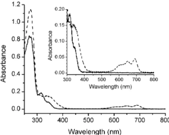
Fig. 2. Electronic absorption spectra of Fe2+-AK (dashed line) (31 µM) and Co2+-AK (solid line) (41 µM) in 50 mM Tris-HCl buffer pH 7.4.
Fe2+-AKgig exhibits three maxima at 275, 311 and 344 nm (ε311 = 5270 M-1cm-1, ε345 = 2390 M-1cm-1) (Fig. 2). The band at 311 nm and shoulder at 340 nm are due to the S- → Fe2+ charge transfer, in good agreement with the UV-Vis spectrum of Fe2+-AK from
Paracoccus denitrificans [15].
The UV-Vis spectrum of Zn2+-AKgig exhibits a maximum at 275 nm. Zinc provides the LMCT band with sulphur atoms at 275 nm [111], which is hidden by the absorption of aromatic amino acid residues of the protein.
Kinetic parameters.
The kinetic properties of recombinant holo-forms of AK are shown in Table 1. Metal-chelated forms of AKgig have much higher affinity for the substrates of the forward reaction with higher Vmax values than those of the backward reaction. Similar differences between the Vmax values of the forward and backward reactions have been reported for the AK from E. coli and
yeast extract, while for the AK from P. denitrificans they are comparable [16].
Table 1. Comparative kinetic parameters of metal-chelated forms of AKgig.1
protein Km(AMP),
M
Km(MgATP),
M
Vmax (AMP, MgATP),
µmol/(min·mg)
Km(ADP),
M
Vmax (ADP)
µmol/(min·mg)
Zn2+-AK 46 4 34 3 1335 165 25 980
Co2+-AK 40 4 49 4 1310 247 30 730
Fe2+-AK 71 4 76 4 1220 128 20 690
Km (ADP) and Vmax (ADP) were calculated according to the assumption that two molecules of ADP bind to the enzyme with the same affinity. The Km for AMP and MgATP was determined at a single fixed concentration of co-substrates (50 µM).
![Fig. 3 Structure of AK from E. coli on the closed conformation; ATP, and AMP are in stick representation; the Mg 2+ atom and its 4 coordinating water molecules are in a ball-and-stick representation [46]](https://thumb-eu.123doks.com/thumbv2/123dok_br/16474989.731970/22.892.341.616.646.979/structure-closed-conformation-stick-representation-coordinating-molecules-representation.webp)
![Fig. 4 Overal structural comparison of human AK4 Lys171Pro mutant with the native AK4 in open and closed form (left is native AK4 open form, middle is native AK4 bound with GP5 in close form and right is the AK4 Lys171Pro [49]](https://thumb-eu.123doks.com/thumbv2/123dok_br/16474989.731970/23.892.138.823.506.813/overal-structural-comparison-mutant-native-closed-native-middle.webp)