w w w . j c o l . o r g . b r
Journal of
Coloproctology
Review Article
Screening anal cancer in women living with
HIV/AIDS
Vanessa Laís Diefenthäler
a,b,∗, Janice de Fátima Pavan Zanella
a,b, Janaina Coser
a,baPrograma de Pós-Graduac¸ão Stricto Sensu em Atenc¸ão Integral à Saúde – Universidade de Cruz Alta (UNICRUZ) e Universidade
Regional do Noroeste do Estado do Rio Grande do Sul (UNIJUÍ), Cruz Alta/Ijuí, RS, Brazil
bLaboratório de Citopatologia, Universidade de Cruz Alta, Cruz Alta, RS, Brazil
a r t i c l e
i n f o
Articlehistory:
Received 6 October 2017 Accepted 24 January 2018
Keywords:
HIV HPV Anal cancer
a b s t r a c t
Aim: Addressing the main methodologies published in the scientific literature and used to screen anal cancer in women living with HIV/AIDS.
Methodology:The current study is an integrative literature review applied to articles pub-lished between 2013 and 2017 in databases such as PUBMED, EBSCO and LILACS.
Results:Eight studies were selected to compose the current review after the inclusion and exclusion criteria were applied. All the articles had evidence level IV. Anal cytology and the DNA-HPV test were the methodologies prevailing in the studies. The number of partic-ipants in the studies ranged from 35 to 863, and all the studies involved women living with HIV/AIDS. The aim of most of the herein reviewed studies was to assess the prevalence of anal cytologic changes or HPV infection in women living with HIV/AIDS (WLHA).
Conclusion: Studies have pointed out that there is concern about high anal cancer and anal HPV infection rates. They also highlighted the importance of the screening procedure for anal cancer prevention through cytology associated, or not, with molecular HPV detection methods.
© 2018 Sociedade Brasileira de Coloproctologia. Published by Elsevier Editora Ltda. This is an open access article under the CC BY-NC-ND license (http://creativecommons.org/ licenses/by-nc-nd/4.0/).
Rastreamento do câncer anal em mulheres vivendo com HIV/AIDS
Palavras-chave:
HIV HPV Câncer anal
r e s u m o
Objetivo:Abordar as principais metodologias que podem ser utilizadas para o rastreamento do câncer anal em mulheres vivendo com HIV/AIDS, que têm sido publicadas atualmente na literatura científica.
Metodologia:Trata-se de uma revisão integrativa de literatura, realizada através de pesquisa de artigos nas bases de dados PUBMED, EBSCO e LILACS, publicados entre os anos de 2013 a 2017.
∗ Corresponding author.
E-mail:vanessa.diefenthaler@yahoo.com.br(V.L. Diefenthäler). https://doi.org/10.1016/j.jcol.2018.01.001
Resultados:A partir da aplicac¸ão dos critérios de inclusão e exclusão, foram selecionados oito estudos para compor essa revisão. Todos possuíam nível de evidência IV. As metodologias que predominaram nos estudos foram a citologia anal e o teste DNA-HPV. O número de participantes nos estudos variou de 35 até 863, e todos envolveram mulheres vivendo com HIV/AIDS. A maioria tinha o objetivo de avaliar a prevalência de alterac¸ões citológicas anais ou infecc¸ão pelo HPV em mulheres vivendo com HIV/AIDS.
Conclusão: Os estudos apontaram que há uma preocupac¸ão com os altos índices de câncer anal e infecc¸ão anal por HPV. Também registram a importância do rastreamento para prevenc¸ão do câncer anal, através da citologia associada ou não a métodos moleculares de detecc¸ão do HPV.
© 2018 Sociedade Brasileira de Coloproctologia. Publicado por Elsevier Editora Ltda. Este ´e um artigo Open Access sob uma licenc¸a CC BY-NC-ND (http://creativecommons.org/ licenses/by-nc-nd/4.0/).
Introduction
Anal cancer development is associated with human papillo-mavirus (HPV) infection.1The advancement in the
antiretro-viral therapy (ART) enables women living with human immunodeficiency virus (HIV) or with acquired immunode-ficiency syndrome (AIDS) to increase their life expectancy. However, it raises their risk of developing comorbidities such as anal cancer.2,3
HIV/HPV coinfection in WLHA is a risk factor for the development of such neoplasm, mainly because of the
immunocompromised condition faced by these patients.4In
addition, WLHA are at increased risk of infections caused by more than one HPV type, mainly those of high oncogenic risk such as 16 and 18.3,5 It is estimated that 70% of anal
can-cer cases are HPV-dependent, since the infection persistence predisposes patients to develop the disease.2,4
The overall incidence of anal cancer is low; however, this incidence has been increasing in WLHA,5,6whose risk of
devel-oping the disease is 5 to 14 times higher than that of women without HIV.7 In addition, the prevalence of anal infections
caused by HPV is higher in WLHA with history of cervical abnormalities.8
The anal cancer screening in some countries is con-ducted through cytological examination, which is followed by anoscopy and/or biopsy, whenever abnormalities are detected. Anal cytology may be performed similarly to that of the cervix, since the anus also has a transformation zone, which
is vulnerable to HPV infection and lesion development.7The
examination is performed through the conventional method,
or liquid medium9; sample collection may be performed
by scraping the anal canal with a brush, or Dacron swab, moistened in physiological solution, introduced 2–4 cm in the canal. The slides are processed through Papanicolaou tech-nique and the results are classified according to the Bethesda System.10–12
Anoscopy is recommended when anal cytology shows abnormal results, since the exam identifies lesion sites to be treated and subjected to biopsy. The results need fur-ther confirmation through histopathological examination.3,4
In addition, the use of the HPV test, both as virus detection method and as viral typing, at low or high oncogenic risk, has been constantly discussed as complementary to cytological screening.13
A screening program may be effective to this at-risk popu-lation, due to increase in the number of WLHA cases, since it would help detecting pre-neoplastic lesions and enable their treatment, thus reducing the incidence of anal cancer.14
Therefore, the aim of the current study is to address the main methodologies available in the scientific literature, which may be used to screen anal cancer in WLHA.
Methodology
The present study is an exploratory, descriptive and inte-grative literature review based on the search, assessment and synthesis of available evidences on the herein addressed subject. The research was conducted in March 2017. It com-prised databases belonging to the US National Library of Medicine/National Institutes of Health (PUBMED), to the Latin-American and Caribbean System on Health Sciences Information (LILACS), and to the Elton B. Stephens Company (EBSCO). The following meshes were used in the search: “Anal cancer in HIV”; “Anal cancer cytology HIV”; and “Anal cancer screening HIV”. The meshes were crossed through the Boolean operator AND.
Table 1 – Characterization of selected articles according to publication data, study type, evidence level, sample size and methodology.
Article Author Place Year
Study type/evidence level
Sample size/recruitment period
Methodology
A1 Gaisa et al.
USA 2016
Retrospective cohort study
IV
745 WLHA 2009 to 2014
Conventional cytology (anal) Anoscopy
A2 Goeieman et al.
South Africa 2017
Cross-sectional study IV
200 WLHAa Conventional cytology (anal and cervical)
Anoscopy
DNA-HPV test: Digene Hybrid Capture 2 and Aptima E6/E7 mRNa to detect HPV
A3 Hessol et al.
USA 2013
Nested cohort cross-sectional study IV
470 WLHAb
2001 to 2003
Liquid-based cytology (anal) Conventional cytology (cervical)
Colposcopy and Anoscopy
DNA-HPV test: PCR and hybridization typing
A4 Pittyanont et al.
Thailand 2014
Prospective descriptive study IV
590 WLHA 2013 to 2014
Conventional cytology (anal)
DNA-HPV test: Cervista human papillomavirus high risk (HPV HR) assays and Cervista HPV 16/18 assays
A5 Sananpanichkul et al.
Thailand 2015
Prospective descriptive study IV
599 WLHA 2013 to 2014
Conventional cytology (anal and cervical)
A6 Bisherwal et al.
India 2016
Cross-sectional study IV
40 WLHAa
2011 to 2013
Conventional cytology (anal and cervical)
A7 Cambou et al.
Brazil 2015
Cohort study IV
863 WLHA 2011 to 2013
Liquid-based cytology (anal and cervical)
Anal DNA-HPV test: PapilloCheck HPV-Screening Test Cervical DNA-HPV test: Linear-Array HPV
Genotyping Test
A8 Heard et al.
France 2015
Nested cohort cross-sectional study IV
171 WLHA 2012
Liquid-based cytology (anal and cervical) Anoscopy
DNA-HPV test: Linear-Array HPV Genotyping Test
WLHA, women living with HIV/AIDS.
a The period when the study was conducted was not described.
b The study also included women without HIV, whose results were not included because they were not relevant to the aim of the integrative
review.
Results
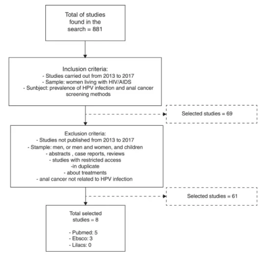
The mesh-based search generated 881 studies in total; eight were selected according to the pre-established inclusion and exclusion criteria. Five out of the eight studies derived from PUBMED, three derived from EBSCO, and no study was selected from LILACS (Fig. 1).
Most of the studies (3 articles – 37.5%) were carried out in 2015, 2 (25%) were conducted in 2016, and only 1 (12.5%) study was performed in 2013, 2014 and 2017. All the studies had evidence level IV. The countries where they were car-ried out were Brazil, India, South Africa and France (1 study each); as well as Thailand and the United States (2 studies each).
The sample size in the studies varied from 35 to 863 WLHA; four different anal lesion screening methodologies were identified: three studies used anal cytology, anoscopy and DNA-HPV test; two used anal cytology associated with DNA-HPV test; two used anal cytology, only; and one study used anal cytology and anoscopy. Studies using conventional cytology prevailed (62.5%); they were followed by 2 (25%) stud-ies that used the liquid-based method; and by 1 (12.5%) study that applied liquid-based cytology to the anal sample and con-ventional cytology to the cervical one. Liquid-based cytology
was the method of choice in most of the studies that per-formed the HPV-DNA test.
With respect to clinical follow-up, only half of the stud-ies performed anoscopy when changes were found in the anal cytology, or colposcopy, when cervical changes were diag-nosed.
The findings were distributed as follows, according to the overall change rates: 26.3% of WLHA whose anal cytology showed some atypia or lesion; 56.5% positivity in DNA-HPV tests; 49.5% underwent anoscopy and 53.7% of them had the anal cytology changes confirmed through histological exami-nation.
Discussion
Screening using anal cytology is the most cost-effective and easiest method, although data about its sensitivity and speci-ficity in the literature remain scarce. One of the arguments in favor of this method lies on the biological similarity between cervix and anal specimens. In addition, the application of con-comitant cytological screening to higher-risk patient groups may help decreasing the number of anal cancer cases.
Table 2 – Synthesis of the results recorded in the articles selected according to the described methodologies.
Article Anal cytology Anal HPV Anoscopy/biopsy Cervical
cytology
A1 292 (39%) changes in baseline cytology 111 (15%) changes in follow-up cytology
a 208 (71.2%) performed anoscopy
147 (50%) in baseline cytology and 61 (55%) in follow-up cytology
138 (94%) of baseline cytology showed changes in biopsy
44 (72%) of follow-up cytology showed changes in biopsy
HSIL: 38 (26%) in baseline cytology and 11 (18%) in follow-up cytology
Anal dysplasia of any degree: 100 (68%) in baseline cytology
33 (54%) in follow-up cytology 1 (0.7%) Anal carcinoma
a
A2 148 (74.5%) with
changes in cytology 32 (16%) ASC-US 97 (49%) LSIL 19 (9.5%) ASC-H/HSIL
High-risk HPV in 43% 148 performed anoscopy 89 (60.1%) performed biopsy: 72 (48%) LSIL
17 (8.5%) HSIL
138 (70%) with changes in cytology 11 (6%) ASC-US
98 (49%) LSIL
29 (15%) HSIL or ASC-H
A3 120 (25%) with changes
in cytology 40 (8.5%) ASC 50 (10.6%) LSIL 30 (6.4%) HSIL
163 (42%) infected with anal and cervical HPV
394 (83.8%) subjected to cytology/anoscopy/biopsyb
134 (28.5%) with changes in cytology 48 (10.2%) ASC
63 (13.4%) LSIL 23 (4.9%) HSIL
A4 13 (2.2%) with changes
in cytology 11 (1.9%) ASC-US 2 (0.3%) HSIL
88.9% presented high-risk HPV (8 out of 9 tested samples)
a a
A5a 14 (2.3%) with changes
in cytology 3 (0.5%) HSIL 11 (1.8%) LSIL or less
a a 116 (19.4%) with changes in cytology
46 (7.6%) HSIL
70 (11.6%) LSIL or lower atypia degree WLHA with abnormal cervical cytology showed 3.8 times higher risk of abnormal anal cytology
A6 3 (7.5%) with changes in
cytology 0 (0%) ASC-US 2 (5%) LSIL 1 (2.5%) HSIL
a a 10 (25%) with changes in cytology
2 (5%) ASC-US 6 (20%) LSIL 2 (5%) HSIL
29.4% presented concomitant cervical and anal dysplasia
A7 267 (31%) with changes
in cytology 124 (14%) ASC-US 97 (11%) LSIL
13 (2%) ASC-H 33 (4%) HSIL or more
51% had at least one high-risk HPV type
a 174 (20%) with changes in cytology
72 (8.4%) ASC-US 86 (10.0%) LSIL 2 (0.2%) ASC-H 14 (1.6%) HSIL AGC 5 (0.6%)
WLHA with cervical LSIL cervical are more likely to present abnormal anal cytology
A8 44 (29.3%) with changes
in cytology ASC-US/LSIL in 28 (18.7%)
ASC-H/HSIL 15 (10.0%) Anal carcinoma 1 (0.6%)
99 (57.9%) presented high-risk HPV 81 (47.4%) presented multiple infections 29 (17%) presented HPV 16 infection
169 (98.8%) performed anoscopy and 69 (34.5%) of them performed biopsy 18 (28.1%) LSIL
10 (15.6%) HSIL 1 (1.6%) anal carcinoma
28 (18%) with changes in cytology 26 (15.3%) ASC-US/LSIL
2 (1.1%) ASC-H/HSIL
WLHA with cervical LSIL cervical are more likely to present abnormal anal cytology
ASC-US, atypical squamous cells of undetermined significance; ASC-H, atypical squamous cells-cannot exclude high-grade squamous intraep-ithelial lesion; LSIL, low-grade squamous intraepintraep-ithelial lesion; HSIL, high-grade squamous intraepintraep-ithelial lesion; WLHA, women living with HIV/AIDS.
a The study did not include tests for this methodology.
b The results were not separately presented in the article.
Figure 1 – Study selection results.
62.5% of the studies, since it is a widely used method that
applies simple and low-cost techniques.15,16 Liquid-based
cytology was performed in 25% of the studies; only one study (12.5%) used the two methodologies: the liquid-based cytology was applied to anal specimens and the conventional cytol-ogy was used in cervical samples. Liquid-based cytolcytol-ogy is applied to increase the sensitivity and specificity of the cyto-logical examination. It has higher cost and requires specific equipment to make the smear; however, it has the follow-ing advantages: a sfollow-ingle collection, greater cell preservation, reduced unsatisfactory cytologies and, mainly, preservation of protein molecules and nucleic acids for molecular studies aimed at identifying and typing the HPV virus.15,16
Although there are conventional and liquid-based cytol-ogy techniques, the literature reports that both can be used
successfully in routine cervical screening.12 This can also
be attributed to anal screening, as the cytology techniques used are analogous. Most of the studies that performed out HPV-DNA testing opted for the collection of samples in liquid-based due to the method to allow cytological examination and molecular testing from a single sample.12
According to the analysis applied to the positivity indices, anal cytology change cases recorded 26.3%, on average; these cases ranged from 2.2% in Thailand14to 74.5% in South Africa.3
This difference may be explained by the health condition and quality of life of the studied population living in the countries where the studies were carried out. According to the “2016
Human Development Report”,17issued by the United Nations
Development Program, Thailand has better life expectancy at birth (74.6 years) in comparison to South Africa (57.7 years), which also shows higher prevalence of people living with HIV/AIDS (19.2%) than Thailand (1.1%).
The prevalent cytological changes found in the WLHA investigated in the herein reviewed studies were classified as atypical squamous cells of undetermined significance (ASC-US) and as low-grade squamous intraepithelial lesion (LSIL), according to the Bethesda System.10Goeieman et al.3recorded
the highest index of cases showing these two changes 16% and 49%, respectively. The second highest index was recorded
by Cambou et al.5 with 14% (ASC-US) and 11% (LSIL). It
is atypical squamous cells-cannot exclude high-grade squa-mous intraepithelial lesion (ASC-H) and high-grade squasqua-mous intraepithelial lesion (HSIL) in case of ASCUS, however, most of the studies condensed the results into a single category; there-fore, it was not possible knowing the exact numbers recorded for each of these changes. The highest prevalence of
ASC-H/HSIL were also recorded in the study by Goieiman et al.3
with 9.5% positivity, it was followed by Heard et al.,1 who
recorded 10% positivity. Heard et al.1also reported the only
et al.3 and Hessol et al.2 performed anoscopy in all WLHA
who showed anal cytology changes, whereas Gaisa et al.4
per-formed anoscopy in 71.2% of them. Heard et al.1performed
anoscopy in 169/171 participants regardless of the cytology result; 69 of these patients underwent biopsy. The number of anal changes found in their study was almost the double in the anoscopy, which was followed by biopsy (45.3%), in com-parison to the results recorded in the anal cytology (29.3%).
HPV infections happen due to virus penetration in epithelial microfissure. The virus prefers the region in the transformation zone, where there is transition between the squamous and the glandular epithelium, and where the reserve cells are also located in.18The defense system in these
cells is not yet developed, fact that facilitates the development of anal neoplasia.19 Most HPV infections are intraepithelial
and asymptomatic. In addition, they are eliminated from the body through the action of the immune system; they may be of subclinical and/or transitory nature.18,20Some of them may be
latent; however, they may be reactivated and lead to the devel-opment of benign clinical lesions such as anogenital warts or intraepithelial lesions. These lesions may regress or persist and develop into anal cancer.18,19
HPV infection recorded 56.5% positivity, on average, in the HPV-DNA tests conducted in the selected studies. Heard
et al.1 found 57.9% prevalence of high-risk HPV infection,
whereas Pittyanont et al.14recorded 88.9%. However,
resource-poor countries show limited anoscopy and HPV-DNA test availability.9Taylor et al.21reported that HPV infection appears
to be higher in WLHA than in non-HIV-positive women, mainly the high oncogenic risk types such as HPV 16 and 18. In addi-tion to immunosuppression, WLHA show increased likelihood of developing anal lesion, which may evolve into anal cancer when it is not diagnosed and treated.6
The natural history of HPV infection may change in case of HIV coinfection; HPV viral load may remain elevated even in
WLHA who adhere to antiretroviral therapy.22Brickman and
Palefsky23 performed a meta-analysis on the epidemiology
and pathogenesis of HIV/HPV coinfection; they found that low CD4+T-cell count over a long period of time is associated with the development of cervical and anal cancer, as well as that it is a characteristic often found at the time these neoplasms are diagnosed.
Six studies also presented cervical cytology results; four of them found that history of cervical lesion is one of the risk factors for the development of anal cancer. According to Bisherwal et al.,9 29.4% of the study participants presented
concomitant cervical and anal dysplasia. Cambou et al.5and
Heard et al.1found that WLHA with cervical LSIL are also prone
to show abnormal anal cytology.
Despite the low sensitivity and high unsatisfactory sample rates found in the anal cytology, WLHA with cervical cytology changes are 3.8 times more likely to present abnormal anal
exam.7Studies indicate that the anus may be an HPV
reser-voir, as well as that the risk of developing anal lesion may increase in the presence of undetected and untreated cervi-cal lesions.8,24 However, according to Kost et al.,6 regardless
of negative results for injury and cervical infection caused by HPV, the anus may present positive HPV infection and consequent risk of developing anal lesion, fact that makes it necessary adopting screening methods. Thus, although
cytology shows limitations, it is a screening method that should be taken into consideration due to the increase in anal
cancer cases, mainly in WLHA.7
According to the National Cancer Institute (INCA – Instituto
Nacional de Câncer),25anal tumors are more often diagnosed
in women. Thus, the “Clinical Protocol and Therapeutic
Guide-lines for HIV Infection Management in Adult Individuals”26
recommends performing annual anal cytology examination
in WLHA, according to the immunological standard (CD4+
T-cell above 200 T-cells/mm3), and twice a year when CD4+T cell are below 200 cells/mm3.
This neoplasm is not included in the “Brazilian Cancer Inci-dence Estimate”27reports due to its low incidence, fact that
hinders the accurate epidemiological knowledge in the coun-try and may be one of the reasons for the lack of planning of anal cancer screening policies. Although these recom-mendations are in place, studies describing anal screening techniques and their results in the country remain scarce.
Moreover, only one study presented data on the sensitivity and specificity of cytology. These are important informa-tion to evaluate the benefits of implementing the method in screening anal cancer. Thus further studies are needed that contemplate this description.
Some limitations were found during the current integrative review. A small number of studies involving only WLHA was found during the article selection stage. Most of them were studies whose sample comprised men and women, or gay men. In addition, during the analysis of the selected studies, it was possible seeing that their methodologies varied, as well as that there was no standardization in the way they described the results.
Nevertheless, the authors of the herein selected studies were unanimous about the concern with high anal can-cer and anal HPV infection rates, as well as about the importance of adopting screening procedures to prevent this neoplasm. Some authors reported anal cytology as screening method,3–5,7,9,14whereas others argued that, due to its
lim-itations, cytology should be associated with HPV detection methods in order to better estimate the risk of developing (or not) anal cancer in the future.1,2
Conflicts of interest
The authors declare no conflicts of interest.
r e f e r e n c e s
1. Heard I, Isabelle E, Valérie P, Isabelle PM, Catherine M, Anne-Carole L, et al. High prevalence of anal human papillomavirus-associated cancer precursors in a
contemporary cohort of asymptomatic HIV-infected women. HIV/AIDS. 2015;60:1559–68.
2. Hessol NA, Holly EA, Efird JT, Minkoff H, Weber KM, Darragha TM, et al. Concomitant anal and cervical human
papillomavirus infections and intraepithelial neoplasia in HIV-infected and uninfected women. AIDS. 2013;27:1743–51. 3. Goeieman BJ, Firnhaber CS, Jong E, Michelow P, Swarts A,
4. Gaisa M, Ita-Nagy F, Sigel K, Arens Y, Hennessy MA, Rodriguez-Caprio G, et al. High rates of anal HSIL in HIV-infected women who do not meet screening guidelines. Clin Infect Dis. 2017;64:289–94.
5. Cambou MC, Luz PM, Lake JE, Levi JE, Coutinho JR, Andrade A, et al. Anal human papillomavirus (HPV) prevalences and factors associated with abnormal anal cytology in
HIV-infected women in an urban cohort from Rio de Janeiro, Brazil. AIDS Patient Care STDS. 2015;29:4–12.
6. Kost BP, Hofmann J, Stoellnberger S, Bergauer F, Blankenstein T, Alba-Alejandre I, et al. Prevalence of human papillomavirus infection of the anal canal in women: a prospective analysis of high-risk populations. Oncol Lett. 2017;13:2495–501. 7. Sananpanichkul P, Pittyanont S, Yuthavisuthi P, Thawonwong
N, Techapornroong M, Bhamarapravatana K, et al. Anal Papanicolaou smear in women with abnormal cytology: a Thai hospital experience. Asian Pac J Cancer Prev. 2015;16:1289–93.
8. Godfrey C, Firnhaber CS, D’Souza G, Heard I. Anal dysplasia in HIV-infected women: a commentary on the field. Int J STD AIDS. 2017;28:543–9.
9. Bisherwal K, Pandhi D, Singal A, Guleria K, Mishra K. Evaluation of cervical and anal intraepithelial neoplasia in women attending a sexually transmitted infection clinic. Indian J Dermatol Venereol Leprol. 2016;82:498–504. 10. Nadal SR, Horta SHC, Calore EE, Nadal LRM, Manzione CRM.
Quanto a escova deve ser introduzida no canal anal para avaliac¸ão citológica mais eficaz? Rev Assoc Med Bras. 2009;55:749–51.
11. Heráclio AS, Pinto FRG, Cahen K, Katz L, Souza ASR. Anal cytology in women with cervical intraepithelial or invasive cancer: interobserver agreement. J Bras Patol Med Lab. 2015;51:315–22.
12. Leeds IL, Fang SH. Anal cancer and intraepithelial neoplasia screening: a review. World J Gastrointest Surg. 2016;8:41–51. 13. Santos LA, Silvério ASD, Messora LB. Comparac¸ão do
desempenho da citopatologia convencional e citologia em meio liquido na detecc¸ão de lesões: uma revisão sistemática. Rev Univ Vale Rio Verde. 2014;12:99–107.
14. Costa RFAC, Longatto-Filho A, Pinheiro C, Zeferino LC, Fregnani JH. Historical analysis of the Brazilian cervical cancer screening program from 2006 to 2013: a time for reflection. PLoS ONE. 2015;10:1–11.
15. Pittyanont S, Yuthavisuthi P, Sananpanichkul P, Thawonwong N, Techapornroong M, Suwannarurk K, et al. Prevalence of
abnormal anal cytology in HIV-infected women: a
hospital-based study. Asian Pac J Cancer Prev. 2014;15:6405–9. 16. Nac¸ões Unidas. United Nations Development Programme.
Human Development Report 2016 [Internet]; 2016. Available from:http://hdr.undp.org/sites/default/files/2016 human development report.pdf[cited August 2017].
17. Solomon D, Nayar R. Sistema Bethesda para citopatologia cervicovaginal. 2nd ed. Rio de Janeiro: Revinter; 2004. 18. Egawa N, Egawa K, Griffin H, Doorbar J. Human
papillomaviruses; epithelial tropisms, and the development of neoplasia. Viruses. 2015;7:3863–90.
19. Burd EM. Human papillomavirus laboratory testing: the changing paradigm. Clin Microbiol Rev. 2016;29:291–319. 20. Cruz SHA, Nadal SR, Nadal CRM, Calore EE. Evaluation of
Langerhans cells counts comparing HIV-positive and negative anal squamous cell-carcinoma patients. Acta Cirurg Bras. 2012;27:720–6.
21. Taylor S, Bunge E, Bakker M, Castellsagué X. The incidence, clearance and persistence of non-cervical human
papillomavirus infections: a systematic review of the literature. BMC Infect Dis. 2016;16:1–21.
22. Gonc¸alves PH, Montezuma-Rusca JM, Yarchoan R, Uldrick TS. Cancer prevention in HIV-infected populations. Semin Oncol. 2016;43:173–88.
23. Brickman C, Palefsky JM. Human papillomavirus in the HIV-infected host: epidemiology and pathogenesis in the antiretroviral era. Curr HIV/AIDS Rep. 2015;12:6–15. 24. Nobre MS, Jacyntho CM, Eleutério J Jr, Giraldo PC, Gonc¸alves
AK. Abnormal anal cytology risk in women with known genital squamous intraepithelial lesion. Braz J Infect Dis. 2016;20:294–7.
25. Brasil. Ministério da Saúde. Protocolo Clínico e Diretrizes Terapêuticas para Manejo da Infecc¸ão pelo HIV em Adultos [Internet]. Brasília; 2015. Available from:
http://www.aids.gov.br/pcdt[updated 2015 July; cited June 2017].
26. Brasil. Ministério da Saúde. Instituto Nacional de Câncer – Câncer Anal [Internet]. Rio de Janeiro; 2017. Available from: http://www2.inca.gov.br/wps/wcm/connect/tiposdecancer/ site/home/anal[cited June 2017].
27. Brasil. Instituto Nacional de Câncer. Estimativa 2016 de Incidência do Câncer no Brasil. [Internet] Rio de Janeiro; 2015. Available from: