http://dx.doi.org/10.1590/s2175-97902017000400242
*Correspondence: N. Özdemir. Department of Pharmaceutical Technology,
Faculty of Pharmacy, Ankara University, 06100 - Tandoğan, Ankara, Turkey.
E-mail: [email protected]
Investigation of the parameters affecting the release of flurbiprofen
from chitosan microspheres
Müşerref Günseli Yüksel Tilkan, Nurten Özdemir*
Department of Pharmaceutical Technology, Faculty of Pharmacy, Ankara University, Tandoğan, Ankara, Turkey
Flurbiprofen (FLB), a NSAID, widely used for preventing pain generally for arthritis or dental problems. In this study, FLB loaded chitosan microspheres were prepared by ionotropic gelation method. In this method, microspheres were formed by dropping chitosan solutions containing FLB into sodium alginate solutions including sodium tripolyphosphate (TPP). A variety of formulation parameters like drug:polymer ratio, drug concentration, polymer’s molecular weight, polymer concentration, pH and the concentration of TPP solutions, drying method and stirring time were analyzed. The dissolution studies were performed in a shaking water bath in pH 7.4 phosphate buffer saline (PBS) at 37 °C. Laser diffractometer was used for particle size analysis, and scanning electron microscope (SEM) was used for morphological properties. Drug loading and loading efficiency were calculated by using UV spectrophotometer. The particles obtained were spherical with 0.7-1.3 mm size range, and the loading efficiency was approximately 21-79%. The dissolution studies conducted revealed that drug:polimer ratio and the polymer type and concentration affected the drug release from microspheres. It was observed that increasing the polymer concentration, polymer’s molecular weight and TPP concentration decreased the FLB release from microspheres, which was according to Higuchi kinetics.
Keywords: Flurbiprofen. Chitosan. Tripolyphosphate. Ionotropic gelation. Microsphere.
INTRODUCTION
Controlled release systems have been developed
against the problems commonly associated with
conventional dosage forms (dosage frequency, side effects etc) (Özalp, Özdemir, 2001; Özdemir, Şahin, 1997). One
of these systems, implant systems, enables targeting in local applications, as well as improving the treatment
effectiveness. Implantable controlled release systems are
basically polymeric implants wherein active substance release is controlled by various polymers or polymeric
membranes (Danckwerts, Fassihi, 1991). Giving the active
substance within a polymeric system to ensure controlled release or targeting has now been quite widespread. Polymers used in implant systems, which are capable of controlled release, are categorized into two: synthetic
and natural (Wood, 1980; Davis, Hunneyball, Ratcliffe, 1985; Bogdansky, 1990; Doppalapudi et al., 2014). An attractive feature of polymers used in the preparation of
implantable dosage forms is their being biocompatible and biodegradable. Although biodegradable synthetic polymers have been developed, natural polymers are
widely used due to their many advantages. For example,
they are not antigenic, can be metabolized, have high stability and allow for high loading for water soluble
active substances (Muller et al., 1996). Chitosan, a
natural biodegradable polymer, is often used in the
preparation of particular dosage forms (Bodmeier, Oh, Pramar, 1989; Aral, Akbuğa, 1998; Shu, Zhu, 2002;
Norkar, Sher, Pawar, 2010; Sharma et al., 2012; Al-Qadi
et al., 2012, Zhang, Wang, Pan, 2014; Vivek et al., 2014; Koppolu et al., 2014; Xue et al., 2015; Chen et al., 2016;
Ganguly, Kulkarni, Aminabhavi, 2016). A cationic linear
bioamino polysaccharide, chitosan, is obtained by means
of alkali distillation of chitin (Illum, 1998). Chitosan,
composed of glucosamine and N-acetyl glucosamine units, is a weak base. Though it is not dissolved in organic solvents, neutral and alkali pH’s, it can be dissolved in
diluted acids (Chandy, Sharma, 1990). Chitosan becomes
which it may react with negatively charged polymers or negatively charged surfaces such as mucosa and substrates such as fats and lipids in gastrointestinal tract that may
affect lipid concentration (Bokura, Kobayashi, 2003; Hejazi, Amiji, 2003; Sumiyoshi, Kimura, 2006; Anraku
et al., 2010). Chitosan is an anti-allergenic polymer
which is biologically compatible with living tissues, and biodegradable. Its biodegradation products are harmless amino sugars that can be absorbed by the body (Hejazi,
Amiji, 2003). Chitosan is widely used to prepare particular
drug delivery systems such as microparticules (Yuan,
Chessnut, Utturkar, 2007; Ma, Liu, 2010; Park, Lee, Lee, 2012; Caetano, Almeida, Gonçalves, 2016; Gadalla et al., 2016; Jeon et al., 2016).
One method used to prepare particular systems with
chitosan is ionotropic gelation. This method involves development of particles by dropping the chitosan solution, prepared in acetic acid, into tripolyphosphate (TPP) or into any anionic solution at various concentrations.
Microspheres form as a consequence of TPP’s, a nontoxic
polianion, reacting with chitosan and getting crosslinked.
Then, they are filtered out, washed with distilled water, and dried (Aral, Akbuğa, 1998, Kawashima et al., 1985a;
Kawashima et al., 1985b; Mi et al., 1999; Shu, Zhu, 2000;
Shu, Zhu, 2001).
In this study, flurbiprofen (FLB), a nonsteroidal
antiinflammatory substance, was selected as the model drug. Our purpose is preparing implantable fluriprofen
containing chitosan microspheres by ionotropic gelation
method for postoperative pains. We studied the effect of
parameters like molecular weight and concentration of chitosan, the concentration of TPP and the drug-polymer
ratio on the encapsulation efficiency, size distribution,
particle morphology, release behavior of the drug and release kinetics.
MATERIAL AND METHODS
Material
Chitosans (low, medium and high molecular weight) were obtained from Aldrich, Germany. Sodium
tripolyphosphate and sodium dioctyl sulphosuccinate
were purchased from Sigma,Germany. Flurbiprofen was obtained from Sanovel Drug Ltd.,Turkey. Other reagents
were all analytical grade.
Formulation
Determination of active substance characteristics
For the in vitro release studies, the solubility of
FLB was determined in pH 7.4 phosphate saline buffer
solution (PBS) (EP 6.0), which is used as the dissolution environment to satisfy the sink condition. For this purpose,
first the stability of the active substance was confirmed in a three-month study conducted in 37 °C PBS medium. Then, excessive amount of FLB was poured into sealed flasks including buffer solution and mixed in a 37 °C
shaker water bath. The active substance content of
samples filtrated out at various times was measured by UV
spectrophotometry at 247 nm. Based on the concentration values recorded when the system reached equilibrium
state, the solubility of FLB was calculated (n=3). The
analytical method used in this study was validated for accuracy, precision, linearity, range, limit of detection and
limit of quantification according to ICH Q2 (R1) (2005)
(Table I).
Preparation of chitosan microspheres
Chitosan microspheres containing flurbiprofen were prepared by ionotropic gelation method (Bodmeier, Oh, Pramar, 1989). FLB was added into chitosan solutions
by varying molecular weights prepared by dissolving
in 1% (v/v) acetic acid and mixed until homogenized.
The chitosan solution with active substance which does not contain any air bubble was dropped into the TPP’s
watery solution, which is in the mean time being mixed.
To improve the surface characteristics and obtain smaller
particles, sodium alginate was added to the external phase (Aral, Akbuğa, 1998). The resultant particles were filtrated, which are then dried in oven at 37 °C for 24 hours
(Table II).
In this study, the effect of chitosan’s molecular
weight (LMw, MMw, HMw) and concentration (1%, 2%, 3 % w/v), pH of TPP solutions (pH 5, pH 7 and pH 8.8), TABLE I - Analytical validation parameters
Parameters Results
PBS Ethanol
Concentration range (µg/mL) 2 12
Slope 0,077 0,077
Intercept 0,012 0,023
Determination coefficient (R2) 0,9997 0,9995
SE of slope 0,001 0,002
SE of intercept 0,006 0,006
Limit of Detection (LOD) (µg/mL) 0,389 1,180 Limit of Quantification (LOQ) (µg/mL) 0,456 1,380
TPP concentration (1% and 2% w/v), drug concentration (15%, 30% and 50% w/w), and mixing duration (0.5,
1 and 2 hours) on the drug release from the particular systems prepared were investigated. The formulations are presented in Table II.
Determination of the amount of drug in the microspheres prepared and incorporation efficiency
10 mg of microspheres milled into powder state
was mixed by the magnetic mixer in ethanol for a certain amount of time (n=3). Then, the solution was filtrated,
and spectrophotometric quantity determination was conducted in 247 nm. The active substance content of the
microspheres was calculated as incorporation efficiency (IE %) by means of the equation below (Leonardi et al.,
2009; Park et al., 2016).
Theoretical drug content (a) = (drug amount/total
solid amount) in the internal phase x 100.
Actual drug content (b) = (drug amount measured/
total solid amount used in the measurement) x100.
Incorporation efficiency (IE%) = (b/a) x 100 or (actual drug content / theoretical drug content) x 100.
Analysis of shape and surface morphologies of the particles
From among the formulations prepared, the suitable
ones were subject to scanned electron microscope (SEM)
analysis before and after the release studies to identify their shape and surface morphologies. To this end, electron micrographical images of dry particles were taken in the
SEM machine after they are gold coated (JSM - 6400, JEOL, JAPAN) (Figures 1- 4).
Determination of particle size and size distribution
Sympatec Laser Diffraction was used (Table III).
Because the polymer and the active substance used is insoluble in water, measurements were made in water
(n=3).
TABLE II - The formulations used in the study
Code Drug conc. (%w/w)
TPP conc.
(%w/v)
pH of the external
phase
Chitosan Type
Chitosan
conc (%w/v)
Crosslinking time (min)
Tween 80 conc.
(%v/v)
Sodium alginate conc.
(%w/v)
Drying method
D1 15 1 5.0 HMw 1 30 - 0.5 Oven, 37°C
D2 15 1 7.0 HMw 1 30 - 0.5 Oven, 37°C
D3 15 1 8.8 HMw 1 30 - 0.5 Oven, 37°C
F1 15 1 5.0 HMw 1 30 - 0.5 Oven, 37°C
F2 15 2 5.0 HMw 1 30 - 0.5 Oven, 37°C
F3 15 2 5.0 HMw 1 30 - 0.5 Under
vacuum, at 37°C
F4 15 2 5.0 HMw 1 60 - 0.5 Oven, 37°C
F5 15 2 5.0 HMw 1 120 - 0.5 Oven, 37°C
F6 30 2 5.0 HMw 1 30 - 0.5 Oven, 37°C
F7 50 2 5.0 HMw 1 30 - 0.5 Oven, 37°C
F8 15 2 5.0 LMw 1 30 - 0.5 Oven, 37°C
F9 15 2 5.0 MMw 1 30 - 0.5 Oven, 37°C
F10 15 2 5.0 MMw 2 30 - 0.5 Oven, 37°C
F11 15 2 5.0 MMw 3 30 - 0.5 Oven, 37°C
F12 15 2 5.0 MMw 3 30 1 0.5 Oven, 37°C
F13 15 2 5.0 MMw 3 30 2 0.5 Oven, 37°C
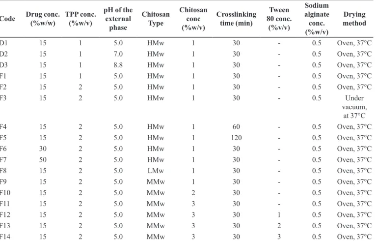
FIGURE 1 - Scanning electron micrographs of F2 coded chitosan microspheres before dissolution test: (A) general appearance and surface morphology (B, C).
FIGURE 2 - Scanning electron micrographs of F6 coded chitosan microspheres (containing 30%FLB)before dissolution test (A,
B) and its cross sectional micrograph (C).
FIGURE 3 - Scanning electron micrographs of cross sections of F2 coded chitosan microspheres: (A), before dissolution test and after 24 hours from dissolution test (B).
In vitro release studies
The static method was used for the in vitro release studies under sink conditions. Particles containing
from which 1,5 mL samples were taken at predetermined
time intervals. Then, the FLB amount released from the particles was calculated spectrophotometrically. The fresh
medium was added back into the flasks in the same amount of samples taken (n=3).
Sterilization
As the formulations prepared are intended to be implanted, they need to be sterilized. However, polymeric structure can be subject to change as a result of sterilization,
which can affect the active substance release. Therefore,
the resultant formulation was sterilized by gamma light, which is the optimum sterilization method for polymeric type drug carrying systems. Sterilization was conducted by 60Co gamma ray source and Issolodovatejl PX-γ-system with 0.51 Mrad/sa-hr dose rate. Microspheres to
be sterilized were placed into the radiation area, which is
in the amber color glass bottles, and 2.5 Mrad was chosen
as the ray dose, commonly accepted to be optimum for
polymeric materials. To identify the effect of sterilization
conditions on the dissolution rate, dissolution rates were measured for the formulations that were sterilized.
RESULTS AND DISCUSSION
Formulation studies
The analytical validation parameters for the UV
spectrophotometric determination of FLB were given in
Table I. Analyzes at a concentration range of 2-12 µg/ ml showed that the final R2 values were close to 1, the
standard error (SE) of the slope and intercept was low,
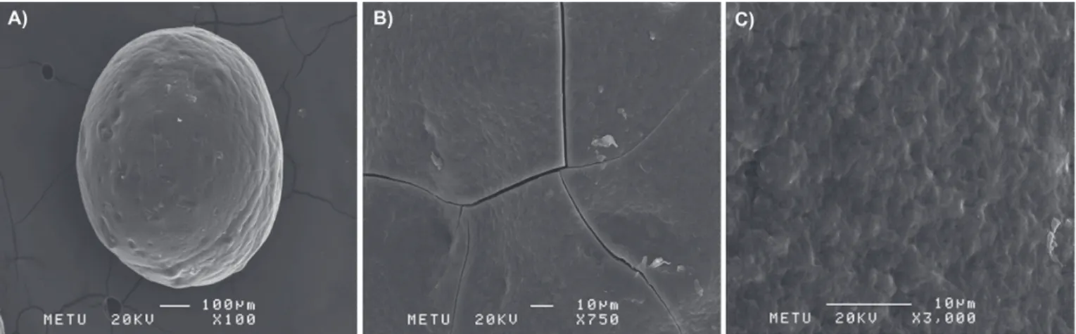
and the relative standard deviation (RSD) values were less FIGURE 4 - Scanning electron micrographs of F2 coded chitosan microspheres after 24 hours in dissolution test: (A) general appearance and surface morphology (B, C).
TABLE III - Particle size analysis and incorporation efficiencies of formulations
Formule Code Mean particle size(µm±SE) Actual drug content(% w/w±SE) Incorporation efficiency(%±SE)
F1 743.0 ± 1.20 5.08 ± 0.002 38.9 ± 0.004
F2 702.0 ± 1.16 8.94 ± 0.006 68.5 ± 0.026
F3 780.0 ± 1.16 7.11 ± 0.003 54.5 ± 0.007
F4 732.0 ± 1.14 8.60 ± 0.004 65.9 ± 0.009
F5 773.0 ± 1.20 7.56 ± 0.003 57.9 ± 0.008
F6 713.0 ± 1.16 18.20 ± 0.001 57.9 ± 0.001
F7 861.0 ± 1.30 22.09 ± 0.001 78.8 ± 0.003
F8 710.1 ± 1.32 5.90 ± 0.002 45.2 ± 0.005
F9 770.0 ± 1.28 6.24 ± 0.001 47.8 ± 0.003
F10 759.0 ± 1.18 3.56 ± 0.004 27.3 ± 0.009
F11 1086.0 ± 1.14 2.79 ± 0.003 21.4 ± 0.006
F12 1069.0 ± 1.16 3.98 ± 0.004 30.5 ± 0.010
F13 976.0 ± 1.17 2.79 ± 0.003 21.4 ± 0.008
than 2. Only the active substance showed peak at 247 nm,
indicating that the chosen method had selectivity for FLB.
Chitosan is a cationic polysaccharide, forming gel
with negatively charged ions. Thus, TPP was used as cross-linking agent, and chitosan microspheres were prepared by
ionotropic gelation method in the present study. Chitosan solution in 1% (v/v) acetic acid was dropped onto TPP
solution, which caused ionic reaction between TPP and chitosan (Hosseinzadeh et al., 2012; Joseph, Sangeetha,
Gomathi, 2016), resulting in the formation of particles. In formulation studies, first, mixing rate speed and external
phase pH were decided on. The formulations prepared are given in Table II.
The effect of mixing speed
The particles were prepared in two different
stirrer (Stirpac) after a mixing process at 100, 300, 500, 1200, 1500, 2000, and 5000 rpm. It was observed that microspheres do not form at such high mixing speed as 5000 rpm and that particles tend to have a perfect spherical form as the mixing speed decreases. As a result, the
optimum microsphere preparation speed was decided to be 100 rpm (Stirpac).
Determination of external phase characteristics
External phase pH, where TPP is located, has a
direct effect on the incorporation efficiency as it will
prevent transition to the external phase also because of the
solubility of the active substance. In this study, the pH of
TPP solutions was set at 5.0, 7.0, and 8.8 to prepare D1, D2 and D3 formulations, respectively. It was observed that the loading activity of 42.8% in the prepared formulation coded as D1 decreased to 16.5% in D2, and to 0.3% in D3. It was found out that, when external phase pH increases, encapsulation efficiency decreases because the solubility of FLB increases in the external phase (Bodmeier, Oh, Pramar, 1989; Bodmeier, Paeratakul, 1989). Therefore, external phase pH was kept constant at 5.0 in all studies to obstruct the passage of the active substance to the external phase and
to keep it within the microspheres that developed.
Volume of the external phase
The internal phase:external phase ratios were changed to 1:10, 1:20, 1:30 in the formulation studies, and it was observed that the incorporation efficiency decreases, though not markedly, when the external phase volume
was increased (Table III). Thus, for the ease of study, the
internal:external phase ratio was decided to be 1:10.
Crosslinking agent effect
Chitosan solutions with varying molecular weights
and concentrations were used to obtain microspheres.
The study intended to use 2% (w/v) HMW, in particular,
to prepare the formulations, yet microspheres could not be made due to the high viscosity of chitosan solution.
It was also observed that, when 1% (v/v) Tween 80 was
added to this solution in order to decrease the viscosity and
increase the flow characteristics, microspheres develop
although they are deformed during the drying process.
For this reason, the HMW chitosan percentage used was found out as 1%.
Sodium alginate was added to the external phase along with the TPP by 0.5% (a/h) since the existing
literature has shown that this allows for particles with narrow particle size distribution and smooth surfaces
(Aral, Akbuğa, 1998).
On the other hand, when microspheres were prepared by using TPP with 1% (F1) and 2% (F2) ratios in the external phase, it was found out that particle size
decreases as TPP ratios increase (Table III) (Jain et al.,
2016). Therefore, TPP concentration was set to 2% in
this study.
Particle size
In this study the particle size of microspheres
prepared varied between 700 and 1300 μm (Table III). The effect of the formulation parameters on the particle
size was analyzed.
The effect of drug concentration
In the formulations prepared by 1% HMW, the particle magnitude obtained were 702.05 μm (F2), 713 μm (F6), and 861 μm (F7) when the existent active substance was 15%, 30%, and 50%, respectively (Table III). These findings are somewhat in concordance with the studies
which report that cross linking density decreases, and thus particle size increases, as a result of increasing drug concentration, which leads to decreasing chitosan amount in the dribble of the same volume (Bodmeier, Paeratakul,
1989; Wan, Lim, Soh, 1994; Jain et al., 2016; Joseph,
Sangeetha, Gomathi, 2016).
The effect of the molecular weight of chitosan
The existing literature has reported that porosity
with TPP decreases. However, in the present study, the
particle sizes were found to be 702 μm, 770 μm, and 710 μm in the formulations prepared with 1% HMW (F2), 1% MMW (F9), and 1% LMW (F8), respectively, which is not indicative of any significant difference (p>0.05) (Shirashi, Imai, Otagiri, 1993).
The effect of polymer concentration
As the polymer concentration used in the prepared
formulations was increased from 1% (F9) to 2% (F 10), the
following particle magnitudes were achieved, respectively:
770 μm and 759 μm. No significant difference was found
between them. However, when the chitosan concentration
was 3% (F11), the particle size increased significantly to 1086 μm (p<0.05). These findings were attributed to the fact that TPP amount proved insufficient due to the increase
in the chitosan viscosity and decrease in the cross linking density due to its being incapable of diffusing into the
particles (Shirashi, Imai, Otagiri, 1993, Gan et al., 2005;
Jain et al., 2016; Joseph, Sangeetha, Gomathi, 2016).
The effect of crosslinking agent concentration
It was observed that the particle size decreased when
TPP concentration was increased from 1% (F1) to 2% (F2), increasing the crosslinking density (p>0.05). The particle size of 743 µm in F1 decreased to 702 µm in F2
(Jain et al., 2016).
The effect of Tween 80
To e l i m i n a t e t h e p r o b l e m s w i t h d r o p p i n g encountered during the preparation of F12 formulation
with 3% MMW, and the agglomeration that occurred in the microspheres that developed, Tween 80 was added to the internal phase, where lies the chitosan by 1% (F12) and 2% (F13). The addition of Tween 80 by 1% and 2% resulted in significant reduction in the particle size. The particle size was 1086 µm in the F11 formulation. It decreased to 1069 µm in the existence of 1% tween 80 (F12), and to 976 µm in the existence of 2% tween 80 (F13) (p<0.05). The agglomeration problem that
developed during the preparation process was also observed to have vanished.
The effect of mixing duration
This duration was fixed at 30 minutes in the
formulations prepared because it was thought that the
mixing duration following the crosslinking agent addition
would affect the crosslinking density, and thus the
particle size. IR analyses of the microspheres prepared found the existence of P=O peaks, which indicates cross
linking between chitosan and TPP. It was evidence to
the sufficiency of this duration. When the duration was
prolonged to 60 minutes (F4) for the preparation of the
formulations, no significant change occurred in terms of the particle size. This also proved that the duration of 30 minutes was sufficient for cross linking (F2). Extension of this duration into 120 minutes (F5) resulted in a significant increase in the particle sizes, which can be explained by the swelling of the microspheres during the mixing process.
Morphological properties
SEM images of chitosan microspheres are presented in Figures 1, 2, and 3. They demonstrate that the active substance is not dissolved in the matrix structure and exists in crystalized form. They also show that the shapes
of the formulations are near-sphere. Figure 1 and Figure 2 indicate that, when the active substance amount was
increased from 15% (F2) to 30% (F6) in the formulations,
the surface roughness increases due to the decrease in the polymer’s crosslinking density. The internal structure density was also observed to have increased (Figure 2 and
Figure 3). Cracks formed both on the surface and in the
cross-sections of the microspheres. At the end of the 24-hour dissolution rate studies, F2 coded formulations were
extracted from the dissolution medium and dried. SEM analysis of these microspheres was conducted to examine
the surface morphology. After the release, the analysis of the surface of the formulation showed that the polymer got loose and swollen and the pores opened. It turned out that the cracks that were observed on the surface and in the cross sections before the release disappeared after the release due to the swelling of the polymer (Figure 1, Figure
3 and Figure 4). Active substance crystals were not seen
after the release (Figure 4).
Release studies
The solubility of FLB was determined in the release
medium (pH 7.4, PBS) was 3.77±0.11 mg/mL (n=3). The effect of formulation parameters on active substance
release from chitosan microspheres were analyzed with release studies under sink conditions.
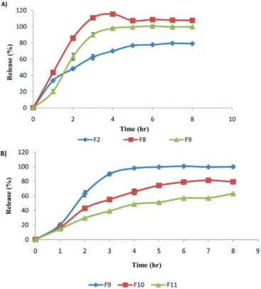
The effect of the molecular weight and concentration of chitosan
and low molecular weight chitosan was used with 1% (w/v) ratio in F2, F8, and F9 formulations, respectively. F2 coded formulation displayed the slowest release (p<0,05) (Figure 5A). As the molecular weight of chitosan increases, the
number of amino groups that go through ionic reaction with TPP increases, which in turn boosts cross linking density. Increased number of crosslinking also increases the hardness of the structure, decreasing the active substance
release (Bodmeier, Oh, Pramar, 1989; Shirashi, Imai, Otagiri, 1993; Ko, Parki Hwang, 2002; Geng, Kwon, Jang, 2005).
Figure 5 B shows the effect of polymer concentration on the active substance release. The F9, F10, and F11 formulations displayed on the figure contain 1%, 2%, and 3% MMW chitosan, respectively. As can be seen
here, active substance release decreased with increased polymer concentration. Density of cross linking with TPP increases when chitosan amount increases, which leads to more intense structure formation in microspheres. This
ultimately decreases active substance release (p<0.05) (Ko, Park, Hwang, 2002; Berthold, Cremer, Kreuter, 1996).
The effect of drug concentration
Figure 6 presents the dissolution profiles of
F2, F6, and F7 formulas prepared with 1% HMW chitosan, which contain 15%, 30%, and 50% drug
concentration, respectively. When the drug amount
in microspheres was increased from 15% to 30%, no significant difference was observed in terms of drug release. Nevertheless, when it was increased to 50%, drug release from microspheres increased significantly (p<0.05). It was predicted that chitosan amount with the
same droplet volume would decrease due to an increase in drug concentration, which in turn would decrease the crosslinking density. The same results were obtained
for F12 and F15 prepared with 3% MMW (Joseph, Sangeetha, Gomathi, 2016).
The effect of TPP concentration and mixing duration
F1 and F2 formulations containing 1% and 2%
TPP, respectively, were prepared in the study (Figure 7). The lowest release rate was attained with the F2 formula. Cross linking was realized through the diffusion
of 5 anions that exist in the TPP molecule into chitosan solutions (El-Gibaly, 2002). Therefore, the crosslinking
duration is important for the strength of dribbles that develop.
The following formulas with varying mixing durations were prepared: F2 (30 minutes), F4 (60 minute), and F5 (120 minutes). The duration increase from 30 minutes to 60 minutes did not yield a significant difference in terms of the dissolution rate values, whereas
an increase to 2 hours caused greater drug release from
the formulas developed (F5) (p<0.05) (Figure 8). This is
attributed to the fact that, although the hardening process
is completed within 30 minutes, the polymer continues FIGURE 5 - Effect of the molecular weight and concentration of
chitosan on the FLB release from chitosan microspheres in PBS: (A) F2 (1% HMw); F8 (1% LMw); F9 (1% MMw); (B) F9 (1% MMw); F10 (2% MMw) and F11 (3% MMw).
FIGURE 6 - Effect of the drug concentration on the FLB release
FIGURE 7 - Effect of the concentration of tripolyphosphate and
drying method on the FLB release from chitosan microspheres in PBS: F1 (1% TPP); F2 (2% TPP); F1 and F2 were dried in the oven and F3 (2% TPP) was dried under vacuum.
FIGURE 8 - Effect of the crosslinking time on the FLB release
from chitosan microspheres in PBS: F2: 30 min. (2% TPP); F4: 60 min. (2% TPP) and F5: 120 min. (2% TPP).
to swell, causing the drug to diffuse over the surface in
the remaining time.
The effect of drying conditions
The existing studies showed that the drying conditions have a major effect on the drug release and that
more surface cracks form with increased drying, which results in increased release. As can be seen in Figure 7, the
drying conditions of the microspheres proved influential in substance release. The analysis of the release profiles of F2 formula dried in the oven (24 hours, 37 °C) and F3 formula dried in the vacuumed oven (24 hours, 37 °C)
reveals that the active substance release in the F2 formula
is less than that in F3 formula (p<0,05) (Figure 7). It was
attributed to the rapid evaporation of water in vacuum
during drying, and the consequent expansion of pores and
quick transmission of the active substance to the surface together with water.
Sterilization effect
Dissolution rate experiment was conducted prior to and after the sterilization process to examine whether
the drug release had changed or not. It was observed that
sterilization process had no effect on the drug release from the 15% FLB containing F2 formulation (p>0.05). This finding led to the use of the sterilization method, which
was considered suitable for microsphere formulations.
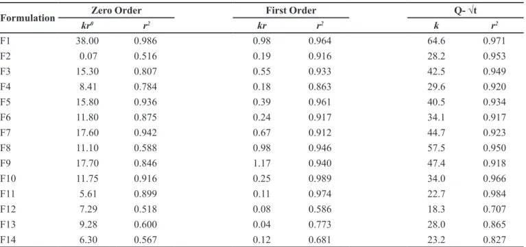
Analysis of the compatibility of kinetic mechanisms
Data obtained in the dissolution rate studies
was analyzed by SPSS 11.5 in terms of compatibility
with various kinetics (Table IV). The majority of the formulations were compatible with Higuchi Q√t kinetics for heterogeneous matrix. It was observed that, as the
chitosan’s molecular weight and concentration and the TPP amount used in the formulations increased, better
compatibility with Q√t kinetics was displayed. An
increase in the crosslinking duration in the formulations
(from 30 to 120 minutes) was associated with greater compatibility with first order kinetics, which could be explained by the swelling of the polymer resulting in the drug migration towards the surface. Being in matrix structure, the particles prepared were expected to be theoretically compatible with Q√t kinetics. The releases displayed compatibility with Q√t kinetics rather than
zero order, which was thought to be an indicator of the fact that dissolution rate was not solubility controlled
and a matrix structure was achieved in the formulation
studies.
CONCLUSION
Chitosan, which is a natural biodegradable
polymer, was used in the study, and the microsphere formulas of flurbiprofen, which is not practically dissolved in water, were prepared by means of the ionotropic gelation method. It was observed that FLB’s rate of release from microspheres decreased parallel to an increase in chitosan’s molecular weight and
concentration, and TPP concentration. SEM analysis
of the microspheres revealed that they are spherical in form, with cracks on their surfaces and cross-sections. The drug release was found to be greatly compatible
REFERENCES
Anraku M, Arahira M, Mady FM, Khaled KA, Yamasaki K, Seo H, et al. Enhancement of dissolution and bioavailibity of flurbiprofen by low molecular weight chitosans. Pharmazie. 2010;65(7):461-467.
Al-Qadi S, Grenda A, Carrion-Recio D, Seijo B. Remunan-Lopez C. Microencapsulated chitosan nanoparticles for pulmonary protein delivery: in vivo evaluation of insulin-loaded formulations. J Control Rel. 2012;157(3):383-90.
Aral C, Akbuğa J. Alternative approach to the preparation of chitosan beads. Int J Pharm. 1998;168(1):9-15.
Berthold A, Cremer K, Kreuter J. Preparation and characterization of chitosan microspheres as drug carrier for prednisolone sodium phosphate as model for anti-inflammatory drugs. J Control Rel. 1996;39(1):17-25.
Bodmeier R, Oh KH, Pramar Y. Preparation and evaluation of drug-containing chitosan beads. Drug Dev Ind Pharm. 1989;15(9):1475-94.
Bodmeier R, Paeratakul O. Spherical agglomerates of water insoluble drugs. J Pharm Sci. 1989;78(11):964-67.
Bogdansky S. Natural polymers as drug delivery systems. In: Chasin M, Langer R, editors. Biodegradable polymers as drug delivery systems. New York, NY: Marcel Dekker; 1990; p. 231-259.
Bokura H, Kobayashi S. Chitosan decreases total cholesterol in women: a randomized, double-blind, placebo-controlled trial. Eur J Clin Nutr. 2003;57:721-725.
Caetano LA, Almeida AJ, Gonçalves LMD. Effect of experimental parameters on alginate/chitosan microparticles for BCG encapsulation. Mar Drugs. 2016;14(5):90-120.
Chandy T, Sharma CP. Chitosan-as a biomaterial. Biomat Artif Cells Artif Org. 1990;18(1):1-24.
Chen P, Xia C, Mei S, Wang J, Shan Z, Lin X. Intra-articular delivery of sinomenium encapsulated by chitosan microspheres and photo-crosslinked GelMA hydrogel ameliorates osteoarthritis by effectively regulating autophagy. Biomaterials. 2016;81:1-13.
Danckwets M, Fassihi A. Implantable controlled release drug delivery systems: a review. Drug Dev Ind Pharm. 1991;17(11):1465-1502.
TABLE IV - The kinetic assesment of release data
Formulation Zero Order First Order Q- √t
kr0 r2 kr r2 k r2
F1 38.00 0.986 0.98 0.964 64.6 0.971
F2 0.07 0.516 0.19 0.916 28.2 0.953
F3 15.30 0.807 0.55 0.933 42.5 0.949
F4 8.41 0.784 0.18 0.863 29.6 0.920
F5 15.80 0.936 0.39 0.961 40.5 0.934
F6 11.80 0.875 0.24 0.917 34.1 0.917
F7 17.60 0.942 0.67 0.912 44.7 0.923
F8 11.10 0.588 0.98 0.946 57.5 0.950
F9 17.70 0.846 1.17 0.940 47.4 0.918
F10 11.75 0.916 0.25 0.989 34.0 0.966
F11 5.61 0.899 0.11 0.974 22.7 0.984
F12 7.29 0.518 0.08 0.586 18.3 0.707
F13 9.28 0.600 0.04 0.773 28.0 0.865
F14 6.30 0.567 0.12 0.681 23.2 0.827
kr0 = zero-order release rate constant; kr = first-order release rate constant; k = rate constant obtained from Q-√t kinetic;
Davis SS, Huneyball IM, Ratcliffe JH. Recent advance in the use of microspheres for targeted therapy. Drugs Exptl Clin Res. 1985;11(9):633-640.
Doppalapudi S, Jain A, Khan W, Domb AJ. Biodegradable polymers: An overview. Polym Adv Technol. 2014;25(5):427-435.
El-Gibaly I. Development and in vitro evaluation of novel floating chitosan microcapsules for oral use: comparison with non-floating chitosan microcapsules. Int J Pharm. 2002;249(1-2):7-21.
European Pharmacopoeia. EP 6.0. Council of Europe. 67075, Strasbourg, Cedex, France, 2008; 512 p.
Gadalla HH, Soliman GM, Mohammed FA, El-Sayed AM. Development and in vitro/in vivo evaluation of Zn-pectinate microparticles reinforced with chitosan for the colonic delivery of progesterone. Drug Deliv. 2016;23(7):2541-2554.
Gan Q, Wang T, Cochrone C, McCarron P. Modulation of surface charge, particle size and morphological properties of chitosan-TPP nanoparticles intended for gene delivery. Colloids Surf B Biointerfaces. 2005;44(2-3):65-73.
Ganguly K, Kulkarni AR, Aminabhavi TM. In vitro cytotoxicity and in vivo efficacy of 5-fluorouracil-loaded enteric-coated PEG-crosslinked chitosan microspheres in colorectal cancer therapy in rats. Drug Deliv. 2016;23(8):2838-2851.
Geng X, Kwon O, Jang J. Electrospinning of chitosan dissolved in concentrated acetic acid solution. Biomaterials. 2005;26(27):5427-32.
Hejazi R, Amiji M. Chitosan-based gastrointestinal delivery systems. J Control Rel. 2003;89(2):151-65.
Hosseinzadeh H, Atyabi F, Dinarvand R, Ostad SN. Chitosan-pluronic nanoparticles as oral delivery of anticancer gemcitabine: preparation and in vitro study. Int J Nanomed. 2012;7:1851-1863.
Illum, L. Chitosan and its use as a pharmaceutical excipient. Pharm Res. 1998;15(9):1326-31.
International Conference for Harmonisation. ICH. Guidelines for Industry, ICH Q2(R1). Validation of analytical procedures: methodology. Geneva. Switzerland: ICH; 2005.
Jain A, Thakur K, Sharma G, Kush P, Jain UK. Fabrication, characterization and cytotoxicity studies of ionically cross-linked docetaxel loaded chitosan nanoparticles. Carbohydr Polym. 2016;137:65-74.
Jeon SJ, Ma Z, Kang M, Galvao KN, Jeong KC. Application of chitosan microparticles for treatment of metritis and in vivo evaluation of broad spectrum antimicrobial activity in cow uteri. Biomaterials. 2016;110:71-80.
Joseph JJ, Sangeetha D, Gomathi T. Sunitinib loaded chitosan nanoparticles formulation and its evaluation. Int J Biol Macromol. 2016;82:952-958.
Kawashima Y, Handa T, Kasai A, Takenaka H, Lin SY, Ando Y. Novel method for the preparation of controlled-release theophylline granules coated with a polyelectrolyte complex of sodium polyphosphate-chitosan. J Pharm Sci. 1985a; 74(3):264-268.
Kawashima Y, Lin SY, Kasai A, Handa T, Takenaka H. Preparation of a prolonged release tablet of aspirin with chitosan. Chem Pharm Bull. 1985b;33(5):2107-13.
Ko JA, Park HJ, Hwang SJ. Preparation and characterization of chitosan microparticles intended for controlled drug delivery. Int J Pharm. 2002;249(1-2):165174.
Koppolu BP, Smith SG, Ravindranathan S, Jayanthi S, Kumar S, Zaharoff DA.Controlling chitosan-based encapsulation for protein and vaccine delivery. Biomaterials. 2014;35(14):4382-89.
Leonardi D, Salomon CJ, Lamas MC, Olivieri AC. Development of novel formulations for Chagas’ disease: Optimization of benznidazole chitosan microparticles based on artificial neural Networks. Int J Pharm. 2009;367(1-2):140-147.
Ma L, Liu C. Preparation of chitosan microspheres by ionotropic gelation under a high voltage electrostatic field for protein delivery. Colloids Surf B: Biointerfaces. 2010;75(2):448-53.
Mi F, Shyu S, Lee S, Wong T. Kinetic study of chitosan-tripolyphosphate complex reaction and acid-resistive properties of the chitosan-tripolyphosphate gel beads prepared by in-liquid curing method. J Polym Sci Part B: Polym Phys. 1999;37(14):1551-64.
M. G. Y. Tilkan, N. Özdemir
Norkar M, Sher P, Pawar A. Stomach-specific controlled release gellan beads of acid-soluble drug prepared by ionotropic gelation method. AAPS PharmSciTech. 2010;11(1):267-77.
Özalp Y, Özdemir N. Controlled release of vancomycin from biodegradable microcapsules. J Microencaps. 2001;18(1):89-110.
Özdemir N, Şahin J. Design of a controlled release osmotic pump system of ibuprofen. Int J Pharm. 1997;158(1):91-97.
Park JM, Lee SY, Lee GH. Design and characterization of doxorubicin-releasing chitosan microspheres for anti-cancer chemoembolization. J Microencapsulation. 2012;29(7):695-705.
Park JW, Yun Y, Park K, Lee JY, Kim H, Kim SE. Ibuprofen-loaded porous microspheres suppressed the progression of monosodium iodoacetate-induced osteoarthritis in a rat model. Colloid Surface B. 2016;147:265:273.
Sharma K, Somavarapu S, Colombani A, Govind N, Taylor K. Crosslinked chitosan nanoparticle formulations for delivery from pressurized metered dose inhalers. Eur J Pharm Biopharm. 2012;81(1):74-81.
Shirashi S, Imai T, Otagiri M. Controlled release of indomethacin by chitosan-polyelectrolyte complex: optimization and in vivo/ in vitro evaluation. J Control Rel. 1993;25(3):217-25.
Shu, XZ, Zhu KJ. A novel approach to prepare tripolyphosphate/ chitosan complex beads for controlled release drug delivery. Int J Pharm. 2000;201(1):51-58.
Shu XZ, Zhu KJ. Novel pH-sensitive citrate cross-linked chitosan film for drug controlled release. Int J Pharm. 2001;212(1):19-28.
Shu XZ, Zhu KJ. Controlled drug release properties of ionically crosslinked chitosan beads:the influence of anion structure. Int J Pharm. 2002;233(1-2):217-25.
Sumiyoshi M, Kimura Y. Low molecular weight chitosan inhibits obesity induced by feeding a high-fat diet long-term in mice. J Pharm Pharmacol. 2006;58(2):201-207.
Vivek R, Thangam R, Nipunbabu V, Ponraj T, Kannan S. Oxaliplatin chitosan noparticles induced intrinsic apoptotic signaling pathway: a “smart” drug delivery system to breast cancer cell therapy. Int J Biol Macromol. 2014;65:289-97.
Wan LSC, Lim LY, Soh BL. Drug release from chitosan beads. STP Pharma Sci. 1994;4(3):195-200.
Wood DA. Biodegradable drug delivery systems. Int J Pharm. 1980;7(1):1-18.
Xue M, Hu S, Lu Y, Zhang Y, Jiang X, An S, et al. Development of chitosan nanoparticles as drug delivery system for a prototype capsid inhibitor. Int J Pharm. 2015;495(2):771-782.
Yuan Y, Chessnut BM, Utturkar G. The effect of cross-linking of chitosan microspheres with genipin on protein release. Carbohyd Polym. 2007;68(3):561-67.
Zhang J, Wang D, Pan J. Efficient resveratrol production by immobilized β- glucosidase oncross-linked chitosan microsphere modified by l-lysine. J Mol Catal B: Enzym. 2014;104:29-34.