ROADMAP TO VASCULITIS
Yrjö T. Konttinen,
Z
ˇiga Rotar,
Tom Pettersson,
Dan C.E. Nordström,
Paul Bacon,
Jörgen Petersen
Department of Medicine/Invärtes medicin, Helsinki University Central Hospital, Helsinki, Finland
ORTON Orthopaedic Hospital of the Invalid Foundation, Helsinki, Finland COXA Hospital for Joint Replacement,Tampere, Finland
Department of Rheumatology, University Medical Centre, Ljubljana, Slovenia Department of Rheumatology, Division of Immunity and Infection,
University of Birmingham, Birmingham, UK Laboratory of Rheumatology,The Finsen Center,
A B S T R A C T
Vasculitis is characterized by vessel wall injury caused by an immunologically initiated inflammatory reac-tion. Vessel wall injury leads to vascular stenosis, aneurysm, bleeding, thrombosis, embolism, vasospasms and ischemia. The vasculitis is clinically important when the patient has general inflammatory and mul-tifocal symptoms, which progress in episodes and can be explained by these vascular lesions. The clinical manifestations of these depend on the size, localization and number of blood vessels involved. This forms the basis of the current vasculitis classification. It is important to recognize the secondary vasculitides, as their treatment is mainly based on elimination of the triggering factor. In primary vasculitides, immuno-suppression alone is the basis of treatment in almost all cases, whereas the management of pseudovascu-litis is dependent on its aetiology. In primary care, basic evaluation should be done: patient history, physi-cal examination, basic laboratory tests and other non-invasive tests to verify suspected surrogate findings. After this, patients should be urgently referred to a specialized centre, where the required histological and radiological tests are performed for diagnosis and immunosuppressive and other necessary treatment is initiated.
Keywords: Vasculitis; Classification; Diagnosis; Treatment.
R E S U M O
As vasculites são caracterizadas por lesão da parede vascular causada por uma reacção inflamatória media-da imunologicamente. Esta lesão causa estenose, aneurismas, hemorragias, tromboses, embolias, vasoes-pasmo e isquémia. A vasculite é clinicamente importante quando o doente tem inflamação sistémica e sintomas multifocais, que progridem por crises e que são explicáveis por estas lesões vasculares. As mani-festações clínicas dependem do tamanho, localização e número de vasos envolvidos. É esta a base da actual classificação das vasculites. É importante reconhecer as vasculites secundárias, porque o seu tra-tamento é baseado na eliminação do factor precipitante. Nas vasculites primárias a imunossupressão é a base do tratamento em quase todos os casos, ao passo que o tratamento das pseudovasculites está depen-dente da etiologia. Nos cuidados primários deve ser feita uma avaliação básica: história clínica, exame objectivo, testes laboratoriais gerais e outros exames não invasivos orientados pela sintomatologia do doen-te. Após esta fase, estes doentes devem ser referenciados urgentemente a centros especializados onde os exames radiológicos e histológicos fundamentais para o diagnóstico serão realizados e a imunossupres-são ou outras eventuais medidas terapêuticas serão iniciadas.
A R T I G O D E R E V I S Ã O
R O A D M A P T O V A S C U L I T I S
Yrjö T. Konttinen
*/**/***, Zˇiga Rotar
****,Tom Pettersson
*,
Dan C.E. Nordström
*, Paul Bacon
*****, Jörgen Petersen
******Vasculitis typically damages the blood vessel wall via an immunologically initiated inflammatory re-action. Damage in the vessel wall can lead to aneurysm and bleeding or, alternatively, obstruc-tion of the vascular lumen, thrombosis/embolism, vasospasm and ischemia. The clinical manifestati-ons depend on what and what size and type of blo-od vessel or vessels are affected. This is also used as the basis in the classification of primary vasculi-tis1-3. In examination of vasculitis patients, it is es-sential to determine fast and accurately the seve-rity of the vasculitic changes and map the affected organs. In the treatment of the patient with serious vasculitis, the clinician has to rapidly determine diagnosis, because delay of aggressive treatment endangers treatment results.
On clinical grounds, vasculitis has to be suspec-ted, when the patient suffers from an illness leading to general symptoms, inflammatory and multifocal disease with unpredictable development of mani-festations, which can be explained on the basis of blood vessel damage4. When suspicion of vasculi-tis has been raised, it is necessary to try to identify any triggering or perpetuating factors and to exclu-de pseudovasculitis. From the point of view of ma-nagement, it is important to consider the possibi-lity of secondary vasculitis and pseudovasculitis. In secondary vasculitis the management is based on the elimination of the triggering or perpetuating factor and in pseudovasculitis on the aetiology, whereas the management of primary vasculitis is
usually based on immunosuppression. Mixed and empirical treatments are sometimes necessary.
In general practice patient history, physical exa-mination, basic laboratory tests and eventually non-invasive diagnostic procedures to identify sur-rogate changes should be done. A patient can de-velop “unstable vasculitic plaques” (pro-thrombo-tic inflammation, which forms initiation sites for thrombi) in blood vessels of vital organs. A patient who is suspected to suffer from vasculitis should be urgently referred to a specialized institution for fur-ther diagnostic work up and management. This enables invasive diagnostic procedures such as bi-opsies and invasive radiology and initiation of ap-propriate immunosuppressive or other initial tre-atment.
STOP Sign
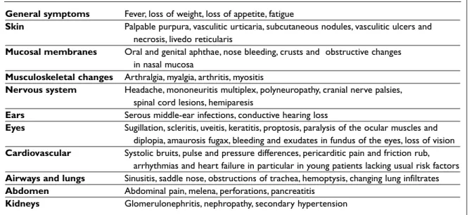
Since vasculitis is a rare disease, the most difficult and important step in tackling vasculitis is to stop and consider the possibility of it. Typically, vascu-litis presents as a suddenly developed and jerk-wise, progressive multiple organ damage in a seve-rely ill patient, the symptoms of whom can be best explained by immune-inflammatory damage of the blood vessel wall (Table I).
First Road Sign: Triggering Factors
Typically, vasculitis is associated with immunolo-gical reactivity such as an immune response lea-ding to antigen specific reaction in form of produc-tion of antibodies or T cell receptor mediated acti-vation of T lymphocytes to produce cytokines. Vas-culitis is not an inherited disease, although a susceptibility to react in a harmful way to antige-nic stimuli and to develop severe disease would be inheritable, e.g. in form of complement or immu-noglobulin subclass deficiencies. Family history does not usually provide much help in definition of *Department of Medicine/invärtes medicin, Helsinki University
Central Hospital, Helsinki, Finland
**ORTON Orthopaedic Hospital of the Invalid Foundation, Helsinki, Finland
***COXA Hospital for Joint Replacement,Tampere, Finland ****Department of Rheumatology, University Medical Centre, Ljubljana, Slovenia
*****Department of Rheumatology, Division of Immunity and Infection, University of Birmingham, Birmingham, UK ******Laboratory of Rheumatology,The Finsen Center,
separated from secondary vasculitic disease (in which some triggering or perpetuating factors can be found) (Table II). Triggering factors comprise bacterial, viral, fungal and parasitic infections, drugs, vaccinations, nutrients and various connec-tive tissue diseases, malignancies and other disea-the type of disease. In vasculitis disea-the triggering and
the perpetuating factors are of central importance. From the point of view of the practising physi-cian, it is important to try to identify triggering fac-tors. Usually vasculitis of unknown cause (in which no triggering or perpetuating factor is known) is
Table I. Typical general and organ and tissue specific changes caused by vasculitis
General symptoms Fever, loss of weight, loss of appetite, fatigue
Skin Palpable purpura, vasculitic urticaria, subcutaneous nodules, vasculitic ulcers and necrosis, livedo reticularis
Mucosal membranes Oral and genital aphthae, nose bleeding, crusts and obstructive changes in nasal mucosa
Musculoskeletal changes Arthralgia, myalgia, arthritis, myositis
Nervous system Headache, mononeuritis multiplex, polyneuropathy, cranial nerve palsies, spinal cord lesions, hemiparesis
Ears Serous middle-ear infections, conductive hearing loss
Eyes Sugillation, scleritis, uveitis, keratitis, proptosis, paralysis of the ocular muscles and diplopia, amaurosis fugax, bleeding and exudates in fundus of the eyes, loss of vision Cardiovascular Systolic bruits, pulse and pressure differences, pericarditic pain and friction rub,
arrhythmias and heart failure in particular in young patients lacking usual risk factors Airways and lungs Sinusitis, saddle nose, obstructions of trachea, hemoptysis, changing lung infiltrates Abdomen Abdominal pain, melena, perforations, pancreatitis
Kidneys Glomerulonephritis, nephropathy, secondary hypertension
Table II. Triggering and/or perpetuating factors in secondary vasculitis
Infections • Hepatitis C
• Hepatitis B • HIV
• Parvovirus B19
• Septicaemia in meningococcal infections • Septicaemia in streptococcal infections • Septicaemia in gonococcal infections
Drugs, vaccinations and • Allopurinol, phenytoin, cefaclor, isotretinoin, methotrexate, NSAIDs,
food items antibiotics, diuretics
• Pneumococcal, influenza or hepatitis B-vaccinations • Food items and additives
Connective tissue diseases and • Rheumatoid arthritis inflammatory bowel diseases • Systemic lupus erythematosus
• Sjögren’s syndrome • Dermato-polymyositis • Crohn’s disease • Ulcerative colitis
Tumours and carcinomas • Lymphoproliferative diseases • Myeloproliferative diseases • Lung cancer
Y R J Ö T. K O N T T I N E N E C O L.
ses associated with the presence of antigens or al-lergens5. Infections as triggering factors form a spe-cial case. Secondary vasculitis and infection always need to be excluded before a specific primary vas-culitis is diagnosed.
Management of secondary vasculitis is based on the elimination or minimization of the triggering stimulus e.g. by the use of antibiotics, antiviral me-dication, interferon, change of the meme-dication, use of anti-inflammatory immunosuppressive drug or surgery, radiation and chemotherapy. Smoking should be stopped. In clinical praxis this treatment aiming to elimination or diminution of the trigge-ring and perpetuating factors is often combined with a simultaneous anti-inflammatory and immu-nosuppressive treatment of the vasculitic compli-cations.
With time, the distinction between primary and secondary vasculitis has become less clear,
becau-se even primary vasculitic dibecau-seabecau-se can often nowa-days be connected with triggering stimuli, for example, hepatitis B virus surface antigen (HBsAg) with polyarteritis nodosa, Staphylococcus aureus
with Wegener’s granulomatosis, A-group strepto-cocci and mycoplasmae with Henoch-Schönlein purpura, hepatitis C-virus with essential cryoglo-bulinaemia and various drugs with cutaneous leu-cocytoclastic vasculitis. On the other hand, in se-condary vasculitis with a clear-cut triggering or per-petuating stimulus it may be necessary to treat the vasculitic changes per se.
The Second Road Sign: The Primary Vasculitis
Because the triggering stimulus according to defi-nition is not known in primary vasculitis, current classification of primary vasculitis is instead basedTable III. Classification of the typical primary and atypical vasculitis diseases has been based on the size of the vessel affected. Classification also takes histological changes and typical clinical manifestations in consideration. Some vasculitic diseases also affect veins (phlebitis). Behçet’s disease, thromboangiitis obliterans, primary angiitis of the central nervous system, panniculitis and Goodpasture’s syndrome have not yet found their final place on the map of vasculitic diseases.
Takayasu arteritis (giant cell arteritis) Large arteries1
Temporal arteritis (giant cell arteritis) Large- and middle-sized arteries2
Behçet’s disease Arteries and veins of all sizes3
Polyarteritis nodosa Middle- and small-sized arteries4
Kawasaki disease Middle- and small-sized arteries5
Thromboangiitis obliterans (Bürger’s disease) Middle- and small-sized arteries, small veins6
Wegener’s granulomatosis Small arteries7
Microscopic polyangiitis Small arteries7
Churg-Strauss syndrome Small arteries7
Primary angiitis of the central nervous system Small arteries8 Henoch-Schönlein purpura Small blood vessels9 Essential cryoglobulinaemic vasculitis Small blood vessels9 Leucocytoclastic vasculitis of the skin10 Small blood vessels9
Lobular panniculitis Small arteries11
Septal panniculitis Small veins11
1Thoracic and abdominal aorta and their branches as well as the pulmonary arteries 2Blood vessels affected have lamina elastica interna
3In particular small veins are affected, can also be regarded as a secondary vasculitis 4Disease can also lead to nephropathy, but not to glomerulonephritis
5Mucocutaneous lymph node syndrome
6Panarteritis and panphlebitis, can also be regarded as pseudovasculitis or secondary vasculitis 7Small arteries, small veins, capillaries and arterioles usually associated with ANCA (ANCA-vasculitis) 8Primary (and isolated) angiitis of the central nervous system or PACNS; in particular small arteries 9Small arteries, capillaries and small veins, leading to palpable leucocytoclastic purpura
10Was earlier known as hypersensitivity vasculitis 11A heterogeneous group of conditions
on the size of the affected blood vessels, the loca-lization of these vasculitic lesions and their histo-pathology1,2(Table III). In autoantibody negative vasculitis, vascular wall damage can be mediated by cell-mediated immune reactions. Nowadays, anti-neutrophilic cytoplasmic autoantibodies (ANCA) are an important new classification crite-rion6. Association with hepatitis or the presence of immune complexes can modify the management approach taken. As no triggering or perpetuating factor is known in primary vasculitides, there is no test-tube test either, which could be used to de-monstrate a specific disease-associated autoanti-body or T-lymphocyte reactivity enabling a speci-fic and unanimous diagnosis. In large blood vessel disease vascular luminal stenosis and occlusions occur upon thickening of the blood vessel wall, whereas in the disease of middle-sized arteries, fo-cal necrotizing aneurysmatic lesions can develop upon weakening of the blood vessel wall. In the di-seases of small arteries, blood vessel wall does usually not contain immunoglobulin precipitati-ons, but the immunological basis of the disease is evident as the patient has circulating ANCA. ANCA vasculitides comprise Wegener’s granulomatosis, microscopic polyangiitis and Churg-Strauss syndrome. ANCA can also be found in many secon-dary vasculitic diseases, but then they usually pos-sess specificity other than proteinase-3 or myelo-peroxidase. Immune complexes or cryoglobulins, which precipitate in the blood vessel wall, are found in the small vessel disease, affecting arteri-oles, capillaries and in particular venules. The typi-cal clinitypi-cal manifestation developing on this basis is a palpable purpura.
Takayasu arteritis
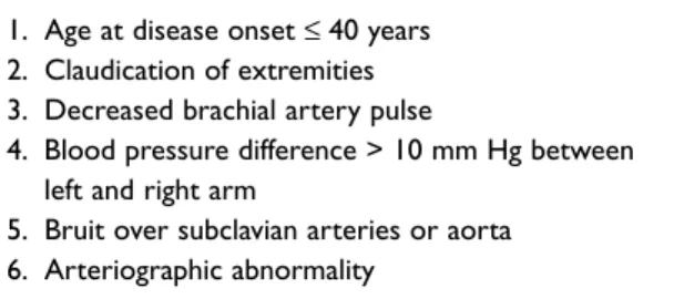
Takayasu panarteritis (Table IV) damages aorta and its large branches as well as pulmonary arte-ries in young women leading to blood vessel ste-nosis and occlusion8. Also thrombosis and aneu-rysms can develop. Takayasu panarteritis is pro-bably based on a cell-mediated autoimmune res-ponse against the smooth muscle cells in the vessel wall media. A febrile and generally sick patient de-velops aortic arch syndrome. Pulses disappear or become weak (“pulseless disease”), systolic bruits can be heard above large blood vessels and blood pressure measurement can demonstrate more than 10 mmHg difference between the arms. Ste-nosis of the coronary arteries, brachiocephalic trunk, left carotid artery and subclavian artery and
mesenteric and renal arteries lead to angina pec-toris, claudication of the upper extremity, fainting, visual disturbances, and stroke, postprandial an-gina, and renovascular hypertension associated with kidney failure. Aneurysmatic dilation of aor-ta can lead to secondary aortic valve insufficiency and involvement of the pulmonary arteries can lead to pulmonary hypertension.
Takayasu arteritis is a typical granulomatous giant cell arteritis, the diagnosis of which can be ra-rely confirmed by histology ante mortem, as it is
dif-ficult to obtain biopsies of the target lesions. Diag-nosis is usually reached by the use of computed to-mography, magnetic resonance imaging and angi-ographies, showing stenosis and poststenotic vessel dilations9-11. The most important differential diagno-ses are atherosclerosis, fibromuscular dysplasia, sar-coidosis, biochemical disturbances of the connec-tive tissue and thrombotic tendencies.
Temporal or giant cell arteritis
A panarteritis can occur in the superficial tempo-ral arteries (Table V), but also in other extracranial, large and middle-sized arteries, leading to thicke-ning of the vascular wall and stenosis and occlusi-on of lumen13-15. Inflammation of the vertebral ar-teries can lead to transient ischemic attack (TIA), stroke and vertigo. Inflammation of the subclavi-an, carotid and brachial arteries can lead to aortic arch syndrome, claudication of the upper extremi-ties and asymmetric pulses or pulselessness. The disease is also known as giant cell arteritis as it can affect also arteries other than temporal arteries. Temporal arteritis is a relatively common primary vasculitis. Its incidence in patients over 50 years of age is estimated at up to 200/million/year. Tempo-ral arteritis and polymyalgia rheumatica follow the rule of 50: both the patient and the erythrocyte se-dimentation rate (ESR) are usually over 50.
Tempo-Table IV. Takayasu arteritis is characterized by at least three of these 1990 ACR criteria7
1. Age at disease onset ≤ 40 years 2. Claudication of extremities 3. Decreased brachial artery pulse
4. Blood pressure difference > 10 mm Hg between left and right arm
5. Bruit over subclavian arteries or aorta 6. Arteriographic abnormality
ral arteritis is not associated with any typical au-toantibody marker. It has been considered a ma-nifestation of cell-mediated immune reaction and granulomatous foreign body reaction against lami-na elastica interlami-na damaged by atherosclerosis and by the pulse pressure16. Therefore, intracranial blo-od vessels, which lack lamina elastica interna, are spared. Arteritis leads to a new headache and ten-derness on palpation or weak and asymmetric pul-ses in the temporal arteries. Masticatory claudica-tion can occur. A much feared complicaclaudica-tion is a sudden and painless loss of vision, which can be preceded by amaurosis fugax and can affect the whole visual field or parts of it. Diplopia can deve-lop. Giant cell arteritis leads to prominent general symptoms, such as fever, loss of weight and polymyalgia rheumatica and even synovitis. Some other vasculitides, such as polyarteritis nodosa and Wegener’s granulomatosis as well as amyloidosis can affect superficial temporal arteries and may mimic the symptoms of giant cell arteritis.
Treatment is initiated with prednisolone, 40-60 mg/day, which has to be, due to the risk for blindness, commenced immediately when the diagnosis is suspected17. If the patient develops ocular symptoms, treatment is initiated with me-thylprednisolone pulses given intravenously. Mus-culoskeletal symptoms usually disappear rapidly upon initiation of prednisolone treatment, and this provides support to the working diagnosis at the initial stages. A small dose of acetosalicylic acid protects against thrombotic complications of the vasculitic lesions. Diagnosis has to be confirmed as soon as possible from temporal artery biopsies, which demonstrate destruction of lamina elastica interna, inflammatory cells in adventitia and giant cell granulomas. False negative biopsies also occur, so the decision to treat is at the end the clinician’s responsibility. Because these inflammatory chan-ges are patchy, it is generally recommended that
about 2–3 cm piece of temporal artery be taken to avoid false negative findings. When involvement of the large vessels is suspected, angiography is re-commended. In spite of the excellent subjective treatment response on short term, blood vessel wall inflammation can gradually in long-term, usually over decades, lead to aortic aneurysms and aortic valve insufficiency.
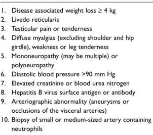
Polyarteritis nodosa
Polyarteritis nodosa (Table VI) is a relatively rare di-sease leading typically to nodular arterial aneurysms13. The classical polyarteritis nodosa af-fects middle-sized arteries. Unlike microscopic polyangiitis (microscopic polyarteritis), polyarte-ritis nodosa does not affect capillaries or venules. Eventually immune complex mediated disease of the middle-sized arteries leads to ischemia and in-farction of the target organs, which can lead to an-gina pectoris, myocardial infarction, melena or gastrointestinal perforation, hypertension, kidney failure and mononeuritis multiplex. Livedo reticu-laris refers to mottled, reticular skin changes of proportions of the extremities or torso caused by involvement of cutaneous blood vessels. Purpura, upper respiratory tract pathology, pulmonary changes and glomerulonephritis are not typical for polyarteritis nodosa. If the patient has these types of changes, she might suffer from small vessel or small artery vasculitis.
There is no serological autoantibody test for polyarteritis nodosa, but the patient can have
he-Y R J Ö T. K O N T T I N E N E C O L.
Table V. Temporal arteritis or giant cell arteritis is characterized by at least 3 of these 1990 ACR criteria12
1. Age at disease onset ≥ 50 years 2. New headache
3. Temporal artery abnormality (tender, pulse changes) 4. Erythrocyte sedimentation rate ≥ 50 mm/hour 5. Abnormal artery biopsy
Table VI. Polyarteritis nodosa is characterized by at least 3 of these 10 1990 ACR criteria18
1. Disease associated weight loss ≥ 4 kg 2. Livedo reticularis
3. Testicular pain or tenderness
4. Diffuse myalgias (excluding shoulder and hip girdle), weakness or leg tenderness 5. Mononeuropathy (may be multiple) or
polyneuropathy
6. Diastolic blood pressure >90 mm Hg 7. Elevated creatinine or blood urea nitrogen 8. Hepatitis B virus surface antigen or antibody 9. Arteriographic abnormality (aneurysms or
occlusions of the visceral arteries)
10. Biopsy of small or medium-sized artery containing neutrophils
patitis-C or human immunodeficiency virus anti-bodies or she can be a carrier of hepatitis-B virus (HBsAg), which apparently all act as some type of triggering and underlying factors. Thrombocyto-penia and leukocytoThrombocyto-penia are not typical for pri-mary polyarteritis nodosa and these changes sug-gest some other associated disease, e.g. hairy cell leukaemia. If electromyography demonstrates ab-normal conduction in the sural nerve or some oth-er poth-eriphoth-eral noth-erve, a noth-erve biopsy can demons-trate necrotizing arteritis or ischemic neuropathy. If the symptoms suggest ischemic muscle disease, muscle biopsy is indicated and can be diagnostic. If no good target for biopsy can be found, it is pos-sible to use angiography to demonstrate microa-neurysms of the middle-sized arteries.
Kawasaki disease
Kawasaki disease (Table VII) usually follows the rule of five: the patient is less than five years of age and has had fever for at least five days20. In Kawa-saki disease fever is associated with mucocuta-neous lymph node syndrome and often also
diar-Echocardiography should always be performed. After the acute phase, thrombocytosis and aneu-rysms develop. In the hospital, anti-inflammatory treatment is provided but the basic treatment com-prises intravenous immunoglobulins (2 g/kg, up to 70 g) in one dose (20). Prednisolone can increase the risk for aneurysms by weakening the blood ves-sel wall and by promoting thromboses of the vas-culitic plaques. It seems to be unnecessary to fol-low the old praxis to order high- (80–100 mg/kg per day as were used in North America) or me-dium-dose (30–50 mg/kg per day as were used in Japan) acetosalicylic acid in these children. Low dose acetosalicylic acid is used in thrombocytosis as a prophylaxis. Prompt diagnosis is critical, since the early administration of intravenous immuno-globulins and aspirin reduces the rate of coronary abnormalities to less than 5 % of patients.
Bürger’s disease
Bürger’s disease is also known as thromboangiitis obliterans. Placement of this vasculitis onto the vas-culitis map is difficult, because it could be also clas-sified as a secondary vasculitis or a pseudovasculi-tis. It is a segmental, inflammatory and thrombotic arteritis of small and middle sized peripheral arte-ries (arteritis) and small veins (migrating superficial phlebitis and venous thrombosis), different from atherosclerosis (21). This disease and its progres-sion are associated with smoking. Bürger’s disease rises somehow as a vasculitic hypersensitivity reac-tion against some component of cigarettes. It usual-ly manifests in patients under 45 years of age, lea-ding to claudication and resting pain in the distal parts of the extremities, ischemic ulcerations, ne-crosis and Raynaud's phenomenon. In contrast to atherosclerosis, these lesions also occur in the up-per extremities and more distally, even in small size arteries. Elderly men with hyperlipidaemia, diabe-tes and hypertension with intermittent leg pain du-ring walking are much more likely to suffer from plain atherosclerosis. To demonstrate involvement of such small peripheral arteries, patients can be asked to raise a hand and make a fist. Both radial and ulnar arteries are then simultaneously com-pressed and the patient is asked to open the fist. By releasing one artery at a time (Allen’s test) it can be demonstrated whether the distal part of this artery can conduct blood to the palm. If the released ar-tery is distally occluded, the palm will remain pale and will not turn red because the blood is not able to flow beyond the occlusion.
Table VII. Kawasaki disease can be diagnosed if in addition to high fever a child has at least four of the following19:
1. Conjunctivits (bilateral, bulbar, non-suppurative) 2. Red cracked lips, strawberry tongue, diffuse
oropharyngeal erythema
3. Lymphadenopathy (cervical lymph nodes > 1.5 cm) 4. Polymorphous rash, not vesicles or crusts
5. Erythema and edema of palms and soles developing to peeling of skin from fingertips
rhoea. It can damage middle-sized arteries in its acute phase (days 1-10), which can lead to the de-velopment of coronary artery aneurysms and rup-tures in the subsequent subacute (days 11-20) and convalescent (days 21-60) phases. The disease is possibly caused by an abnormal immune respon-se against some as yet unrecognized virus or bac-terium. Febrile upper respiratory tract symptoms are accompanied with those enumerated in the fact box above. Laboratory tests disclose signs of acute inflammation. Myocarditis can lead to arrhythmi-as. These patients must be urgently referred to a specialist for further diagnostic work up and ma-nagement.
There are no disease specific and diagnostically useful autoantibodies in this disease. Angiography of the extremities discloses non-atherosclerotic oc-clusions, which, in long lasting disease, are often surrounded by corkscrew like collaterals. Smoking must be stopped. Calcium channel blockers and prostacyclin analogues can be used to avoid am-putations.
Wegener’s granulomatosis
Wegener’s granulomatosis (Table VIII), Churg-Strauss syndrome and microscopic polyangiitis are the three ANCA vasculitides, defined by the presen-ce of ANCA23-25. Wegener’s granulomatosis affects mainly small arteries and is characterized by c--ANCA (cytoplasmicc--ANCA) with proteinase-3 specificity (Table IX). In Wegener’s granulomatosis the upper airways are affected in over 90 % of the patients. Lungs are affected by necrotizing granu-lomatosis and kidneys by glomerulonephritis.
Wegener’s granulomatosis can be limited to up-per airways, lungs or kidneys, but this limited form of the disease can transform into a more wides-pread disease. The upper airways, sinuses, nose and middle ear should be examined. Patients have changes in the upper airways, such as sinusitis, crusts on nasal mucosa, nose bleeds and saddle nose, middle ear inflammations and conductive hearing loss or obstruction of the trachea. Lung di-sease can lead to cough, haemoptysis and dysp-noea and can progress to alveolar bleeding. In glo-merulonephritis, which presents with hematuria, proteinuria, and cylindruria, renal failure can de-velop very rapidly. Other disease manifestations include purpura, infarctions of nail beds and ocu-lar disease, such as conjunctival bleeding, scleritis, uveitis, proptosis and ocular muscle paralysis
cau-Y R J Ö T. K O N T T I N E N E C O L.
sed by retrobulbar inflammation. Patients can also develop melena, myocardial ischemia, sensory neuropathy or mononeuritis multiplex. Wegener’s granulomatosis is characterized by c-ANCA with antiproteinase-3 specificity.
Diagnosis should be confirmed radiologically and histologically. Computed tomography can de-monstrate changes in sinuses and lungs even in ca-ses where regular x-ray images appear normal. Bi-opsies may contain so much necrotic tissue, that demonstration of vasculitis can be difficult. Biop-sies are taken from nasal mucosa, upper airways or kidneys. The best samples from the diagnostic po-int of view are obtained by open lung biopsy, which demonstrates vasculitis, necrotizing inflammation and giant cell granulomas. Granulomatous inflam-mations caused by microbes should be excluded. Renal biopsy determines the type and severity of the renal involvement. Non-specific focal and ne-crotizing glomerulonephritic changes without im-mune complexes are typical findings.
Churg-Strauss syndrome
Churg-Strauss syndrome (Table X) is one of the Table VIII. Wegener's granulomatosis is
characterized by at least 2 of the 1990 ACR criteria22
1. Nasal or oral inflammation leading to oral ulcers or purulent or bloody nasal discharge
2. Abnormal chest radiograph showing nodules, fixed infiltrates or cavities
3. Microhematuria or red cell casts in urine sediment 4. Granulomatous inflammation in the arterial wall or
peri- or extravascular tissue around arteries or arterioles
Table IX. Use of cytoplasmic anti-neutrophil--antibodies in the diagnosis of vasculitis
• Immunofluorescence (IF) for c-ANCA and p-ANCA (this refers to cytoplasmic or perinuclear staining respectively) is a good screening method. • Positive ANCA has to be confirmed using ELISA
for the demonstration of potential proteinase-3 (PR3) and myeloperoxidase-(MPO) autoantibodies. • c-ANCA/PR3 are usually seen in Wegener's
granulomatosis, but also in microscopic polyangiitis, and Churg-Strauss syndrome.
• Wegener's granulomatosis can be diagnosed without histology, only in clinically typical c-ANCA/PR3-positive disease.
• p-ANCA/MPO is usually seen in microscopic polyangiitis and Churg-Strauss syndrome. • p-ANCA without positive PR3- or MPO-ELISA
result is a nonspecific finding, typical for many infections and autoimmune diseases.
• Negative ANCA does not exclude vasculitis. • Continuously high ANCA is associated with
increased risk of relapse.
• Selection of IF- or ELISA-assay in the follow up is done based on findings at the time of diagnosis.
three vasculitides, which can be defined by the pre-sence of ANCA27. It affects mainly small arteries and is characterized by p-ANCA with myelopero-xidase specificity. These patients always have atopy and asthma, which can become milder upon de-velopment of vasculitic changes. Patients suffer from chronic respiratory tract symptoms and have lung infiltrates. Glomerulonephritis is rarer and usually milder than in Wegener’s granulomatosis, even though the kidney biopsy can demonstrate si-milar changes. Mononeuritis multiplex and coro-nary artery lesions can develop. Churg-Strauss syndrome should always be suspected on clinical grounds, when the patient has peripheral eosinop-hilia. Blood test demonstrates p-ANCA (perinu-clear ANCA) due to myeloperoxidase antibodies. Diagnosis can be confirmed by demonstration of granulomatous and necrotizing vasculitis with eo-sinophilic infiltrates.
Microscopic polyangiitis
Microscopic polyangiitis (microscopic polyarteri-tis) (Table XI) is one of the three ANCA vasculiti-des29. It affects small blood vessels (venules, capil-laries, arterioles and small arteries). Microscopic polyangiitis is a better name than microscopic polyarteritis because some patients have no evi-dence of arterial involvement. Usually patients
have p-ANCA with myeloperoxidase specificity or less often c-ANCA with proteinase-3 specificity. These features clearly differentiate it from polyar-teritis nodosa, which affects middle-sized arteries and lacks ANCA. After exclusion of hepatitis B, C and HIV, in a patient with no or only mild upper air-way involvement, who has alveolar bleeding and glomerulonephritis, the diagnosis of microscopic polyangiitis should be suspected. However, this form of vasculitis would have earlier been diagno-sed as polyarteritis nodosa. To recapitulate, mi-croscopic polyangiitis differs from polyarteritis no-dosa as microscopic polyangiitis often affects lung tissue, also affects veins and rarely leads to difficult hypertension, is usually ANCA positive, almost al-ways requires cyclophosphamide treatment, and recurs more often after remission. Overlap syndro-mes between microscopic polyangiitis and polyar-teritis nodosa are also known.
Microscopic polyangiitis can also be clearly differentiated from immune complex-media-ted leukocytoclastic vasculitides of small blood vessels since the focal necrotizing glomeru-lonephritis is not associated with immune com-plex deposition. Giant cells do not occur in this vasculitis.
Primary angiitis of the central nervous system
Some patients with neurological changes without any clecut reasons disclose vasculitis of small ar-teries of the central nervous system in angiography or histology in the absence of systemic vasculitic changes30,31. This condition is then known as primary angiitis of the central nervous system (PACNS). Patients with such symptoms should be studied for an eventual underlying myeloprolife-rative disease, HIV-infection or vasospastic ten-dency. This condition can be classified as benign (BACNS), granulomatous (GACNS) and atypical.
The vasculitic lesion of the wall of the artery leads to thrombosis, ischemia and necrosis. Clini-cally this leads to neurological symptoms, such as headaches, mental changes and focal or systemic neurological defects. These can at the beginning be reminiscent of transient ischemic attacks. Benign disease is usually acute in onset and monophasic, whereas granulomatous disease evolves slowly, is chronic and tends to remit. Diagnostic work up usually begins with magnetic resonance imaging of the brain, which is usually abnormal, and by examination of the cerebrospinal fluid, which usu-ally demonstrates inflammatory changes in the Table X. Churg-Strauss syndrome is characterized
by at least 4 of the 1990 ACR criteria26
1. Asthma
2. Eosinophilia >10% 3. Mono- or polyneuropathy 4. Pulmonary infiltrates, non-fixed 5. Paranasal sinus abnormality 6. Extravascular eosinophils
Table XI. Microscopic polyangiitis (MPA) is characterized by three changes28
1. Presence of rapidly progressive glomerulonephritis and/or alveolar haemorrhages
2. Histologic demonstration of small-sized vessel vasculitis or segmental pauci-immune necrotizing glomerulonephritis
granulomatous disease. Diagnosis of the benign form of the disease is based on angiography, and of granulomatous disease on biopsy and associa-ted inflammatory changes in the cerebrospinal fluid. ESR and CRP are usually within reference li-mits and the ANCA test is negative.
Treatment is based on prednisolone used in combination with cyclophosphamide in granu-lomatous disease, and with calcium channel bloc-kers in the benign disease. PACNS has not yet found its final place in the official classification of vasculitis.
Henoch-Schönlein purpura
Henoch-Schönlein purpura (Table XII), cryoglo-bulinaemias and leukocytoclastic vasculitis of the skin are characterized by immune complex depo-sition in the walls of the small vessels leading to leukocytoclasis, rupture of the vascular wall, and palpable purpura33. Leukocytoclastic vasculitis can also occur in severe systemic ANCA-associated vasculitides that must be excluded. Henoch--Schönlein purpura occurs usually in children as an IgA-dominated febrile hypersensitivity reaction in response to an upper respiratory tract infection. Small artery disease leads to arthralgias, arthritis, microscopic haematuria and abdominal pain. Hypotension, melena and hematemesis can deve-lop. CRP and ESR increase. Anaemia, leukocytope-nia or thrombocytopeleukocytope-nia can occur.
Patients presenting with general symptoms and abdominal pain should be sent to hospital for fol-low up. Diagnosis is confirmed by skin biopsy, which demonstrates leukocytoclastic vasculitis and IgA and C3 deposits in the vessel wall. These can also be found in renal biopsies. Meningococ-cal sepsis should be considered in differential diag-nosis, as it can also lead to petechiae and joint symptoms.
Cryoglobulinaemia
Cryoglobulinaemia (Table XIII) refers to cold pre-cipitating immune complexes or aggregates of mo-noclonal antibody leading to complement con-sumption, leukocytoclastic immune complex vas-culitis of small blood vessels and palpable purpu-ra35. Simple type I cryoglobulinaemia is caused by lymphoproliferative diseases in which the neoplas-tic clone produces monoclonal cryoglobulin. Mixed type II and III cryoglobulinaemias are associated with rheumatoid factor, which in type II diseases is monoclonal and in type III diseases polyclonal. These forms are more common in adults, who suf-fer from lymphoprolisuf-ferative disorders, chronic in-fections (in type II particularly hepatitis C) or other autoimmune diseases such as rheumatoid arthri-tis, systemic lupus erythematosus (SLE) or Sjögren’s syndrome. Cryoglobulinaemia is called essential if the patient does not have any underlying infectious, immunological or neoplastic disorders. Cryoglobu-lins accumulate in the wall of small blood vessels, where they fix complement and lead to leukocyto-clastic vasculitis and clinically to palpable purpu-ra, but also to cold urticaria, glomerulonephritis, arthralgias, abdominal pain, neuropathy, Ray-naud’s phenomenon, ulcerations and necrosis.
Potential association with the lymphoprolifera-tive diseases, infections or autoimmune diseases should be looked for. Cryoglobulins, the hallmark of this condition, are always found in blood and should be analyzed for their composition.
Hypo-Y R J Ö T. K O N T T I N E N E C O L.
Table XII. Henoch-Schönlein purpura is characterized by at least 2 of these 1990 ACR criteria32
1. Palpable purpura (hemorrhagic skin lesions not related to thrombocytopenia)
2. Age ≤ 20 years at disease onset
3. Bowel angina worsening after meals or bowel ischemia usually in form of bloody diarrhea 4. Leukocytoclastic vasculitis on biopsy
Table XIII. Mixed cryoglobulinaemia can be diagnosed if all three major criteria are fulfilled. One major criterion is enough if it occurs together with at least two minor clinical criteria and two minor serological or histopathological criteria34
Major criteria
Clinical: palpable purpura
Serological: cryoglobulins and hypocomplementemia Histopathological: leukocytoclastic vasculitis
Minor criteria
Clinical: chronic hepatitis, membranoproliferative glomerulonephritis, peripheral neuropathy or skin ulcers Serological: rheumatoid factor, hepatitis C, hepatitis B positivity
Histopathological: clonal B-cell infiltrates in the liver and/or bone marrow
complementemia is common. Skin biopsies de-monstrate leucocytoclastic vasculitis and cryoglo-bulin deposits, which are also found in glomeruli.
Leukocytoclastic vasculitis of the skin (previously hypersensitivity vasculitis)
The most benign form of the immune complex-mediated small vessel vasculitis family is the one without any manifestations from the gastrointes-tinal tract, kidneys or joints, solely restricted to the skin in the form of leucocytoclastic vasculitis and palpable purpura. Thus, these patients do not suf-fer from abdominal pain and do not have any blo-od in their stools, do not have hematuria or protei-nuria and have no arthralgias. Patients of all ages can develop vasculitis of small vessels restricted to the skin24. Drugs and infections can lead to leukocytoclastic vasculitis, which can also occur as a secondary manifestation of, for example, rheu-matoid arthritis and SLE. Systemic involvement must be excluded and eventual triggering factors looked for.
Goodpasture’s syndrome
Goodpasture’s syndrome is a rare but clinically im-portant syndrome, the classical syndrome triad consisting of pulmonary haemorrhages, glomeru-lonephritis and anti-basement membrane antibo-dies36. It is characterized by pathogenic autoanti-bodies usually called anti-glomerular basement membrane autoantibodies, but these antibodies actually react with the noncollagenous 1 (NC1) do-main of the α3chain of basement membrane type IV collagen. They lead to rapidly progressive and eventually fatal inflammation in alveolar (alveolar bleeding) and glomerular (glomerulonephritis) ba-sement membranes, where the corresponding au-toantigen is relatively exposed to antibody binding. Locally formed antigen-antibody complexes fix complement, which leads to basement membra-ne damage. Serum samples contain anti-glomeru-lar basement membrane antibodies and renal and lung biopsies disclose linear deposits of (auto)an-tibodies in these basement membranes. Rapid di-agnosis is essential as prednisolone and cyclop-hosphamide are necessary to suppress anti-glo-merular basement membrane autoantibody pro-duction, combined with plasmapheresis to remove the pathogenic autoantibodies.
Panniculitis
Panniculitis is inflammation of fat tissue, which is
divided into lobules by septa. It leads to red sub-cutaneous nodules occasionally associated with vasculitis of small or even middle-sized blood ves-sels37. If arteries are involved, the entire fat lobule is affected (lobular panniculitis), but if only veins are involved the inflammation is confined to the septum (septal panniculitis). The most common
form of panniculitis is erythema nodosum in which involvement of both arteries and veins can coexist, and which can be triggered by a delayed hypersensitivity reaction. These patients have of-ten had Streptococcus or Yersinia infection, have used sulfonamides or birth control pills, or suffer from Crohn’s disease, tuberculosis or sarcoidosis. The most common vasculitic panniculitis is cau-sed by cutaneous polyarteritis nodosa which leads to nodules, ulcerations and livedo recticularis in the legs, but also to myalgias and arthralgias or even involvement of middle-sized arteries. Classi-fication of panniculitis is not straight-forward, be-cause in many cases a triggering stimulus can not be identified.
The biopsy taken for diagnosis should be deep enough to extend to subcutaneous fat. Mana-gement is based on the elimination of the trigge-ring factor, if any, and symptomatic treatment. Cu-taneous polyarteritis nodosa is treated with pred-nisolone and occasionally even with cyclop-hosphamide.
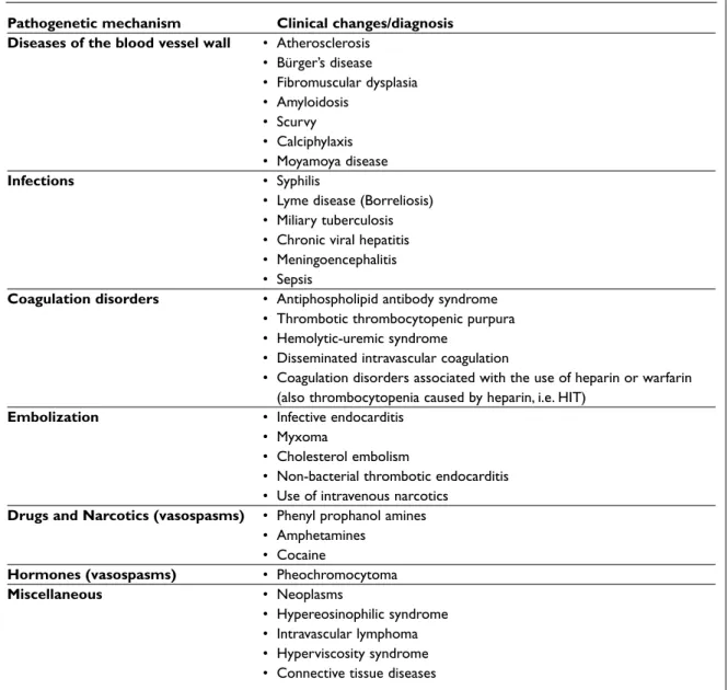
The Third Road Sign: Pseudovasculitis
In addition to vascular wall inflammation and da-mage caused by immunological-inflammatory vasculitis, a large number of conditions has been described which on some other basis lead to ste-nosis, occlusion, thromboembolism, vasospasm, infection or inflammation, all of which can lead to a wrong road38(Table XIV). These conditions are named pseudovasculitis and they are characteri-zed by disturbances in the vascular circulation in the absence of vasculitic changes in the blood ves-sel wall. In spite of this, clinical symptoms and signs, laboratory and radiological findings can be very similar to those found in systemic vasculitides. Pseudovasculitides are not rare and should always be kept in mind when systemic vasculitis is suspec-ted. As a matter of fact, the road sign to secondary vasculitis and to pseudovasculitis should be checked before the one pointing to primary vascu-litis is followed. It is always important to look for
triggering factors and to exclude pseudovasculitis. Management of pseudovasculitides is very diffe-rent from that of primary systemic vasculitides, which emphasizes the importance of differential diagnosis. Different specific and even curative tre-atments are often available for pseudovasculitis. On the other hand, treatment for vasculitis in a pa-tient with pseudovasculitis can result in irrepara-ble damage.
Atherosclerosis
Atherosclerosis is a common disease of large
and middle-sized arteries, in which atheros-clerotic plaques cause stenosis of the arteries and may lead to their occlusion as a result of plaque rupture and thromboembolic disease. Ischemic pain and skin alterations in severe cases resemble those caused by vasculitides. Atherosclerosis is one of the most common differential diagnostic alternatives for vasculitis. Also, atherosclerotic pla-ques contain inflammatory cells. However, patients with atherosclerosis do not usually deve-lop prominent acute phase responses although slightly elevated CRP and fibrinogen are risk factors.
Y R J Ö T. K O N T T I N E N E C O L.
Table XIV. Pseudovasculitis can be classified as blood vessel damaging, obstructing, thromboembolic or vasospastic conditions and other pseudovasculitis.
Pathogenetic mechanism Clinical changes/diagnosis Diseases of the blood vessel wall • Atherosclerosis
• Bürger’s disease • Fibromuscular dysplasia • Amyloidosis • Scurvy • Calciphylaxis • Moyamoya disease Infections • Syphilis
• Lyme disease (Borreliosis) • Miliary tuberculosis • Chronic viral hepatitis • Meningoencephalitis • Sepsis
Coagulation disorders • Antiphospholipid antibody syndrome • Thrombotic thrombocytopenic purpura • Hemolytic-uremic syndrome
• Disseminated intravascular coagulation
• Coagulation disorders associated with the use of heparin or warfarin (also thrombocytopenia caused by heparin, i.e. HIT)
Embolization • Infective endocarditis • Myxoma
• Cholesterol embolism
• Non-bacterial thrombotic endocarditis • Use of intravenous narcotics
Drugs and Narcotics (vasospasms) • Phenyl prophanol amines • Amphetamines
• Cocaine
Hormones (vasospasms) • Pheochromocytoma
Miscellaneous • Neoplasms
• Hypereosinophilic syndrome • Intravascular lymphoma • Hyperviscosity syndrome • Connective tissue diseases
Infections do predispose to atherosclerotic plaque rupture, which can lead to medical calamities like myocardial infarction or stroke and to differential diagnostic problems. Usually atherosclerotic chan-ges are stable so that a certain degree of physical exercise always and consistently leads to ischemic pain, such as angina pectoris or intermittent clau-dication. Atherosclerosis as such is slowly progressi-ve and the patients typically haprogressi-ve a long history spanning back for even a few decades.
Antiphospholipid antibody syndrome (slightly modified criteria)
Antiphospholipid antibody syndrome (Table XV) is characterized by arterial and venous thrombo-sis, thrombocytopenia and recurrent abortions as well as cardiolipin antibodies, β2-glycoprotein I antibodies and/or positive lupus anticoagulant40. Antiphospholipid antibody associated clinical symptoms form a wide spectrum and can include stroke, livedo reticularis and varicose ulcers, which can be similar to those seen in systemic vasculiti-des. Antiphospholipid antibody syndrome asso-ciated Libman-Sacks endocarditis can be a source of peripheral emboli. The so called catastrophic antiphospholipid syndrome is caused by occlusion of the microvasculature and can manifest itself as a progressive renal and multi-organ failure. SLE patients can have antiphospholipid antibodies and, therefore, non-inflammatory vascular occlu-sions and secondary immune complex vasculitis. Antiphospholipid antibody syndrome can be ag-gravated by e.g. estrogens and glucocorticoids.
Infective endocarditis
Infective endocarditis is caused by inflammation of the endocardium and heart valves, which con-tain aggregated platelets, fibrin, microbes and in-flammatory cells in form of vegetations. Heart val-ve pathology, such as bicuspid aortic valval-ve or mi-tral valve insufficiency, as well as immunocompro-mising states predispose to endocarditis. Patient history can reveal an iatrogenic or accidental trau-ma, but often the source of bacteraemia remains unclear. The clinical symptoms and findings, such as petechiae, hematuria, arthralgias and arthritis can resemble a systemic connective tissue disease or a vasculitis. Infective endocarditis can lead to pseudovasculitis as a result of microembolisms, which can also be caused by secondary vasculitis associated with immune complex formation. Even fungal endocarditis has to be considered as a
cau-se of large vescau-sel stenosis recau-sembling vasculitis. Typically, auscultation reveals heart murmurs, which shift in character as the vegetations change as they grow or embolize. Transthoracic or transe-sophageal echocardiography can be used to visu-alize vegetations and underlying or endocarditis-associated valve pathology and blood cultures can be used to disclose bacteraemia. Acute phase reac-tion, hypergammaglobulinemia and rheumatoid factor can be found by laboratory tests in both en-docarditis and vasculitis.
Table XV. A condition can be classified as a definite antiphospholipid antibody syndrome if at least one of the following clinical and one of the laboratory criteria are met39:
Clinical criteria
1. Vascular thrombosis
One or more clinical episodes of arterial, venous, or small vessel thrombosis, in any tissue or organ. Thrombosis must be confirmed by imaging or doppler studies or histopathology, with the excep-tion of superficial venous thrombosis. For
histopathologic confirmation, thrombosis should be present without significant evidence of inflamma-tion in the vessel wall.
2. Pregnancy morbidity
– One or more unexplained deaths of a
morphologically normal foetus at or beyond the 10th week of gestation, with normal foetal morphology documented by ultrasound or by direct examination of the foetus, or
– One or more premature births of a
morphologically normal neonate at or before the 34th week of gestation because of severe preeclampsia or eclampsia, or severe placental insufficiency, or
– Three or more unexplained consecutive spontaneous abortions before the 10th week of gestation in the absence of maternal anatomic or hormonal abnormalities and paternal and maternal chromosomal causes.
Laboratory criteria (on at least two occasions, at least 6 weeks apart, measured from a blood test using methods fulfilling international standards)
– Anticardiolipin antibody of IgG and/or IgM isotype, present in medium or high titer
– Lupus anticoagulant
Myxoma of the heart
Myxoma of the heart is a benign intracardial tu-mour, which can occur at all ages, more commonly in women than in men41. Myxoma can lead to car-diac and extra-carcar-diac symptoms, such as embo-lization to peripheral tissues and lungs, arthritis, petechiae and Raynaud’s phenomenon. Haema-turia and proteinuria occur. Embolic manifestati-ons can simulate vasculitis as they develop sud-denly and lead to severe ischemic changes. Appro-ximately half of these patients have fever and lose weight depending on the production of pro-in-flammatory cytokines in myxomatous tissue. Anaemia, leukocytosis, thrombocytosis and in-creased ESR are common laboratory findings, which are in part caused by interleukin-6 produc-tion by the myxomatous cells.
Biopsies and the embolic lesions demonstrate myxomatous cells, but not vasculitic changes. Transthoracic and transesophageal echocardio-graphies are useful, although differential diagno-sis between myxoma and intracardial thrombodiagno-sis can be difficult. Diagnosis of myxoma is confirmed with computed tomography or magnetic resonan-ce imaging. Resection of myxoma is usually a cu-rative treatment.
Cholesterol embolism
Occasionally the atherosclerotic plaque ulcerates as a result of endovascular procedures (mechani-cal manipulation), anticoagulation (exposing the plaque surface below the thrombus mass) or spon-taneously, leading to cholesterol crystal emboliza-tion42. Small, needle-like cholesterol crystals are disseminated into various tissues, usually kidneys. The typical patient is an elderly man suffering from advanced atherosclerosis, who has recently under-gone an invasive vascular procedure, for example an angiography. In some cases the condition has been reported after warfarin or thrombolytic tre-atment. Also, spontaneous cholesterol emboliza-tions can occur. The manifestaemboliza-tions greatly resem-ble those caused by vasculitis, for example eczema, petechiae, ulcerations and livedo reticularis are common. As the cholesterol emboli are so small, the peripheral pulses of the larger blood vessels re-main easily palpable in spite of the colour of the toes in a typical blue or purple toe syndrome. The most serious manifestations of cholesterol embo-lism are amaurosis fugax and permanent blind-ness, myocardial infarction, bowel infarction, pe-ripheral neuropathy and progressive renal failure.
Laboratory tests usually demonstrate increased ESR, leukocytosis and often eosinophilia. Throm-bocytopenia and hypocomplementemia can also occur. Rheumatoid factor, antinuclear antibodies and even ANCA, usually proteinase-3, non--myeloperoxidase type, can be found. Diagnosis is histopathological and is based on the demonstra-tion of cholesterol crystals, which leave slits in the damaged tissue as they are dissolved away during regular tissue sample processing. There is no spe-cific medical treatment for cholesterol embolism and the mortality of the reported cases has been re-latively high. If a patient using warfarin is diagno-sed suffering from cholesterol embolisms, warfa-rin may have to be stopped. Without surgical tre-atment, cholesterol embolism is a recurrent pro-cess with a high mortality rate.
The First Milestone: Patient History and
Physical Examination
Careful patient history and physical examination have been emphasized as central in establishing the diagnosis. The more complicated the situation seems to be, the more important these are so that invasive (=potentially dangerous and expensive) examinations can be correctly targeted.
In vasculitis of aorta and large arteries, transient ischemic attack and vision disturbances in young women or new headache and polymyalgia rheu-matica in elderly patients are important clues. Non-symmetrical pulse and difference in the blood pressure in the upper extremities or tenderness of the temporal arteries and pulse difference between the right and the left side are useful findings. When middle-sized arteries are affected in polyarteritis nodosa, this often leads to a serious generalized di-sease, in which multiple organ lesions and mono-neuritis multiplex lead the thoughts to the possibi-lity of vasculitis. Kawasaki disease is typically a disease of small children and manifests in form of mucocutaneous lymph node syndrome, which can lead to formation of coronary artery aneurysms and in which immunomodulatory treatment is dif-ferent from the usual regime. When the small arte-ries are diseased in Wegener’s granulomatosis, up-per respiratory tract, pulmonary and renal chan-ges develop; in microscopic polyangiitis severe re-nal disease and palpable purpura; and in Churg-Strauss syndrome a patient with atopy and asthma develops eosinophilia and pulmonary
filtrates. In diseases of the very smallest blood ves-sels, in particular immune complex mediated ve-nulitis, palpable purpura is the cardinal finding, but it can also occur in vasculitis of small-size ar-teries. Henoch-Schönlein purpura usually deve-lops in a child after an upper respiratory tract in-fection and also leads to arthralgia, haematuria and melena, but in the somewhat similar cryoglo-bulinaemic vasculitis melena is usually absent. In-flammation of the superficial and deep veins can lead to thrombophlebitis and phlebothrombosis. Hydrostatic pressure increases and leads to local peripheral oedema. The most feared complication in this condition is pulmonary embolism.
The Second Milestone: Laboratory Tests and
Surrogate Markers
Inflammation leads to cytokine production and acute phase reaction, which lead to general symp-toms like fever, fatigue, anorexia, weight loss, night sweats and myalgias and arthralgias associated with typical laboratory findings (Table XVI).
Already relatively few, but well directed labora-tory tests can be helpful in the diagnosis of vascu-litis. In almost all primary vasculitic diseases the ESR and CRP are increased. Complete blood count with white cell differential is one of those basic la-boratory tests to be ordered in suspected vasculi-tis cases. Primary vasculivasculi-tis is usually associated with normocytic anaemia, leukocytosis and
thrombocytosis. In contrast, thrombocytopenia is not usually seen in primary vasculitis, but is rather suggestive of SLE, infiltration of malignant disea-ses into the bone marrow, thrombotic throm-bocytopenic purpura, disseminated intravascular coagulation or antiphospholipid antibody syndro-me. Leukocytopenia is not typical for vasculitis, but can be suggestive of SLE, sepsis or some other severe or viral infections, aleukaemic leukaemia, myelodysplasia or various drugs. Eosinophilia is part of the Churg--Strauss syndrome, but it can also be seen in Wegener’s granulomatosis and in rheumatoid vasculitis. Chemical screening of the urinary sample and/or urine cytology are patholo-gical in glomerulonephritis.
Autoantibody tests are to a large extent perfor-med in the referral hospital. All tests are not neces-sary in the evaluation of all patients suspected of having vasculitis, but the selection of the tests is de-pendent on the clinical symptoms. Antinuclear an-tibodies and anti-DNA anan-tibodies are recommen-ded when SLE is suspected or has to be exclurecommen-ded. Glomerular basement membrane antibodies are important when the cause for alveolar bleeding and glomerulonephritis is sought. Antiphospholi-pid antibodies are important when unclear arterial or venous thrombosis or thrombocytopenia are evaluated and are also necessary when catastro-phic antiphospholipid antibody syndrome is suspected. Serum complement components C3 and C4 decrease upon consumption in leukocyto-clastic, immune complex-mediated vasculitides and are sometimes useful in the follow-up of glo-merulonephritis. Complement concentrations are low in cryoglobulinaemia and they can be decrea-sed in endocarditis, but are usually normal in polyarteritis nodosa, Wegener’s granulomatosis, microscopic polyangiitis, Churg-Strauss syndro-me and Henoch-Schönlein purpura and can in-crease in infections as they are acute phase reac-tants. Serological tests for the demonstration of hepatitis B and C can be recommended if the pa-tient suffers from polyarteritis nodosa, cryoglobu-linaemia, polyarthritis or cutaneous vasculitis.
ANCA, which has proteinase-3 or myeloperoxi-dase specificity, are seen in a large proportion of patients, who have generalized small artery vascu-litis. c-ANCA and proteinase 3 antibodies are strongly suggestive of Wegener’s granulomatosis43. p-ANCA with myeloperoxidase specificity rather imply microscopic polyangiitis or Churg-Strauss syndrome. ANCA are seen in many other diseases Table XVI. Laboratory tests and findings in basic
health care when vasculitic diseases are suspected
• Erythrocyte sedimentation rate and C-reactive protein are increased
• Complete blood count with white cell differential demonstrates anaemia, leucocytosis, eosinophilia and thrombocytosis
• X-ray of the sinuses demonstrates shadowing or fluid levels
• Chest X-ray demonstrates pulmonary infiltrates • Urinary sample demonstrates haematuria • ANCA suggest small artery vasculitis
• Tests are ordered based on the clinical picture and they can also be used to demonstrate the triggering or perpetuating factor in secondary vasculitis and to exclude pseudovasculitis
such as infections, vasculitic reactions caused by drugs and in many inflammatory diseases other than vasculitides. The reactivity is then usually against something else than proteinase-3 or mye-loperoxidase. Therefore, the vasculitis diagnosis can never be based solely on demonstration of ANCA positivity.
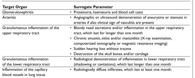
Immunological events induce vasculitic inflam-mation. This can be directly observed in biopsies taken from symptomatic organs or tissues. Histo-pathological diagnosis forms the basis of exact diagnosis, but because such procedures are inva-sive, attempts have been made to develop surro-gate changes for histopathology (Table XVII).
General practitioners can easily take skin biop-sies to confirm leukocytoclastic vasculitis, but as palpable purpura is also clinically easily recogni-zable, such patients should be sent without biopsy to a specialist, if elimination of the triggering sti-mulus has not corrected the situation and suspici-on of vasculitis has been csuspici-onfirmed or still remains after the basic tests, except in trivial and mild ca-ses, often restricted to the skin. Patients with unsta-ble angina pectoris are urgently referred to hospi-tal and the same principle should naturally apply if progressive and unpredictable vasculitic changes develop in potentially dangerous sites since these are “unstable vasculitic plaques” and can lead to severe vital organ damage and failure of heart, kid-neys, brain, and lungs. In the diagnosis and treat-ment of vasculitis an early diagnosis is important, also because the use of platelet drugs or anti-coagulants is difficult in vasculitis, which is
asso-ciated with bleeding tendency. In fact, such drugs may be contraindicated in spite of a prothrombo-tic inflammatory condition associated with unsta-ble vasculitic plaques.
The Third Milestone: Tests in Specialized
Centers
Histopathology
When the patient has been referred to a speciali-zed centre after the basic work up on the primary health care level including patient history, physi-cal examination and basic laboratory tests, biop-sies of symptomatic organs are performed. Recog-nition of targets for biopsies is based on clinical fin-dings, laboratory tests and other objective exami-nations, as blind biopsies easily lead to false negative results. If several organs are affected by the disease, choice of the site of the biopsy is de-termined by the potential risks and the likeliness to obtain a disease specific diagnosis. The biopsy sample has to be large enough since vasculitis does not affect the blood vessels and tissues in a uniform and predictable way, but in a haphazard manner. Biopsies can demonstrate immunoglobulin and complement depositions, T-lymphocytes, ma-crophages, granulomas and necrosis. For example, in giant cell arteritis giant cells and granulomas are seen, in necrotizing polyarteritis nodosa the neu-trophilic granulocyte is the main cell and in Churg-Strauss syndrome associated with atopy and asth-ma the eosinophil is the asth-main cell. In sasth-mall vessel
Y R J Ö T. K O N T T I N E N E C O L.
Table XVII. Surrogate markers in the diagnosis of vasculitis
Target Organ Surrogate Parameter
Glomerulonephritis • Proteinuria, haematuria and blood cell casts
Arteritis • Angiographic or ultrasound demonstration of aneurysms or stenosis in arteries if also clinical sign of vasculitis are present
Granulomatous inflammation of the • Bloody nasal secretions and/or inflammation in the upper respiratory upper respiratory tract tract, which last for longer than one month
• Chronic sinusitis, otitis and/or mastoiditis (X-ray examination, computerized tomography or magnetic resonance imaging) • Sudden hearing loss without trauma
• Destruction of the skull bones and/or cartilage
Granulomatous inflammation • Radiological demonstration of inflammation in lower respiratory tract of the lower respiratory tract (shadowing or cavitations), which last longer than one month
Inflammation of the capillary • Radiologically diffuse infiltrates, which last at least one month. blood vessels in lung tissue
leukocytoclastic vasculitis the dominating cell is a neutrophil associated with nuclear dust. Tissues affected by vasculitis may demonstrate ischemic lesions and necrosis without apparent vasculitic changes. Sometimes it can be difficult to differen-tiate vasculitis, for example, from infections or an-giocentric lymphoma. The phenotypic examinati-on of the cells and other special techniques can be helpful.
Angiographies
Lesions in the vascular wall can lead to stenosis, oc-clusion, vasospasm, thrombosis, embolism, ische-mia, aneurysm or bleeding. Angiography and Dop-pler ultrasound examination can demonstrate stenosis, occlusions, vasospasms, aneurysms, thrombosis and bleedings. X-rays, computed to-mography and magnetic resonance imaging are suitable for the demonstration of bleedings, le-sions, complications and assessment of the extent of the disease.
Angiographies are useful when lesions in the aorta and its main braches are suspected or if the patient has aneurysms. It can also be useful in the diagnosis and follow-up of vasculitis of the central nervous system and of Kawasaki disease and arte-ritis of the coronary arteries. Magnetic resonance angiography (MRA) is useful for the evaluation of the size of the lumen of the large blood vessels and the thickness of their wall. Angiography of the abdominal arteries is used when arteritis of the middle-sized arteries is suspected and histological diagnosis has not been obtained. Polyarteritis no-dosa and other middle-sized artery diseases can lead to microaneurysms and stenosis, which can be visualized using angiography. Polyarteritis no-dosa has microaneurysms in 60-90 % of cases. Aneurysms are more likely to develop with time and are not always found in the early stages of the disease. Microaneurysms of internal organs are not diagnostic of polyarteritis nodosa, as they can also be found in other diseases affecting middle-sized or slightly smaller arteries, for example Wegener’s granulomatosis and Churg-Strauss syndrome, as well as in non-vasculitic conditions, such as myxo-ma of the heart atria and in infectious endocardi-tis. Angiography has no place in the diagnosis of small vessel vasculitis.
Empiric treatment with glucocorticoids
Empiric treatment with glucocorticoids is someti-mes necessary as a part of the diagnostic work-up,
but even though patients would benefit from such medications, this is not enough for diagnosing vas-culitis. A complete diagnostic work up should al-ways be performed if at all possible.
Management of Vasculitis
In the hospital, the activity and extent of vasculitis is examined. In this workout, structured instru-ments such as The Birmingham Vasculitis Activity Score (BVAS)44and The Vasculitis Damage Index (VDI)45are useful. They enable clinicians to iden-tify an active disease, including the level of its ac-tivity, and to separate it from irreversible but al-ready stable damage. This can be of great impor-tance in justifying the use of immunosuppressive treatment. Some new tests like measurement of von Willebrand factor or procalcitonin can be help-ful in this respect. In many centres vasculitis pa-tients are enrolled into international controlled cli-nical trials on the treatment of vasculitis.
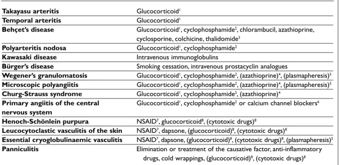
If and when the triggering and perpetuating fac-tor can not be identified, the management of pri-mary vasculitis is based on immunosuppression46 (Table XVIII). Immunosuppression is also used in the management of the secondary vasculitis with poor response to treatment targeted at the elimi-nation of the suspected triggering factor47. Treat-ment is tailor-made, but some general manage-ment strategies have been created.
In arteritis of the large vessels treatment is ini-tiated with prednisolone alone in high immuno-suppressive doses16. In Takayasu arteritis azathio-prine or weekly methotrexate can add to the effi-cacy and enable tapering of the glucocorticoids48. In temporal arteritis occasionally azathioprine or methotrexate is added to the glucocorticoid treat-ment as a glucocorticoid sparing agent.
Treatment of vasculitis of the middle and small sized arteries is based on prednisolone combined with cyclophosphamide, either per os (more
effec-tive) or intravenously (safer)46,49. The dose of the immunosuppressants is adjusted so that blood leukocytes remain over 3.0–3.5 x 10⁹/l and neu-trophils over 1.0–1.5 x 10⁹/l. To avoid irritation and eventual development of cancer of the urinary bladder, fluid intake should be at least three litres per day and can be combined with prophylactic treatment with mesna. In Kawasaki disease in chil-dren, glucocorticoids should not be used as the vasculitic plaques of the coronary arteries in