Comparison of Gleason upgrading rates in transrectal
ultrasound systematic random biopsies versus US-MRI
fusion biopsies for prostate cancer
_______________________________________________
Paulo Priante Kayano ¹, Arie Carneiro ¹, Tiago Mendonça Lopez Castilho
1, Arjun Sivaraman ², Oliver Rojas
Claros ¹, Ronaldo Hueb Baroni ¹, Rodrigo Gobbo Garcia ¹, Guilherme Cayres Mariotti ¹, Oren Smaletz ¹,
Renne Zon Filippi ¹, Gustavo Caserta Lemos
11 Hospital Israelita Albert Einstein, São Paulo, SP, Brasil; 2 Memorial Sloan Kettering Cancer Center -
USA, New York, NY, EUA
INTRODUCTION
Brazilian data shows that prostate cancer (PCa) is the most common non-cutaneous
malig-ABSTRACT
Purpose: Ultrasound-magnetic resonance imaging (US-MRI) fusion biopsy (FB) im-proves the detection of clinically significant prostate cancer (PCa).
We aimed to compare the Gleason upgrading (GU) rates and the concordance of the Gleason scores in the biopsy versus final pathology after surgery in patients who underwent transrectal ultrasound (TRUS) systematic random biopsies (SRB) versus US-MRI FB for PCa.
Materials and Methods: A retrospective analysis of data that were collected prospec-tively from January 2011 to June 2016 from patients who underwent prostate biopsy and subsequent radical prostatectomy. The study cohort was divided into two groups: US-MRI FB (Group A) and TRUS SRB (Group B).
US-MRI FB was performed in patients with a previous MRI with a focal lesion with a Likert score ≥3; otherwise, a TRUS SRB was performed.
Results: In total, 73 men underwent US-MRI FB, and 89 underwent TRUS SRB. The GU rate was higher in Group B (31.5% vs. 16.4%; p=0.027). According to the Gleason grade pattern, GU was higher in Group B than in Group A (40.4% vs. 23.3%; p=0.020). Analyses of the Gleason grading patterns showed that Gleason scores 3+4 presented less GU in Group A (24.1% vs. 52.6%; p=0.043).
The Bland-Altman plot analysis showed a higher bias in Group B than in Group A (-0.27 [-1.40 to 0.86] vs. -0.01 [-1.42 to 1.39]).
In the multivariable logistic regression analysis, the only independent predictor of GU was the use of TRUS SRB (2.64 [1.11 – 6.28]; p=0.024).
Conclusions: US-MRI FB appears to be related to a decrease in GU rate and an increase in concordance between biopsy and final pathology compared to TRUS SRB, sug-gesting that performing US-MRI FB leads to greater accuracy of diagnosis and better treatment decisions.
ARTICLE INFO
Keywords:
Prostatic Neoplasms; Magnetic Resonance Spectroscopy; Image-Guided Biopsy
Int Braz J Urol. 2018; 44: 1106-113
_____________________
Submitted for publication: October 22, 2017
_____________________
Accepted after revision: August 05, 2018
_____________________
Published as Ahead of Print: September 10, 2018
(TRUS)-guided random biopsies. The Gleason sco-re of PCa has been shown to be an important crite-rion to predict tumour behaviour and to determine the appropriate course of treatment (2). However, the randomness and non-targeted nature of TRUS biopsy can result in inaccurate sampling of the cancer and misclassification of cancer risk. Rese-archers have demonstrated discrepancies in the Gleason score of TRUS biopsy compared to the final surgical specimens, with under-estimation reported in approximately 30% of cases (3).
Recently, multiparametric magnetic reso-nance imaging (mpMRI) of the prostate has been shown to be valuable in the detection, localization and characterization of prostatic tumour foci (4). Target biopsy of the abnormality detected by MRI was initially performed by cognitive guidance of the topographic location of the cancer, but in re-cent years, devices were developed to combine ul-trasound and mpMRI images. The US-MRI fusion images can guide biopsy and improve the detec-tion of clinically significant prostate cancer (5, 6). A more accurate diagnostic method is desirable to avoid misclassification, which is particular-ly important in appropriate decision-making for the treatment of PCa (active surveillance or focal therapy or radical treatment). It is plausible that the Gleason score misclassification and upgrading noted in radical prostatectomy specimens can be reduced by employing more accurate biopsy tech-niques.
The aim of this study was to compare the Gleason upgrading rates and the concordance of the biopsy versus final pathology Gleason scores in patients who underwent TRUS systematic ran-dom biopsies (SRB) versus US-MRI fusion biopsies (FB) for prostate cancer.
MATERIALS AND METHODS
Patient selection and data collection
We included all consecutive patients who underwent prostate biopsy and subsequent radical prostatectomy at our institution. The study cohort was divided into those who had US-MRI FB (from June 2013 to July 2015) (Group A) and TRUS SRB (from June 2010 to February 2015) (Group B). Pathological analyses of the prostatectomy
spe-cimens were reviewed by a single, experienced pathologist and were considered as the standard of reference. Patients who did not undergo biopsy and radical prostatectomy at our institution and those with pathologic specimens that were not re-viewed by the same pathologist were excluded to avoid bias. IRB approval and a waiver for infor-med consent were obtained for this retrospective study using prospectively collected data from our institution database.
US-MRI FB was performed in all patients with mpMRI-detected abnormalities and Likert scores ≥3. Patients with a normal mpMRI or Li-kert scores <3 underwent TRUS SRB. All radical prostatectomies were performed by our institution Urology staff either by robot-assisted radical pros-tatectomy or by an open approach.
Data related to clinical, biopsy, histopa-thological and MRI characteristics were collected.
Multiparametric MRI
MRIs were performed on 3T scanners (Sie-mens Prisma 3T, Sie(Sie-mens PetRM 3T, GE 750W 3,0T, Philips 3,0T) with a phased-array coil and included high-resolution T2-weighted imaging, diffusion-weighted imaging and dynamic con-trast-enhanced imaging. A Likert scale score, that is a subjective assessment on the likelihood of the presence of prostate cancer on a 5 point scale (7), was assigned by one of our uro-radiologist with years of experience in interpreting prostate MRI (median of 7 years of experience; range 5 to 15 years) and every exam were reviewed by other experienced radiologist, and if there was a discre-pancy in the analyses, the score was assigned after a consensus. Only lesions classified with scores ≥3 were defined as targets for US-MRI fusion biopsy.
US-MRI Fusion Biopsy
Each biopsy was performed with the pa-tient in a left lateral decubitus position, using endocavitary 4 to 9 MHz broadband curved ar-ray end-fire transducers and an 18-gauge side--notch cutting core biopsy needle (20-mm stroke length). Patients first underwent systematic 14-core biopsies (six from each lobe and one more from each transitional zone), followed by targe-ted biopsies generally consisting of 2 or 3 cores from each target.
Histopathology
Gleason scoring was performed according to the 2005 International Society of Urological Pathology consensus recommendations (8). We classified the patients according to the 2014 ISUP consensus meeting held in Chicago in 2014, which classify the Gleason scores into grade groups (Gle-ason score ≤6 = ISUP 1; Gleason score 3+4 = ISUP 2; Gleason score 4+3 = ISUP 3; Gleason score 4+4 = ISUP 4; Gleason score 9 or 10 = ISUP 5) (9).
Cores from each lesion were numbered and labelled according to the target, enabling radiology-pathology correlation in patients with multiple targets.
Surgical specimens were processed using a modified Stanford technique; 3- to 5-mm trans-verse sectioned samples were taken from the apex to the base and from the sagittal section of the distal 5 to 8 mm of the apex and base.
Statistical analysis
The primary endpoint of this study was to compare the rate of any Gleason score upgra-de of RP compared to US-MRI fusion biopsy and random biopsy alone. Descriptive statistics were used for patient characteristics. An independent Student’s t, Mann-Whitney, chi-square or Fisher’s exact test was used to compare characteristics of the patients when appropriate. Gleason upgrading was compared by comparison of proportions. A multivariable logistic regression using forced en-try was carried out to assess the independent dictors of Gleason upgrading. The results are pre-sented as odds ratios (ORs) and 95% confidence intervals. Agreement between Fusion US-MRI and histopathology and Random Biopsy and
histopa-thology was assessed using a Bland-Altman plot, and bias was calculated with their respective 95% confidence intervals.
All analyses were conducted with SPSS v.20 (IBM SPSS Statistics for Windows, Version 20.0. Armonk, NY: IBM Corp.) or R v.2.12.0 (R Foundation for Statistical Computing, Vienna, Austria). For all analyses, two–sided p < 0.05 were considered significant.
RESULTS
Characteristics of the cohort
A total of 73 men who underwent US--MRI fusion biopsies and 89 who underwent TRUS systematic random biopsies were included in our analyses. In both groups, the patient demographics were similar (Table-1). There were no differences according to prostate volume (histopathology), PSA, clinical and pathologic staging and number of lymph nodes. However, there was a significant difference in biopsy Gleason score between both groups. Patients from Group A had fewer Gleason score 6 tumours (11% vs. 28%), and patients from Group B had greater total tumour volumes (15% vs. 10%) and fewer clinically significant tumours (70.8% vs. 89%) (Table-1), defined as patients with Gleason score greater or equal to 3+ 4 or greater than ISUP 1.
Primary endpoint
Table 1– Patients characteristics.
US- MRI fusion Random
p value (n = 73) (n = 89)
Age (years), median (IQR) 65.0 (57.5 – 69.0) 64 (59 – 69) 0.838
PSA (ng/mL), median (IQR) 4.8 (3.7 – 6.4) 5.5 (4.2 – 7.2) 0.060
Prior biopsy status, n (%) 9 / 73 (12.3) 10 / 89 (11.2) 0.829
Biopsy Gleason grade group, n (%)
0.007 Less or equal to 6 8 / 73 (11.0) 25 / 89 (28.1)
3+4 = 7 29 / 73 (39.7) 19 / 89 (21.3)
4+3 = 7 21 / 73 (28.8) 23 / 89 (25.8)
8 8 / 73 (11.0) 18 / 89 (20.2)
9-10 7 / 73 (9.6) 4 / 89 (4.5)
Number of total cores, median (IQR) 18.0 (12.0 - 19.5) 15.0 (14.0 - 17.5) 0.144
Number of random cores, median (IQR) 14.0 (11.0 - 18.0) 14.0 (14.0 - 17.0) 0.171
Number of targeted cores, median (IQR) 4.0 (3.0 – 5.0) ND
Positive cores, n (%) 61 / 71 (85.9) 13 / 20 (65) 0.007
Positive targeted cores, median (IQR) 6.0 (4.5 – 10.0) ND
Prostate volume at histopathology (grams), median (IQR) 42.0 (30.0 – 56.0) 40.0 (32.5 – 47.0) 0.223
Surgical specimen Gleason grade group, n (%)
0.205 Less or equal to 6 2 / 73 (2.7) 9 / 89 (10.1)
3+4 = 7 32 / 73 (43.8) 29 / 89 (32.6)
4+3 = 7 28 / 73 (38.4) 31 / 89 (34.8)
8 5 / 73 (6.8) 8 / 89 (9.0)
9-10 6 / 73 (8.2) 12 / 89 (13.5)
Total tumor volume (%), median (IQR) 10.0 (7.0 – 20.0) 15.0 (10.0 – 20.0) 0.024
Bilateral tumor, n (%) 54 / 73 (74.0) 67 / 89 (75.3) 0.848
Multifocal tumor, n (%) 60 / 73 (82.2) 76 / 89 (85.4) 0.580
Positive lymph node, n (%) 1 / 70 (1.4) 3 / 87 (3.4) 0.424
Clinically significant tumor, n (%) 65 / 73 (89.0) 63 / 89 (70.8) 0.004
Time between biopsy and surgery (days), median (IQR) 30.0 (30.0 – 60.0) 60.0 (30.0 – 60.0) 0.224
---submitted to US-MRI fusion biopsy (-0.27 [-1.40 to 0.86] vs. -0.01 [-1.42 to 1.39]) (Figure-1). In the multivariate logistic regression, the use of TRUS systematic random biopsy, compared to US-MRI fusion biopsy, was the only independent predictor of Gleason upgrading (2.64 [1.11 – 6.28]; p=0.024) (Table-3).
The comparison analysed by the Bland--Altman plot of Group A and Group B showed
that the agreement bias between Gleason score on biopsy and Gleason score on surgical specimen was lower in Group A (Figure-1).
DISCUSSION
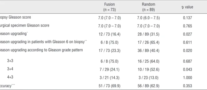
In our study, we found a significantly lo-wer rate of Gleason upgrading using US-MRI FB, showing that this method can improve prosta-Table 2 – Gleason upgrading.
Fusion (n = 73)
Random
(n = 89) p value
Biopsy Gleason score 7.0 (7.0 – 7.0) 7.0 (6.0 – 7.5) 0.137
Surgical specimen Gleason score 7.0 (7.0 – 7.0) 7.0 (7.0 – 7.0) 0.765
Gleason upgrading* 12 / 73 (16.4) 28 / 89 (31.5) 0.027
Gleason upgrading in patients with Gleason 6 on biopsy**
6 / 8 (75.0) 17 / 26 (65.4) 0.611
Gleason upgrading according to Gleason grade pattern 17 / 73 (23.3) 36 / 89 (40.4) 0.020
3+3 6 / 8 (75.0) 16 / 25 (64.0) 0.687
3+4 7 / 29 (24.1) 10 / 19 (52.6) 0.043
4+3 3 / 21 (14.3) 3 / 23 (13.0) 1.000
Accuracy*** 51 / 73 (69.9) 56 / 89 (62.9) 0.353
* defined as the number of patients with Gleason score in surgical specimen greater then biopsy sample.
** defined as the number of patients with Gleason score 6 in biopsy sample (clinically non-significant disease) that presented a Gleason score greater than 6 in surgical specimen (clinically significant disease). Patients with Gleason score greater than 6 were excluded from this analyses.
*** defined as the number of patients who presented the same Gleason score in biopsy sample and surgical specimen.
Figure 1 - Bland - Altman Plot Group A vs. Group B.
Mean ([Surgical specimen Gleason + Biopsy Gleason]/2)
Difference (Surgical specimen Gleason - Biopsy Gleason) Difference (Surgical specimen Gleason - Biopsy Gleason)
Group A Group B
Mean ([Surgical specimen Gleason + Biopsy Gleason]/2)
Fusion: Bias (red line) e 95% confidence limits (red dot line): -0.014 (-1.42 to 1.39) Random: Bias (red line) e 95% confidence limits (red dot line): -0.270 (-1.40 to 0.86)
3
2
1
0
-1
-2
-3
2
1
0
-1
-2
te cancer characterization at biopsy. All mpMRI, biopsies and surgical specimens were evaluated by the same team and methods to keep the pattern and to reduce bias in the Gleason and Likert score classifications.
The use of US-MRI FB was associated with a lower rate of Gleason upgrading compared to the use of TRUS SRB (16.4% vs. 31.5%; p=0.027). In the clinical setting, a diagnostic tool that can determine the “true” Gleason score plays a cru-cial role in guiding the clinician in making the best therapeutic decision, mainly for low- and intermediate-risk PCa. In the active surveillance scenario, it is imperative to decrease the risk of missing a high-grade disease and delaying a ra-dical treatment, providing more confidence to the urologist and patient with conservative manage-ment of PCa.
For patients who will be treated by prosta-tectomy or radiation therapy, the biopsy Gleason score is considered a key point in most nomogra-ms for determining the indication of extended lymphadenectomy and changes in the irradiation field or the time of hormone therapy in patients under radiation therapy, thus changing the impact on the morbidity (10-14).
Data reported in the literature show that the Gleason score is frequently lower for TRUS guided biopsies compared to that for surgical specimens, with under-estimation reported in about 30% of cases. In patients with low-grade prostate biopsies, the risk of upgrading may in-crease up to 50% (3).
Our study showed that patients with ini-tial Gleason scores of 6 presented more Glea-son upgrading in the US-MRI FB group (75% vs. 65.4%; p=0.611); however, this difference was not statistically significant, probably due to the relatively small number of patients. Biopsies with Gleason scores of 3+4 presented less Glea-son upgrading in the US-MRI FB group (24.1% vs. 52.6%; p=0.043). The concordance was hi-gher when the highest Gleason grading pattern was analysed between biopsy and surgical spe-cimens, consistent with our hypothesis that US--MRI FB increases the detection of the highest--grade tumour (3, 15, 16).
Arsov et al. reported an MRI FB and TRUS random biopsy upgrading of 21.2% and 32.7%, respectively (17). In this study, Gleason upgrading was twice as frequent in patients who underwent US-MRI FB compared with TRUS SRB (31.5% ver-sus 16.4%).
Multiparametric MRI of the prostate has shown its value in the detection, localization and characterization of prostatic tumour foci (4) and plays an important role to avoid unnecessary biopsies in patients with previously negative ones, showing accuracies of approximately 90% for the diagnosis of significant prostate cancer (18-20).
In our institution, most urologists are using mpMRI in biopsy-naïve patients, avoiding some biopsies in patients with low probability of clinically significant prostate cancer, which can explain the lower rate of Gleason 6 in patients who underwent US-MRI FB (11% vs. 28.1%) Table 3 – Logistic Regression (outcome: Gleason upgrading).
Univariate Multivariate
OR (95% CI) p value OR (95% CI) p value
Age 1.01 (0.97 – 1.06) 0.596 ---
---Baseline PSA 1.05 (0.94 – 1.19) 0.371 1.03 (0.91 – 1.17) 0.632
Prostate volume 0.99 (0.97 – 1.01) 0.277 0.99 (0.96 – 1.01) 0.282
Time between biopsy and surgery 1.00 (0.99 – 1.01) 0.358 0.99 (0.97 – 1.01) 0.173
LIKERT score 1.26 (0.56 – 2.81) 0.575 ---
---Random biopsy 2.33 (1.09 – 5.01) 0.030 2.64 (1.11 – 6.28) 0.028
Prostate mpMRI and target biopsy could be incorporated into active surveillance selection criteria, having a higher accuracy for risk strati-fication (21). Prostate mpMRI can also reduce the need for repetitive biopsies by as much as 68% through non-invasive serial monitoring for those on active surveillance (22). Disease reclassifica-tion on those in active surveillance with normal mpMRI appears to be very low, with negative pre-dictive value ranges from 81–90% (23-25).
One limitation of this study is the non--randomized retrospective study design and the heterogeneous population studied, which might lessen the generalizability of our results because of potential selection bias. Another possible selec-tion bias is the small number of patients with Gle-ason 6 biopsy scores, because we perform mpMRI in biopsy naïve patients and avoid the biopsy on those with LIKERT 1 or 2. A prospective rando-mized study might eliminate this bias and might confirm our hypothesis.
CONCLUSIONS
US-MRI FB appears to be associated with a lower Gleason upgrading rate and a higher con-cordance between biopsy and final pathology compared to TRUS SRB, leading to greater accu-racy of diagnosis and therefore better treatment decisions. The routine use of MRI before biopsy is associated with a decrease in the detection of clinically insignificant tumours.
COMPLIANCE WITH ETHICAL STANDARDS
For this type of study, formal consent is not required. This study was approved by our local ethical committee (registered in number (CAAE): 61372916.4.0000.0071).
FINANCIAL SUPPORT
This study received financial support from “Sociedade Beneficente Israelita Brasileira Albert Einstein – Amigo H”.
CONFLICT OF INTEREST
None declared.
REFERENCES
1. [No Authors]. Estimativa 2016: incidência de câncer no Brasil / Instituto Nacional de Câncer José Alencar Gomes da Silva – Rio de Janeiro: INCA, 2015. Available at. < http://santacasadermatoazulay.com.br/wp-content/ uploads/2017/06/estimativa-2016-v11.pdf>
2. Fine SW, Amin MB, Berney DM, Bjartell A, Egevad L, Epstein JI, et al. A contemporary update on pathology reporting for prostate cancer: biopsy and radical prostatectomy specimens. Eur Urol. 2012;62:20-39.
3. Cohen MS, Hanley RS, Kurteva T, Ruthazer R, Silverman ML, Sorcini A, et al. Comparing the Gleason prostate biopsy and Gleason prostatectomy grading system: the Lahey Clinic Medical Center experience and an international meta-analysis. Eur Urol. 2008;54:371-81.
4. Delongchamps NB, Rouanne M, Flam T, Beuvon F, Liberatore M, Zerbib M, et al. Multiparametric magnetic resonance imaging for the detection and localization of prostate cancer: combination of T2-weighted, dynamic contrast-enhanced and diffusion-weighted imaging. BJU Int. 2011;107:1411-8.
5. Siddiqui MM, Rais-Bahrami S, Truong H, Stamatakis L, Vourganti S, Nix J, et al. Magnetic resonance imaging/ ultrasound-fusion biopsy significantly upgrades prostate cancer versus systematic 12-core transrectal ultrasound biopsy. Eur Urol. 2013;64:713-9.
6. Sonn GA, Natarajan S, Margolis DJ, MacAiran M, Lieu P, Huang J, et al. Targeted biopsy in the detection of prostate cancer using an office based magnetic resonance ultrasound fusion device. J Urol. 2013;189:86-91.
7. Rosenkrantz AB, Kim S, Lim RP, Hindman N, Deng FM, Babb JS, et al. Prostate cancer localization using multiparametric MR imaging: comparison of Prostate Imaging Reporting and Data System (PI-RADS) and Likert scales. Radiology. 2013;269:482-92.
8. Epstein JI, Allsbrook WC Jr, Amin MB, Egevad LL; ISUP Grading Committee. The 2005 International Society of Urological Pathology (ISUP) Consensus Conference on Gleason Grading of Prostatic Carcinoma. Am J Surg Pathol. 2005;29:1228-42.
10. Stephenson AJ, Scardino PT, Eastham JA, Bianco FJ Jr, Dotan ZA, Fearn PA, et al. Preoperative nomogram predicting the 10-year probability of prostate cancer recurrence after radical prostatectomy. J Natl Cancer Inst. 2006;98:715-7. Erratum in: J Natl Cancer Inst. 2012;104:423.
11. Cooperberg MR, Hilton JF, Carroll PR. The CAPRA-S score: A traightforward tool for improved prediction of outcomes after radical prostatectomy. Cancer. 2011;117:5039-46. 12. Carneiro A, Sasse AD, Wagner AA, Peixoto G, Kataguiri A, Neto
AS, et al. Cardiovascular events associated with androgen deprivation therapy in patients with prostate cancer: a systematic review and meta-analysis. World J Urol. 2015;33:1281-9. 13. Briganti A, Larcher A, Abdollah F, Capitanio U, Gallina A,
Suardi N, et al. Updated nomogram predicting lymph node invasion in patients with prostate cancer undergoing extended pelvic lymph node dissection: the essential importance of percentage of positive cores. Eur Urol. 2012;61:480-7. 14. Bolla M, de Reijke TM, Van Tienhoven G, Van den Bergh
AC, Oddens J, Poortmans PM, et al. Duration of androgen suppression in the treatment of prostate cancer. N Engl J Med. 2009;360:2516-27.
15. King CR, Long JP. Prostate biopsy grading errors: a sampling problem? Int J Cancer. 2000;90:326-30.
16. San Francisco IF, DeWolf WC, Rosen S, Upton M, Olumi AF. Extended prostate needle biopsy improves concordance of Gleason grading between prostate needle biopsy and radical prostatectomy. J Urol. 2003;169:136-40.
17. Arsov C, Becker N, Rabenalt R, Hiester A, Quentin M, Dietzel F, et al. The use of targeted MR-guided prostate biopsy reduces the risk of Gleason upgrading on radical prostatectomy. J Cancer Res Clin Oncol. 2015;141:2061-8. 18. Kim JY, Kim SH, Kim YH, Lee HJ, Kim MJ, Choi MS.
Low-risk prostate cancer: the accuracy of multiparametric MR imaging for detection. Radiology. 2014;271:435-44. 19. Baco E, Ukimura O, Rud E, Vlatkovic L, Svindland A, Aron
M, et al. Magnetic resonance imaging-transectal ultrasound image-fusion biopsies accurately characterize the index tumor: correlation with step-sectioned radical prostatectomy specimens in 135 patients. Eur Urol. 2015;67:787-94.
20. Thompson J, Lawrentschuk N, Frydenberg M, Thompson L, Stricker P; USANZ. The role of magnetic resonance imaging in the diagnosis and management of prostate cancer. BJU Int. 2013;112(Suppl 2):6-20.
21. Bjurlin MA, Mendhiratta N, Wysock JS, Taneja SS. Multiparametric MRI and targeted prostate biopsy: Improvements in cancer detection, localization, and risk assessment. Cent European J Urol. 2016;69:9-18.
22. Siddiqui MM, Truong H, Rais-Bahrami S, Stamatakis L, Logan J, Walton-Diaz A, et al. Clinical implications of a multiparametric magnetic resonance imaging based nomogram applied to prostate cancer active surveillance. J Urol. 2015;193:1943-9.
23. Mullins JK, Bonekamp D, Landis P, Begum H, Partin AW, Epstein JI, et al. Multiparametric magnetic resonance imaging findings in men with low-risk prostate cancer followed using active surveillance. BJU Int. 2013;111:1037-45.
24. Margel D, Yap SA, Lawrentschuk N, Klotz L, Haider M, Hersey K, et al. Impact of multiparametric endorectal coil prostate magnetic resonance imaging on disease reclassification among active surveillance candidates: a prospective cohort study. J Urol. 2012;187:1247-52.
25. Park BH, Jeon HG, Choo SH, Jeong BC, Seo SI, Jeon SS, et al. Role of multiparametric 3.0-Tesla magnetic resonance imaging in patients with prostate cancer eligible for active surveillance. BJU Int. 2014;113:864-70.