Other uses, including reproduction and distribution, or selling or
licensing copies, or posting to personal, institutional or third party
websites are prohibited.
In most cases authors are permitted to post their version of the
article (e.g. in Word or Tex form) to their personal website or
institutional repository. Authors requiring further information
regarding Elsevier’s archiving and manuscript policies are
encouraged to visit:
Reproducing stone monument photosynthetic-based
colonization under laboratory conditions
Ana Zélia Miller
a,⁎
, Leonila Laiz
b, Juan Miguel Gonzalez
b, Amélia Dionísio
c,
Maria Filomena Macedo
a, Cesareo Saiz-Jimenez
baDepartamento de Conservação e Restauro, Faculdade de Ciências e Tecnologia, Universidade Nova de Lisboa, Monte de Caparica,
2829-516 Caparica, Portugal
bInstituto de Recursos Naturales y Agrobiologia, CSIC, Av. Reina Mercedes 10, 41012 Sevilla, Spain
cCentro de Petrologia e Geoquímica, Instituto Superior Técnico, Av. Rovisco Pais, 1049-001, Lisboa, Portugal
A R T I C L E I N F O
A B S T R A C T
Article history:
Received 13 December 2007 Received in revised form 26 June 2008
Accepted 28 June 2008
Available online 2 September 2008
In order to understand the biodeterioration process occurring on stone monuments, we analyzed the microbial communities involved in these processes and studied their ability to colonize stones under controlled laboratory experiments. In this study, a natural green biofilm from a limestone monument was cultivated, inoculated on stone probes of the same lithotype and incubated in a laboratory chamber. This incubation system, which exposes stone samples to intermittently sprinkling water, allowed the development of photosynthetic biofilms similar to those occurring on stone monuments. Denaturing gradient gel electrophoresis (DGGE) analysis was used to evaluate the major microbial components of the laboratory biofilms. Cyanobacteria, green microalgae, bacteria and fungi were identified by DNA-based molecular analysis targeting the 16S and 18S ribosomal RNA genes. The natural green biofilm was mainly composed by the Chlorophyta Chlorella,
Stichococcus, andTrebouxia, and by Cyanobacteria belonging to the generaLeptolyngbyaand
Pleurocapsa. A number of bacteria belonging to Alphaproteobacteria, Bacteroidetes and Verrucomicrobia were identified, as well as fungi from the Ascomycota. The laboratory colonization experiment on stone probes showed a colonization pattern similar to that occurring on stone monuments. The methodology described in this paper allowed to reproduce a colonization equivalent to the natural biodeteriorating process.
© 2008 Elsevier B.V. All rights reserved.
Keywords:
Biodeterioration Laboratory chamber Limestone
Phototrophic microorganisms Colonization experiment
1.
Introduction
Stone monuments represent an important part of our world's cultural heritage. Natural stones are weathered by physical, chemical and biological factors. One of the most complex problems in monument conservation is to contrast biological deterioration or biodeterioration (Ortega-Calvo et al., 1995; Herrera et al., 2004; Miller et al., 2006). The interactions between environmental factors affecting stone monuments (e.g. light intensity, air pollution, humidity) and microorgan-isms are not well understood. Among the components of the
microbial communities, phototrophic organisms are the primary producers and play an important role in the coloniza-tion and deterioracoloniza-tion of stone monuments, causing extensive aesthetic, physical and chemical damages (Tomaselli et al., 2000; Ciferri, 2002; Crispim and Gaylarde, 2005). In order to understand the biodeterioration process, laboratory experi-ments present the advantage of controlling environmental variables which simplifies the answering of important ques-tions. A number of different laboratory-based experimental biofilm model systems have been developed (Guillitte and Dreesen, 1995; Tiano et al., 1995; Tomaselli et al., 2000; Monte,
⁎Corresponding author.Tel./fax: +351 21 294 83 22.
E-mail address:[email protected](A.Z. Miller).
0048-9697/$–see front matter © 2008 Elsevier B.V. All rights reserved. doi:10.1016/j.scitotenv.2008.06.066
a v a i l a b l e a t w w w . s c i e n c e d i r e c t . c o m
2003; Prieto and Silva, 2005; Miller et al., 2006). In most cases, these systems were used to study single-species biofilms or a predefined mixed community, which cannot give a complete idea of the complex process involved in stone deterioration. In nature, microbes develop in more or less complex commu-nities (Sand, 1997). Laboratory studies using natural microbial biofilms are scarce in biodeterioration studies.
The main goal of this work is the assessment of the ability of natural phototrophic communities colonizing stone probes under laboratory conditions.
2.
Materials and methods
2.1. Site description and environmental sampling
Santa Clara-a-Velha Monastery is a gothic monument con-structed in 1283, on the left bank of the Mondego River, with a yellowish, oolitic and dolomitic limestone of Sinemurian age (Aires-Barros et al., 2000), and a fine-grained compact and homogeneous limestone of Bathonian–Bajocian age – Ançã limestone–used for decorative architecture elements (Dionísio, 1997). Since the beginning of the 14th century, part of the monastery was buried by 5 to 7 m of alluvial sediments due to several river flooding occurrences. The monastery was aban-doned in the 17th century, triggering a progressive deterioration process leading to a state of ruin. An exhaustive restoration campaign of Santa Clara-a-Velha Monastery started in 1995, entailing the removal of accumulated sediments in the cloister and in the church. It also included the construction of a peripheral water retention barrier. In June 2006, samples of a natural green biofilm, covering Ançã limestone columns of the Monastery's North façade, were collected at 50 cm above the ground, under aseptic conditions by scraping the biofilms into sterile tubes. Samples for culturing procedures were stored at 4 °C while those for nucleic acid analyses were preserved in the laboratory at−80 °C until being processed (samples AL1, AL2,
and AL3).
2.2. Experimental setup
In order to reproduce in laboratory conditions the develop-ment of a natural phototrophic biofilm present on biodeter-iorated stone surfaces, a natural green biofilm from Santa Clara-a-Velha Monastery, in Coimbra (central Portugal) was collected, its microbial community analyzed, and cultivated in liquid medium BG11 during three months. The last experi-mental step was the inoculation of this cultivated microbial community on lithic probes of similar composition in a laboratory chamber. The methodology used to monitor the composition of microbial community from environmental samples, in the cultural medium BG11 and on the stone probes is a culture-independent technique described below.
2.3. Analysis of microbial communities
Microbial communities DNA present in the natural green biofilm were extracted using the Nucleospin Food DNA Extraction Kit (Macherey-Nagel, Düren, Germany). The 16S and 18S rRNA genes were used for the identification of
prokaryotes (bacteria and cyanobacteria) and eukaryotes (fungi and microalgae), respectively. Amplification of DNA was carried out by PCR in a BioRad iCycler iQ thermal cycler (BioRad, Hercules, CA). Bacterial 16S rRNA genes were amplified using the general primer pair 616F (5′-AGA GTT TGA TYM TGG CTC AG) (corresponding to position 27 of
Escherichia coli16S rRNA gene) (Zimmermann et al., 2005) and 907R (5′-CCC CGT CAA TTC ATT TGA GTTT) (corresponding to position 907 ofE. coli 16S rRNA gene) (Schabereiter-Gurtner et al., 2001), comprising a denaturing step of 2 min at 95 °C, 35 cycles of denaturing (95 °C for 15s), annealing (55 °C for 15s) and elongation (72 °C for 2 min), following a terminal elongation step of 10 min at 72 °C. Cyanobacterial and chloroplasts 16S rRNA genes were amplified using the primer pair Cya106F (5′-CGG ACG GGT GAG TAA CGC GTG A) and Cya781R (5′-GAC TAC TGG GGT ATC TAA TCC CWT T) and PCR conditions as described byNübel et al. (1997). Eukaryotic 18S rRNA genes were amplified with the primer pair EukA (5′-AAC CTG GTT GAT CCT GCC AGT) and EukB (5′-TGA TCC TTC TGC AGG TTC ACC TAC) (Diez et al., 2001). This amplification was carried out with a denaturing step of 2 min at 95 °C, 35 cycles of denaturing (95 °C for 15s), annealing (50 °C for 12s) and elongation (72 °C for 2 min), following a terminal elongation step of 10 min at 72 °C. TaKaRa ExTaq Polymerase (TaKaRa Shuzo Co., Otsu, JP) was used throughout this study.
Amplification products were used for two different proto-cols. The first analysis consisted in obtaining prokaryotic community fingerprints by Denaturing Gradient Gel Electro-phoresis (DGGE) from all amplification products obtained using the primer pairs 616F-907R and Cya106F-Cya781R. A nested-PCR reaction was performed using the primer pair 341F-GC (5′-CC TAC GGG AGG CAG CAG, with a GC-rich tail added at its 5′-end) and 518R (5′-ATT ACC GCG GCT GCT GG), following the method described by Muyzer et al. (1993)and
Gonzalez and Saiz-Jimenez (2004). Amplification conditions were as described for bacterial 16S rRNA genes with the exception of an extension step of only 30 s. Four references strains were used as migration markers for comparison of DGGE patterns:Pseudomonas sp.,E. coli, Paenibacillussp. and
Streptomycessp.
(http://www.ncbi.nlm.nih.org/blast/). Nucleotide sequences obtained in this study have been deposited in the GenBank database under accession number from EU333004–EU333026.
The phototrophic microorganisms constituting the biofilm that colonizes Ançã limestone were also cultured, isolated and identified by light microscopy. The natural green biofilm samples (AL1, AL2, AL3) were inoculated on agar BG11 culture medium and incubated at 20 °C. Features of individual species were observed with an optical microscope equipped with a photographic camera. Taxonomic identification was carried out according to
Bourrelly (1990)andKomárek and Anagnostidis (1999).
2.4. Liquid culture enrichment
The aim of this study was to simulate photosynthetic colonization on stone samples in the laboratory. BG11 culture medium was selected to promote the growth of these microorganisms (NORMAL 9/88). An aliquot of the natural biofilm sample was suspended in 15 ml of sterile water and homogenised; the suspension was inoculated in 500-ml flask containing 400 ml of liquid BG11 medium and incubated, during three months, at room temperature (22 °C ± 2 °C) under natural light and with air circulation provided by an air pump. Every two weeks (days 15, 30, 45, 60, 75 and 90), 200 ml of this culture was collected and stored at−80 °C for DGGE
monitor-ing. This volume was replaced by fresh BG11 medium.
2.5. Stone colonization experiment
Quarry stone samples of Ançã limestone were used in the colonization experiment. Petrophysical characteristics of Ançã limestone, open porosity and capillarity, were previously determined and described inMiller et al. (2006). Six replicate stone probes (parallelepipeds of 4 × 1 × 4 cm) were washed with sterile water and sterilized at 120 °C and 1 atm, for 20 min. After cooling, the probes were placed in a non-commercial
incubator system (Fig. 1). This laboratory chamber allows the inoculation of materials with an active growing microbial culture using an automatic irrigation system with a pre-established periodicity.
During the first two weeks of this experiment, the stone probes within the chamber were inoculated with the culti-vated green biofilm, which passed through sprinkling rails and projected on the top of the stone probes for 5 min every 8 h. After each sprinkling, the liquid deposited in the chamber was collected, recycled by a pump and sent towards the sprinkling rails. During the following two weeks, the stone probes were sprinkled with 1000× diluted BG11 medium for 5 min every 8 h. After this period, the probes have been sprinkled for 5 min every 12 h, in order to simulate outdoor environmental conditions.
The development of photosynthetic biofilms on stone probes was monitored by collecting, every two weeks, small samples of the biofilm covering the surface of two probes and analyzed by DGGE. The samples were obtained by scraping the biofilm into sterile tubes and immediately stored at−80 °C
until processing.
3.
Results and discussion
3.1. Identification of microbial communities of the natural green biofilm
DNA-based molecular analysis of the green biofilm collected from Santa Clara-a-Velha Monastery revealed a complex microbial community. 16S and 18S rRNA genes libraries were used to identify the microbial components of the community. The number of sequenced clones during this study was 39 for prokaryotes and 22 for eukaryotes.
Fig. 1–Scheme of the laboratory chamber used to assess the ability of phototrophic communities to colonize stone probes: 1—liquid culture; 2—silicon tubing; 3—automatic pumps; 4—sprinklers; 5—outlet valve device; 6—control unit; 7—stone probes.
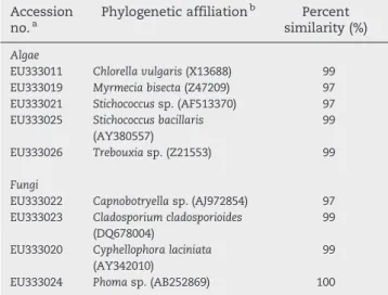
Table 1–Phylogenetic affiliations of eukaryotic
microorganisms derived from a natural green biofilm of Santa Clara-a-Velha Monastery
Accession
no.a Phylogenetic affiliation
b Percent
similarity (%)
Algae
EU333011 Chlorella vulgaris(X13688) 99 EU333019 Myrmecia bisecta(Z47209) 97 EU333021 Stichococcussp. (AF513370) 97 EU333025 Stichococcus bacillaris
(AY380557)
99
EU333026 Trebouxiasp. (Z21553) 99
Fungi
EU333022 Capnobotryellasp. (AJ972854) 97 EU333023 Cladosporium cladosporioides
(DQ678004)
99
EU333020 Cyphellophora laciniata
(AY342010)
99
EU333024 Phomasp. (AB252869) 100
a Accession numbers of the sequences obtained in this study by
cloning, are available at the NCBI database. Screening of clones was performed by restriction enzymatic digestion.
b Closest relatives obtained by comparison with the NCBI database.
The phototrophic microorganisms detected in this study were Chlorophyta and Cyanobacteria. Within Chlorophyta,
Chlorella,Stichococcus,TrebouxiaandMyrmeciawere the genera identified. Among the Cyanobacteria, two genera were detected,LeptolyngbyaandPleurocapsa.
The bacteria identified belong to Bacteroidetes, Alphapro-teobacteria, Actinobacteria and Verrucomicrobia. Fungi were also identified:Cyphellophora sp., Phomasp., Cladosporium sp. andCapnobotryellasp. According toWollenzien et al. (1995), the most abundant fungal genera on calcareous stone from monu-ments areCladosporium,Penicillium,Trichoderma, Fusariumand
Phoma. As reported (Burford et al., 2003), fungi play an important role in rock weathering and also in the biodeterioration of stone monuments as suggested by their presence in the studied samples. Using sequence analysis of 18S rRNA gene, high similarity (≥99%) was found with NCBI database entries of the
algaeChlorella vulgaris,Stichococcus bacillarisandTrebouxiasp., and the fungiCladosporium cladosporioides,Cyphellophora laciniata
and Phoma sp. (Table 1). Fig. 2 presents the phylogenetic affiliations of the phototrophic microorganisms and bacteria obtained from the natural green biofilm.
Most of the microorganisms detected in this study are very widespread on building stones according to several studies carried out by other authors in samples taken from stone
monuments located in Europe (Ortega-Calvo et al., 1993; Tomaselli et al., 2000; Bellinzoni et al., 2003; Ascaso et al., 2004; Miller and Macedo, 2006).
For an enhanced assessment of the microbial communities present in samples from Santa Clara-a-Velha Monastery, DNA-based molecular analysis were complemented with cultivation procedures as recommended byElsas et al. (1998)
and Gonzalez and Saiz-Jimenez (2006). The cultivation approach allowed the assessment of the major phototrophic microorganisms present in the natural green biofilm samples. Enrichments in BG11 of these samples revealed the microalga genus Chlorella as well as the filamentous cyanobacterium
Leptolyngbya sp. and the unicellular and nitrogen-fixing cyanobacterium Pleurocapsa sp. These results are in fair agreement with the data obtained by molecular techniques. All the three strains identified by microscopy were detected in the natural green biofilm corresponding to the strains analyzed by molecular techniques.
3.2. Liquid culture monitoring
The phototrophic biofilm growing in a flask containing BG11 medium was monitored during the three-month incubation period. Macroscopically, visible growth of the phototrophic
Fig. 2–DGGE patterns of 16S rRNA gene from a natural green biofilm collected from Santa Clara-a-Velha Monastery (A) and its liquid culture growing in BG11 medium over three months (B). Lanes AL1bact and AL1cya (A) correspond to DGGE patterns obtained after amplification using general bacterial primers and cyanobacteria-specific primers, respectively. Lanes 15, 30, 45, 60, 75, and 90, corresponding to the sampling days of the liquid culture (B), were obtained after amplification using
community started one week after inoculation, presenting a brightly green colour. After one month, homogeneously distributed photosynthetic cells were observed showing a dense green colour, which remained constant until the end of the incubation period.
The development of the photosynthetic microbial com-munity over the three-month period was monitored by DGGE to reach a steady state representing an establishment of the microbial components of the community. DGGE profiles showed the presence of up to ten distinguishable bands (Fig. 2). The microalgaChlorella (Fig. 2, bands 1–3, 9 and 10) was the predominant microorganism present in the cultured community, being detected in all samples along the incubation time as well as the cyanobacteria Leptolyngbya
sp. (Fig. 2, bands 11 and 13) andPleurocapsasp. (Fig. 2, band 8). In addition, two bands corresponding to the Verrucomi-crobia was detected in cyanobacteria-specific DGGE (Fig. 2, band 12 and 14). According to band migration during DGGE analysis, Bacteroidetes and Alphaproteobacteria (Fig. 2, bands 4 and 5, respectively) were present in the liquid culture profiles (Fig. 2, B). Band number 6 was mostly
present in the original sample but was not significant in the liquid culture. The microbial development suggested that the culture reached a mature stage 45 days after inoculation, remaining constant. The DGGE profile of the liquid culture after 75 days incubation showed the highest similarity with the inoculum DGGE profile. This culture was chosen as a standardized phototrophic community ade-quate for the stone colonization experiment.
Molecular methods were very useful for this purpose, although microscopy techniques can also be used. DGGE analysis has become a useful and elegant method for profiling and monitoring bacterial communities (Schabereiter-Gurtner et al., 2001). By applying molecular methods we attempted to understand both the microorganisms growing easily on culture media and the uncultured microbial majority in the natural green biofilms. Microscopy allowed visualisa-tion of spatial distribuvisualisa-tion but it was not easy to identify the phylogenetic lineage of its microbial components. Nevertheless, microscopy and molecular techniques are complementary for a complete assessment of the micro-bial communities.
3.3. Stone colonization experiment
Ançã limestone samples were rapidly and highly colonized by phototrophic microorganisms. After one month of incubation, the stone probes presented a thick and homogeneously distributed green layer on the top face of the stones (Fig. 3). The colonization process also led to increased photosynthetic biomass on the vertical sides of the samples, showing preferential growth on rough areas.
At two months of exposure in the laboratory chamber, the setup had been exposed to more desiccating conditions (lower sprinkling frequency). One week later, the photo-trophic layer covering the substrates started to detach from the top of the probes. The relative humidity fluctuation in the chamber might have been an essential factor in this detachment and this can also be a factor under natural conditions. Monuments experience large fluctuations in water circulation, including repeated desiccation and wet-ting, which was reproduced in the laboratory chamber. However, new re-colonization occurred on these stone surfaces, presenting a light green colour, which remained constant until the end of the experiment. In contrast, lateral and rear sides of the stone slabs were widely covered by photosynthetic biomass, showing dark green and dense biofilms (Fig. 3). This showed a similar pattern with that occurring in monuments where a great biomass growth is observed in humid areas protected from the rain.Guillitte and Dreesen (1995) in laboratory chamber studies also noticed that very rapidly the phototrophic biofilm covering the materials had cracked, forming partially curled-up scales, after decreasing sprinkling frequency. Detailed observations in different monuments suggest that the formation of an algal film developed on the stone surface depends strongly on the length of the period of wetness and the spatial orientation of the substratum. If conditions favour water evaporation, the algal layers may shrink as a result of desiccation, and remove stone grains which detach from the surface (Ortega-Calvo et al., 1991). If the stone remains wet long enough, the growth of the biofilm, production of organic matter and humification processes may produce protosoils, which may favour the settlement of mosses and higher plants (Ortega-Calvo et al., 1995).
After three months, it was observed that stone samples presented white colonies on the green biofilms covering the stone surfaces (Fig. 3). This colonization was attributed to fungal growth. The algal communities contributed through photosynthetic activity to a significant increase in the organic carbon content of the stone, favouring the growth of hetero-trophic microorganisms. This result confirmed interactions between photosynthetic microorganisms (algae and cyano-bacteria) and heterotrophic microorganisms (fungi and bacteria). For instance, Saiz-Jimenez et al. (1995) observed that heterotrophic microorganisms, within samples from Belgian monuments, can live either at the expense of the extracellular organic matter synthesized by living algae.
As judged by visual examination, the inoculated probes developed biofilms similar in appearance to those occurring on outdoor stone monuments. The major colonizers of the stone during the laboratory experiment were represented by the unicellular microalga Chlorella and the filamentous
cyanobacterium Leptolyngbya (Fig. 4). However, one of the
Chlorella sp. showed less ability to adapt to the changes of exposing conditions than Leptolyngbya sp., as revealed by decreasing of intensity of the corresponding band (Fig. 4). The nitrogen-fixing cyanobacterium Pleurocapsa was a minor representative in the laboratory stone colonization, suggesting that this microorganism was not a major colonizer of the stone surface. An unknown band was observed after 15 days of stone incubation which remained during all of the exposure period and likely corresponds to a microbial contamination.
The photosynthetic components of the natural green biofilm colonized the stone probes during the three months of incubation, and thereby demonstrated stability with time. Comparison of the microorganisms present in the environ-mental sampled biofilm, the liquid culture, and the colonizing community on stone probes showed great similarities and the major components were common to these three cases suggesting that the selected microbial assemblage dominated by phototrophs is a good inoculum for stone colonizations to be performed under laboratory conditions above all when climatic conditions are simulated.
Additionally, the bioreceptivity (Guillitte, 1995) of Ançã limestone to photosynthetic microorganisms was also studied. The stone colonization experiment showed that this lithotype is highly bioreceptive to phototrophic colonization. According toAlbertano et al. (2000), carbonate rocks have a high risk of colonization by microorganisms and are extremely susceptible to cyanobacteria and algae. Previous reports proposed that a high porosity, linked to important open capillarity, allows the rapid development of colonizing organisms (Guillitte and Dreesen, 1995). The petrophysical characteristics of the lithotype used in this study, high porosity and capillary absorption, and its chemical composition were the keys to the establishment and development of the microbial biofilms (Miller et al., 2006). The high bioreceptivity presented by
this limestone may induce severe biodeterioration on the substrate, which should be assessed by using environmental scanning electron microscopy and confocal scanning laser microscopy (Roldán et al., 2006). Further work complementing molecular methods and microscopy will contribute to decipher the actual role and effects of specific microbial cells on stone monuments. One of the drawbacks of molecular methods is that no morphological and physiological information is extracted. Since microbial ecology involves the study of the relationships between microorganisms and their environ-ment, microscopy techniques are necessary to study the interaction between the microbial communities and the mineral substrate, including both taxonomical and morpho-logical characterizations.
4.
Conclusions
The microbial community obtained during this study repre-sents a complexity comparable to natural communities and the procedure using a laboratory chamber was able to reproduce colonization of stone in the laboratory. This study shows that reproducible colonization experiments under accelerated laboratory conditions can be performed using complex microbial communities, presenting the advantage of simulating the existence of competition and/or synergy between colonizing microorganisms.
Acknowledgments
This is a paper from the project CONSOLIDER CSD2007-00058. This work was also supported by the Ministério da Ciência, Tecnologia e Ensino Superior, Portugal, with a doctoral grant (SFRH/BD/21481/2005). LL acknowledges support from the Consejo Superior de Investigaciones Científicas, project 200740/011. The facilities provided by Santa Clara-a-Velha Monastery are gratefully acknowledged.
R E F E R E N C E S
Aires-Barros L, Basto MJ, Charola AE. Black crusts:“fossil”and modern. A comparison from two case studies in Coimbra Monuments (Central Portugal). Int Z Bauinstandsetzen Baudenkmalpflege 2000;6:63–72.
Albertano P, Bruno L, Bellezza S, Paradossi G. Polysaccharides as a key step in stone bio-erosion. In: Fassina V, editor. Ninth Int. Cong. deterioration and conservation of stone vol. 1. Venice: Elsevier; 2000. p. 425–32.
Altschul SF, Gish W, Miller W, Myers EW, Lipman DJ. Basic local alignment search tool. J Mol Biol 1990;215:403–10.
Ascaso MA, del Cura G, de Los Ríos A. Microbial biofilms on carbonate rocks from a quarry and monuments in Novelda (Alicante, Spain). In: Clair S, Seaward M, editors.
Biodeterioration of stone surfaces. Dordrecht: Kluwer Academic Publishers; 2004. p. 79–98.
Bellinzoni AM, Caneva G, Ricci S. Ecological trends in travertine colonization by pioneer algae and plant communities. Int Biodeterior Biodegrad 2003;51:203–10.
Bourrelly P. Les algues d'eau douce–Initiation à la systématique, I: Les Algues Vertes. Paris: N. Boubée; 1990. 572 pp.
Burford EP, Kierans M, Gadd GM. Geomycology: fungi in mineral substrata. Mycologist 2003;17:98–107.
Ciferri O. The role of microorganisms in the degradation of cultural heritage. Rev Conserv 2002;3:35–45.
Crispim CA, Gaylarde CC. Cyanobacteria and biodeterioration of cultural heritage: a review. Microb Ecol 2005;49:1–9.
Diez B, Pedros-Alio C, Marsh TL, Massana R. Application of denaturing gradient gel electrophoresis (DGGE) to study the diversity of marine picoeukaryotic assemblages and comparison of DGGE with other molecular techniques. Appl Environ Microb 2001;67:2942–51.
Dionísio, A. A Pedra de Ançã: características minero-químicas e petrofísicas. MscE Thesis, Universidade Técnica de Lisboa, Instituto Superior Técnico 1997, Portugal.
Elsas JD, Duarte GF, Rosado AS, Smalla K. Microbiological and molecular biological methods for monitoring microbial inoculants and their effects in the soil environmental. J Microbiol Methods 1998;32:133–54.
Gonzalez JM, Saiz-Jimenez C. Microbial activity in biodeteriorated monuments as studied by denaturing gradient gel
electrophoresis. J Sep Sci 2004;27:174–80.
Gonzalez JM, Saiz-Jimenez C. Current advances in the molecular characterization of microbial communities on cultural heritage. In: Fort R, Alvarez de Buergo M, Gomez-Heras M, Vazquez-Calvo C, editors. First Int Conf heritage, weathering and conservation. London: Taylor and Francis; 2006. p. 343–8. Gonzalez JM, Ortiz-Martinez A, Gonzalez-delValle MA, Laiz L,
Saiz-Jimenez C. An efficient strategy for screening large cloned libraries of amplified 16S rDNA sequences from complex environmental communities. J Microbiol Methods 2003;55:459–63. Guillitte O. Bioreceptivity: a new concept for building ecology
studies. Sci Total Environ 1995;167:215–20.
Guillitte O, Dreesen R. Laboratory chamber studies and petrographical analysis as bioreceptivity assessment tools of building materials. Sci Total Environ 1995;167:365–74. Herrera LK, Arroyave C, Guiamet P, Gómez de Saravia S, Videla H.
Biodeterioration of peridotite and other constructional materials in a building of the Colombian cultural heritage. Int Biodeter Biodegr 2004;54:135–41.
Komárek J, Anagnostidis K, Cyanoprocaryota I. In: Ettl H, Gerloff J, Heynig H, Mollenhauer D, editors. Süßwasserflora von Mitteleuropa, Band 19/1. Paris: Gustav Fischer Verlag; 1999. 548 pp.
Miller A, Macedo MF. Mapping and characterization of a green biofilm inside of Vilar de Frades Church (Portugal). In: Fort R, Alvarez de Buergo M, Gomez-Heras M, Vazquez-Calvo C, editors. First Int Conf heritage, weathering and conservation. London: Taylor and Francis; 2006. p. 329–35.
Miller A, Dionísio A, Macedo MF. Primary bioreceptivity: a comparative study of different Portuguese lithotypes. Int Biodeterior Biodegrad 2006;57:136–42.
Monte M. Oxalate film formation on marble caused by fungus. J Cult Herit 2003;4:255–8.
Muyzer G, de Waal EC, Uitterlinden AG. Profiling of complex microbial populations by denaturing gradient gel electrophoresis analysis of polymerase chain
reaction-amplified genes coding for 16S rRNA. Appl Environ Microb 1993;59:695–700.
NORMAL 9/88. Microflora autotrofa ed eterotrofa: tecniche di isolamento in coltura. Raccomandazioni Normal, Doc. Normal 9/88, Istituto Centrale del Restauro-Commissione Normal (ICRCNR), Rome: ICR-CNR; 1998. 26 pp.
Nübel U, Garcia-Pichel F, Muyzer G. PCR primers to amplify 16S rRNA genes from cyanobacteria. Appl Environ Microbiol 1997;63:3327–32.
Ortega-Calvo JJ, Sanchez-Castillo PM, Hernandez-Marine M, Saiz-Jimenez C. Isolation and characterization of epilithic chlorophyta and cyanobacteria from two Spanish cathedrals (Salamanca and Toledo). Nova Hedwig 1993;57:239–53. Ortega-Calvo JJ, Ariño X, Hernandez-Marine M, Saiz-Jimenez C.
Factors affecting the weathering and colonization of monuments by phototrophic microorganisms. Sci Total Environ 1995;167:329–41.
Prieto B, Silva B. Estimation of the potential bioreceptivity of granitic rocks from their intrinsic properties. Int Biodeterior Biodegrad 2005;56:206–15.
Roldán M, Oliva F, Gónzalez del Valle MA, Saiz-Jimenez C, Hernández-Mariné M. Does green light influence the fluorescence properties and structure of phototrophic biofilms? Appl Environ Microbiol 2006;72:3026–31.
Saiz-Jimenez C, Ariño X, Ortega-Calvo JJ. Mechanisms of stone biodeterioration by photosynthesis-based epilithic biofilms. In: De Cleene M, editor. Interactive physical weathering and bioreceptivity study on building stones, monitored by Com-puterized X-Ray Tomography (CT) as a potential non-destruc-tive research tool. Protection and Conservation of the European
Cultural Heritage Research Report N° 2. European Commission, Brussels: Directorate-General XII; 1995, pp. 25–62.
Sand W. Microbial mechanisms of deterioration of inorganic substrates—a general mechanistic overview. Int Biodeterior Biodegrad 1997;40:183–90.
Schabereiter-Gurtner C, Piñar G, Lubitz W, Rölleke S. An advanced molecular strategy to identify bacterial communities on art objects. J Microbiol Methods 2001;45:77–87.
Tiano P, Accolla P, Tomaselli L. Phototrophic biodeteriogens on lithoid surfaces: an ecological study. Microb Ecol
1995;29:299–309.
Tomaselli L, Lamenti G, Bosco M, Tiano P. Biodiversity of photosynthetic micro-organisms dwelling on stone monuments. Int Biodeterior Biodegrad 2000;46:251–8. Wollenzien U, deHoog GS, Krumbein WE, Urzi C. On the isolation
of microcolonial fungi occurring on and in marble and other calcareous rocks. Sci Total Environ 1995;167:287–94. Zimmermann J, Gonzalez JM, Saiz-Jimenez C. Detection and