* Corresponding author.
E-mail: saude@ef.ufop.br (D.A.S. Guimarães).
0102-695X/$ - see front matter © 2014 Sociedade Brasileira de Farmacognosia. Published by Elsevier Editora Ltda. All rights reserved. http://dx.doi.org/10.1016/j.bjp.2014.09.005
Original article
Assessment of acute toxicity of the ethanolic extract of
Lychnophora pinaster (Brazilian arnica)
Simone A. Ferreira
a, Andrea Grabe Guimarães
b, Fernanda Cristina Ferrari
a,
Cláudia Martins Carneiro
c, Nívia Carolina Nogueira de Paiva
c,
Dênia Antunes Saúde Guimarães
a,*
aLaboratório de Plantas Medicinais, Escola de Farmácia, Universidade Federal de Ouro Preto, Ouro Preto, MG, Brazil bLaboratório de Farmacologia Experimental, Escola de Farmácia, Universidade Federal de Ouro Preto, Ouro Preto, MG, Brazil cLaboratório de Imunopatologia, Universidade Federal de Ouro Preto, Ouro Preto, MG, Brazil
Introduction
The genus Lychnophora, family Asteraceae, comprises species native to the Brazilian savanna (Cerrado), mostly in Goiás,
Bahia and Minas Gerais states (Semir, 1991). The aerial parts of many species of Lychnophora are widely used in Brazilian folk medicine, macerated in ethanol or “cachaça” and administered by oral or topical route, as anti-inflammatory, and to treat
A R T I C L E I N F O
Article history: Received 3 June 2014 Accepted 6 September 2014
Keywords:
Lychnophora pinaster Acute oral toxicity
Hematological and biochemical parameters
A B S T R A C T
Species of the Lychnophora genus are plants native to Brazil, popularly known as “Brazilian arnica” and used in folk medicine as alcoholic and hydro-alcoholic preparations for the treat-ment of bruises, inflammation, pain, rheumatism and insect bites. The present study aimed to evaluate the safety of the use of Lychnophora pinaster Mart., Asteraceae. Acute toxicity of the crude ethanolic extract was evaluated by administration of the extract by oral route to male and female Swiss mice. A single extract dose of 125, 250 or 500 mg/kg was administered and the effects on spontaneous locomotor activity, exploratory behavior, muscle strength, body weight, food and water consumption, relative organ weight, histology, as well as hematologi-cal and biochemihematologi-cal parameters were evaluated. The three doses administered to the animals did not cause muscle tone alterations, but doses of 250 and 500 mg/kg induced a significant inhibition of the spontaneous locomotor activity and exploratory behavior of the animals in open-field test. There was no alteration to hematological parameters and consumption of water and food, body weight variation and organs relative weight. Changes were observed in AST and ALT during assessment of biochemical parameters. The histopathological evaluation showed that the extract provoked cellular alterations, such as vacuolar degeneration and in-flammation in kidneys and liver at all doses. Liver morphometric analyses of male and female mice showed that the extract did not have dose-dependent effects. Although females showed a significant increase in inflammatory cells, the effect was not dose-dependent.
bruises, pain and rheumatism (Cerqueira et al., 1987; Saúde et al., 1998).
Extracts of several Lychnophora species, including
Lychnophora pinaster Mart., significantly decreased the number of writhing induced by acetic acid, as well as the carrageenan-induced paw oedema, evidencing the anti-inflammatory activity of the topical administration of Lychnophora species (Guzzo et al., 2008).
According to Ferrari et al. (2012) the extract of Lychnophora trichocarpha elicited serious toxicity signs to both female and male mice. A significant inhibition of the spontaneous locomotor activity and exploratory behavior in mice were observed in the open-field and traction tests; moreover, the same dose (0.75 g/kg) reduced the muscle strength 1 h after administration. Furthermore, histopathological changes were observed such as kidney and liver congestion and inflammation.
Taking in consideration the previously observed analgesic and anti-inflammatory activities of L. pinaster (Guzzo et al., 2008), the aim of present study was to evaluate the safety of this species when administered orally to mice.
Materials and methods
Plant material
The aerial parts of Lychnophora pinaster Mart., Asteraceae, were collected in March 2007, in Minas Gerais state, Brazil with permission of Instituto Chico Mendes de Conservação da Biodiversidade/Sistema de Autorização e Informação em Biodiversidade (License nº 20113). The plants were identified by Dr. Julio Antonio Lombardi. A voucher specimen was deposited at the Herbarium of the Instituto de Ciências Biológicas, UFMG, Belo Horizonte, Brazil, under the reference number 19.520.
Preparation of plant extracts
The aerial parts of the L. pinaster were dried at 40°C for one week and subsequently powdered. About 2.139 g of the plant was extracted with ethanol at room temperature, for two weeks. The solvent was removed under reduced pressure (below 40°C) to obtain the dried crude ethanolic extract (265 g). The extract yield was 12.4% (w/w). For the toxicological evaluation, the extract was dissolved in dimethylsulfoxide (DMSO), tween and distilled water (1:1:8) to get the concentrations of 12.5, 25 and 50 g/ml, which were used to administer 125, 250 e 500 mg/kg, respectively. The maximum volume of the solutions administered to the animals was 0.05 ml.
Oral acute toxicity
Animals
Adult Swiss mice (30 ± 5 g) of both genders were used for acute toxicity evaluation. The animals were housed in groups of five and maintained on 12 h light:dark cycle under controlled temperature (22 ± 2°C), with standard pellet diet and water ad libitum. Prior to the administration of solutions p.o., animals were kept without food for 8 h but free access to water was
allowed. The experimental protocols were approved by the Ethical Committee of Universidade Federal de Ouro Preto (nº 2009/06) and are in accordance with the Guide for the Care and Use of Laboratory Animals, published by the US National Institute of Health (NIH Publication, revised in 1985).
Short-term toxicity evaluation
The experimental protocol was carried out in accordance to specifications published by Anvisa (2004) determining the evaluation of acute toxicity in both genders of one mammal species. Each single oral dose of the extract (125; 250 or 500 mg/ kg) or vehicle was administered to eight males and six females. The volume administered was about 10 ml/kg of body weight. General behavior as locomotion, exploratory behavior and muscle tone of the animals were determined as described below for each test. The spontaneous locomotor activity and exploratory behavior were evaluated using the open field test as described by Turner (1972) with modifications; and the muscle strength variation was evaluated using the traction test described by Rudzik et al. (1973). These parameters were evaluated after 1, 4 and 24 h, after 7 and 14 days from extract administration. The number of survivors was recorded up to 24 h and after that, daily. Body weight variations of males and females were recorded one day before the administration of the extract and four, seven, eleven and fourteen days after . The food and water consumption was recorded one day before the administration of the extract and every three days afterwards, and the proportion by group (gender/dose) was counted enrolling the medium values of each group. After 7 and 14 days, the surviving animals were anesthetized with pentobarbital (45 mg/kg, intraperitoneal) and blood samples were collected from each animal in order to determine biochemical and hematological parameters. The liver, kidneys, lung and brain, were retrieved, dissected, washed with saline and preserved in a formalin solution (10%) for further histological analysis.
Hematological analysis
Blood samples (300 µl) were collected in eppendorf tubes with 5 µl of 10% EDTA (Laborlab®, São Paulo, Brazil) (Barros et al.,
2005; Abbès et al., 2006). The collected samples were immersed immediately in an ice bath to preserve the morphology and cell number. The following parameters were evaluated using an automatic hematological analyzer (ABX diagnostics, micros 60, France) with adapted dilutions: red blood cells count (RBC), hemoglobin (Hb), hematocrit (Hct), mean corpuscular volume (MCV), mean corpuscular hemoglobin (MCH), mean corpuscular hemoglobin concentration (MCHC), platelets number, white blood cells count (WBC) (Silva et al., 2005; Costa-Silva et al., 2008). The differential leukocyte count was analyzed using optical microscopy; 100 cells were analyzed and counted for each assay (40×).
Biochemical analysis
Blood samples without anticoagulant were centrifuged at 13,000
(CRE) for renal function; and amylase (AMY) for pancreatic function. The colorimetric tests were obtained from Bioclin® (Brazil) and the analyses were carried out using the Random Access Clinical Analyzer (Wiener lab, model CM-200, Argentina).
Organs weight
Euthanization of animals was done by exsanguination with anesthesia on the 7th or 14th day after treatment. The liver,
kidneys, lung and brain were quickly removed soon after the animals’ death, cleaned with saline and their wet weight was determined. The relative organ weight of each animal was then calculated relating the absolute organ weight and body weight of the animal on the day of sacrifice (Mabeku et al., 2007).
Histopathological analysis
Samples of liver, kidneys, lungs and brain were collected for histopathological analysis. After fixation, the tissues were processed routinely and embedded in paraffin. Sections stained with HE were evaluated qualitative and quantitatively. The qualitative analyses of the different organs (liver, kidneys, lungs and brain) were performed using an Olympus BX50 microscope (10 and 40×) in order to verify alterations in the tissues, as well as presence of cells from the parenchymal polymorphonuclear inflammatory infiltrate (PMNII). The inflammatory infiltrate in the liver was quantified using twenty fields taken randomly (total area traveled equal to 1.5 × 106 µm2, 40×). The morphometric analysis of the inflammatory
infiltrate in this organ was performed from digital images obtained using the micro-camera Leica DFC340FX associated to the microscope Leica DM5000 B (Germany), and all the
images were analyzed using the image processing software, Leica Qwin.
Statistical analysis
The results are expressed as mean ± SEM. Variance of data for body weight, hematology, blood biochemistry and relative organ weight was checked for homogeneity using Bartlett’s method. If the variance was homogeneous, the data were tested using one-way analysis of variance (ANOVA) followed by Tukey’s test. For spontaneous locomotor activity and exploratory behavior, the unpaired t test was used. For the muscle strength, the Fisher’s test was used to detect statistical differences between controls and treated groups. A probability level of p ≤ 0.05 was considered significant. The statistical analysis was made using the GraphPad Prism© 5.0 software.
Results and discussion
The results showed that the acute treatment with crude ethanolic extract of Lychnophora pinaster Mart., Asteraceae, by oral route at doses de 125, 250 or 500 mg/kg did not cause death in male and female mice for 7 or 14 days during observation time. However, doses of 250 and 500 mg/kg impaired locomotion, observed in the open field test 4 and 24 h after administration (Table 1) without altering the muscle strength (Table 2), indicating some toxicity of the higher doses, orally administered. The literature demonstrated that the reduction of the spontaneous locomotor activity reflects a reduced excitability, originated from disturbances of the central nervous system (Ozturk et al., 1996; Perez et al., 1998).
Time n Control 125 mg/kg 250 mg/kg 500 mg/kg
0 hour 28 88.2 ± 9.43 81.9 ± 5.50 88.8 ± 5.78 92.2 ± 5.32
1 hour 28 81.6 ± 9.04 70.7 ± 5.18 87.1 ± 5.28 83.2 ± 6.07
4 hours 28 73.9 ± 9.98 69.0 ± 4.64* 69.2 ± 5.57* 76.5 ± 5.76*
24 hours 28 94.4 ± 12.86 76.4 ± 6.80 69.7 ± 5.81* 72.6 ± 6.62*
7 days 28 100.5 ± 10.23 91.9 ± 6.24 101.6 ± 9.11 97.1 ± 7.08
14 days 14 103.1 ± 13.63 89.6 ± 9.67 97.1 ± 14.29 94.6 ± 9.77
Each value represents the mean ± S.E.M. *p ≤ 0.05 compared to the control time (t test), n, number of animals used in the experiment.
Time n Control 125 mg/kg 250 mg/kg 500 mg/kg
0 hour 28 3.57 0 10.71 0
1 hour 28 7.14 21.43 14.29 14.29
4 hours 28 0 14.29 14.29 28.57
24 hours 28 3.57 10.71 10.71 14.29
7 days 28 3.57 7.14 14.29 10.71
14 days 14 0 7.14 0 21.43
The data represent the percentage of animals that failed the test. The groups were compared to the control group (Fisher’s test); n, number of animals used in the experiment.
Table 1
Effect of the crude ethanolic extract of Lychnophora pinaster on the number of squares covered by the animals in the open-field test, for 5 min.
Table 2
The hematological profile of control and treated groups are presented in Table 3. There were no significant differences in the hematological parameters analyzed. The hematological evaluation provides important indication of local and systemic intoxication manifestations induced by drugs (Wyllie and Wyllie, 1991). However, changes in these parameters, along with biochemical parameters, occur relatively slowly and the length of the experiment probably was not enough to identify these alterations, as previously observed by Ferrari et al. (2012).
The acute oral treatment of mice with the crude ethanolic extract of L. pinaster, in a general, did not induce significant modifications of the biochemical profile when compared to the control group. However, a decrease in AST and ALT serum blood levels were observed in the animals that received the three doses (125, 250 or 500 mg/kg) in comparison to the control group (Table 4). ALT is a more specific marker of liver cell damage, because it occurs more frequently in the liver while AST is also found in heart, skeletal muscle, kidneys, brain, pancreas and blood cells (Filippin et al., 2004). In the liver, ALT is confined to cytoplasm, while AST is found in both mitochondria (80% of total intracellular enzyme) and cytoplasm (20%) (Kew, 2000). In patients with kidney failure, the AST and ALT are significantly lower than in healthy individuals, due to decreased free pyridoxal-5-phosphate, which reduces the enzymatic activity (Allman et al., 1992). Changes in these enzymatic levels may suggest liver disease too, but the absence of these changes does not exclude the occurrence of a clinical disorder, as it is the case of the advanced stages of chronic hepatitis C with normal ALT levels (Van Thiel et al., 1995).
Water consumption as well as food intake in all groups that received 125, 250 or 500 mg/kg was similar to that of the control group. The determinations of water and food consumption are important parameters to evaluate the safety of a product with potential therapeutic activity (Iversen and Nicolaysen, 2003).
The body weight analysis, assessed for 7 or 14 days of observation showed that L. pinaster did not induce significant changes in either male or female animals. The animal body weight is also an important factor to evaluate the toxicity of a substance (Jahn and Günzel, 1997). The reduction in body weight and internal organ weight can be a simple and sensitive index of toxicity after exposure to a toxic substance (Raza et al., 2002; Teo et al., 2002). Changes in organ weight have long been accepted as indicators of test-induced changes, which are often associated with treatment-related effects (Sellers et al., 2007). In the present work, the extract of L. pinaster did not induce significant changes to the relative weight of the organs of mice of both genders (liver, kidneys, lungs and brain).
Histopathological analysis revealed no alterations of the organs from control animals, as well as the lung and the brain of the treated animals of both genders with the three doses of the extract. The histological analysis of the kidneys of control animals presented normal aspect (Fig. 1A and B). The kidneys’ histological analysis of mice, of both genders, treated with three doses (125, 250 or 500 mg/kg) of the extract revealed time and dose-dependent alterations. Doses of 125 and 250 mg/kg induced tubular degeneration without glomerular loss in treated mice kidneys, seven days after the extract administration (Fig. 1C). After 500 mg/kg of extract, the kidneys, besides tubular degeneration, presented glomerular congestion
Group WBC
(103/mm3)
RBC (106/mm3)
Hb
(g/dl) Hct (%)
Platelets (103/mm3)
MCV (mm3)
Control D7 2.8 ± 0.28 8.9 ± 0.23 14.1 ± 0.33 41.8 ± 1.10 1007.0 ± 33.27 46.6 ± 0.33 D14 3.4 ± 0.45 9.3 ± 0.10 14.5 ± 0.27 42.9 ± 0.87 1006.0 ± 27.75 46.8 ± 0.37
125 mg/kg D7 3.2 ± 0.22 9.0 ± 0.11 14.0 ± 0.18 41.5 ± 0.54 1009.0 ± 32.70 46.1 ± 0.29 D14 4.2 ± 0.63 9.0 ± 0.12 14.4 ± 0.16 42.7 ± 0.41 1081.0 ± 30.42 47.3 ± 0.30
250 mg/kg D7 3.7 ± 0.26 8.9 ± 0.13 13.9 ± 0.33 41.0 ± 0.88 956.0 ± 39.02 46.9 ± 0.32 D14 3.8 ± 0.32 9.3 ± 0.09 14.8 ± 0.12 43.9 ± 0.63 1010.0 ± 29.30 47.4 ± 0.58
500 mg/kg D7 3.3 ± 0.36 8.8 ± 0.17 14.0 ± 0.18 41.6 ± 0.66 1119.0 ± 38.04 47.1 ± 0.35 D14 3.3 ± 0.37 8.9 ± 0.22 14.2 ± 0.21 42.0 ± 1.01 1086.0 ± 30.59 47.1 ± 0.25
Group MCH
(pg)
MCHC (g/dl)
Eosinophils (%)
Neutrophils (%)
Lymphocytes (%)
Monocites (%)
Control D7 15.7 ± 0.13 33.7 ± 0.20 1.1 ± 0.14 22.7 ± 2.32 73.4 ± 2.30 2.8 ± 0.27 D14 15.8 ± 0.09 33.6 ± 0.20 1.3 ± 0.13 16.9 ± 2.35 78.6 ± 2.33 3.2 ± 0.45
125 mg/kg D7 15.6 ± 0.13 33.8 ± 0.26 1.2 ± 0.13 18.1 ± 1.82 77.6 ± 1.73 2.9 ± 0.19 D14 15.9 ± 0.09 33.7 ± 0.11 1.3 ± 0.13 12.6 ± 1.42 82.2 ± 1.64 3.9 ± 0.35
250 mg/kg D7 15.8 ± 0.17 33.6 ± 0.14 1.1 ± 0.13 15.0 ± 1.16* 81.2 ± 1.05** 2.8 ± 0.15 D14 15.9 ± 0.12 33.3 ± 0.18 1.2 ± 0.12 9.4 ± 1.05** 86.8 ± 1.19** 2.4 ± 0.26
500 mg/kg D7 15.7 ± 0.10 33.2 ± 0.21 1.2 ± 0.13 18.1 ± 1.76 77.8 ± 1.77 2.9 ± 0.18 D14 15.8 ± 0.04 33.6 ± 0.15 1.3 ± 0.12 12.1 ± 1.27 83.3 ±12.32 2.7 ± 0.16 Each value represents the mean ± S.E.M., n = 14 animals (ANOVA followed by Tukey’s test with significant difference at *p ≤ 0.05; **p < 0.01). WBC, white blood cells count; RBC, red blood cells count; Hb, hemoglobin; Hct, hematocrit; MCV, mean corpuscular volume; MCH, mean corpuscular hemoglobin; MCHC, mean corpuscular hemoglobin concentration. D7, after 7 days of the administration of the extract; D14, after 14 days of the administration of the extract.
Table 3
Figure 1 – Photomicrographs of Swiss mice kidney sections submitted to the toxicity evaluation of the crude ethanolic extract of Lychnophora pinaster (LPiE), administered orally at three different doses (125; 250 or 500 mg/kg of bodyweight), in accordance to gender and time after treatment: A and B. Animals of the control group display kidneys with normal aspect; C. Animals that received 125 or 250 mg/kg of body weight of LPiE (seven days) show kidneys with tubular degeneration; D. Animals that received 500 mg/kg of body weight of LPiE (7 days) show kidneys with congestion and discrete inflammation; E. Animals that received 125 or 250 mg/kg of body weight of LPiE (fourteen days) display kidneys with tubular degeneration and dilation; F. Animals that received 500 mg/kg of body weight of LPiE (fourteen days) show kidneys with tubular degeneration, dilation and moderate inflammatory infiltrate (Hematoxylin and Eosin, 400× original magnification, A 200× original magnification).
Group (mg/dl)BUN (mg/dl)AUR (mg/dl)CRE (mg/dl)AMY AST (U/l) ALT (U/l) ALP (U/l) (g/dl)PT
Control D7 62.2 ± 1.72 2.1 ± 0.20 0.4 ± 0.04 839.4 ± 29.26 112.6 ± 10.79 57.7 ± 9.54 124.3 ± 9.99 5.5 ± 0.08 D14 60.4 ± 2.16 1.5 ± 0.13 0.4 ± 0.06 739.2 ± 21.74 110.1 ± 11.87 54.2 ± 5.07 163.2 ± 14.87 5.6 ± 0.32
125 mg/kg D7 57.1 ± 2.38 3.0 ± 0.37 0.4 ± 0.03 767.2 ± 32.57 78.3 ± 8.69* 36.2 ± 3.72* 118.5 ± 7.63 5.5 ± 0.40 D14 52.0 ± 2.15 1.7 ± 0.20 0.4 ± 0.04 739.7 ± 27.19 111.0 ± 10.77 62.0 ± 8.81 165.9 ± 11.55 5.3 ± 0.18
250 mg/kg D7 56.5 ± 2.61 2.3 ± 0.19 0.4 ± 0.08 826.2 ± 21.77 80.3 ± 7.69* 35.7 ± 4.99* 121.0 ± 5.28 5.3 ± 0.13 D14 52.8 ± 2.74 1.6 ± 0.17 0.4 ± 0.03 770.2 ± 28.79 91.4 ± 4.63 30.5 ± 2.16 * 142.5 ± 8.34 5.3 ± 0.16
500 mg/kg D7 62.1 ± 3.12 2.1 ± 0.23 0.4 ± 0.04 831.9 ± 22.60 81.9 ± 7.19* 26.0 ± 4.02** 132.4 ± 11.04 5.3 ± 0.16 D14 59.6 ± 3.78 1.4 ± 0.14 0.4 ± 0.03 767.4 ± 34.32 96.2 ± 4.65 47.7 ± 5.16 142.2 ± 8.52 5.4 ± 0.12 Each value represents the mean ± S.E.M., n = 14 animals (ANOVA followed by Tukey’s test with significant difference at *p ≤ 0.05; **p < 0.01). BUN: serum urea nitrogen; AUR: uric acid; CRE: serum creatinine; AMY: amylase; AST: aspartate aminotransferase; ALT: alanine aminotransferase; ALP: alkaline phosphatase; PT: total proteins. D7: after 7 days of the administration of the extract; D14: after 14 days of the administration of the extract.
Table 4
and a slight inflammation (Fig. 1D). After fourteen days of administration, the animals treated with 125 or 250 mg/kg of
L. pinaster extract, presented tubular vacuolar degeneration, glomerular congestion (Fig. 1E), discrete glomerular loss and renal tubules dilation with eosinophilic deposition into the tubules’ lumen. Additionally, at the same time, for a dose of 500 mg/kg of the extract, a moderate inflammatory infiltrate was observed (Fig. 1F). These histological changes did not alter CRE and BUN levels in plasma, probably due to the insufficient time of experimentation.
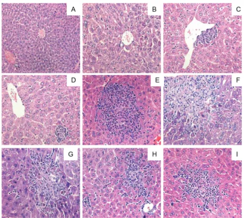
The liver of the control animals presented normal aspect (Fig. 2A). The liver histological analysis of L. pinaster (125 or 250 mg/ kg) treated mice, seven days after the extract administration, showed vacuolar degeneration, mainly of perivascular hepatocytes without inflammatory process (Fig. 2B and C). Nevertheless, the liver parenchyma had a similar aspect as the controls. Seven days after the administration of 500 mg/ kg of extract, the liver presented moderate venular congestion, sinusoidal congestion and the presence of parenchymal
polymorphonuclear inflammatory infiltrate cells (PMNII) (Fig. 2D and E). After fourteen days of extract administration (125 or 250 mg/kg), the liver presented a moderate parenchymal and perivascular inflammatory infiltrate; focal hepatic necrosis (Fig. 2F and G) surrounded by PMNII cells and hydropic degeneration. Fourteen days after the higher dose extract (500 mg/kg) was administered, the liver analysis revealed an intense inflammatory process besides the alterations found at lower doses (Fig. 2H and I). Histological changes in the liver can sustain the biochemical changes observed for AST and ALT in animals that received the extract at the three evaluated doses.
The morphometric analysis of the liver showed that the extract does not present hepatotoxic effect time and dose dependent manner, but the hepatotoxic effect of the extract in the females was larger than in the males (Table 5). These observations demonstrate that the females are more susceptibility than the males when a single dose of the crude ethanolic extract of Lychnophora pinaster is administered orally. According to Stickel et al. (2005) several medicinal plants can
Group n Gender Number of cell nuclei /area
Control
8 Males D7 194.8 ± 4.65
D14 233.3 ± 5.64 **
6 Females D7 264.4 ± 3.48 **
o
D14 248.2 ± 2.91 **
125 mg/kg
8 Males D7 158.3 ± 3.52 *
D14 196.2 ± 3.64 * **
6 Females D7 302.6 ± 3.39 * **
o
D14 340.1 ± 3.46 * ** o •
250 mg/kg
8 Males D7 208.8 ± 3.43
D14 167.7 ± 3.68 * **
6 Females D7 310.1 ± 3.43 * **
o
D14 355.0 ± 8.29 * ** o •
500 mg/kg
8 Males D7 204.4 ± 3.72
D14 183.2 ± 3.97 *
6 Females D7 349.0 ± 6.67 * **
o
D14 340.6 ± 3.99 * ** o
The values represent the mean ± S.E.M. of the 8 and 6 slides for males and females respectively; n, number of animals used in the experiment (ANOVA followed by Tukey’s test). D7, after 7 days of the administration of the extract; D14, after 14 days of the administration of the extract. *p < 0.001 compared to your respective control groups; **p < 0.05 compared to males D7; op < 0.001 compared to males D
14; •p < 0.001 compared to females D7.
Table 5
Frequency of cell nuclei per area (1,6 × 107 µm2) in the liver of males and females Swiss mice treated with vehicle or different
doses of the crude ethanolic extract of Lychnophora pinaster after 7 or 14 days after the administration of the extract.
cause hepatotoxicity and most of those plants affect more female individuals. This gender difference does not reflect a higher likelihood of women to use these preparations, but they have a higher susceptibility towards herb-induced liver damage (Kessler et al., 2001), as it was observed for the majority of adverse hepatic reactions induced by conventional drugs.
Conclusion
Additional studies will be necessary to verify if the observed alterations are reversible and if it is possible tissues recovery. In any case, some precautions have to exist when the extract is given orally, since the integral safety does not seem to exist for this extract. Besides, the present study evaluated the short-term toxicity, by a single-dose administration. In order to attend completely the Resolution nº 90 of 2004 of the National Agency of Sanitary Surveillance of Brazil; it will also be necessary the evaluation of multiple-dose administration, for instance, for thirty days, on a daily basis. In conclusion, the acute oral administration of the crude ethanolic extract of Lychnophora pinaster at the three doses evaluated induced toxicity to the renal and hepatic cells, evaluated by histological analysis.
Authors’ contributions
SAF (MSc) contributed in running the laboratory work and analysis of the data. FCF drafted the paper. AGG contributed to toxicological studies in vivo and to critical reading of the
manuscript. CMC and NCN contributed to histological analysis. DASG designed the study, supervised the laboratory work and contributed to critical reading of the manuscript. All authors have read the final manuscript and approved the submission.
Conflicts of interest
The authors declare no conflicts of interest.
Acknowledgements
The authors thank Fundação de Amparo à Pesquisa do Estado de Minas Gerais for the financial suport, Rede TOXIFAR FAPEMIG, CNPq, Universidade Federal de Ouro Preto and FAPEMIG for the scholarships of scientific initiation and CAPES for the master’s degree scholarship.
R E F E R E N C E S
Abbès, S., Ouanes, Z., Ben Salah-Abbès, J., Houas, Z., Oueslati, R., Bacha, H., Othman, O., 2006. The protective effect of hydrated sodium calcium aluminosilicate against haematological, biochemical and pathological changes induced by Zearalenone in mice. Toxicon 47, 567-574.
Anvisa 2004. Resolução no 90 de 16 de março de 2004. Determina a publicação da “Guia para a realização de estudos de toxicidade pré-clínica de fitoterápicos”. Agência Nacional de Vigilância Sanitária. Ministério da Saúde. Diário Oficial da União (DOU), 18 de março de 2004.
Barros, S., Ropke, C.D., Sawada, T.C.H., Silva, V.V., Pereira, S.M.M., Barros, S.B.M., 2005. Assessment of acute and subchronic oral toxicity of ethanolic extract of Pothomorphe umbellata L. Miq (Pariparoba). Rev. Bras. Cienc. Farm. 41, 53-61.
Cerqueira, M.B.S., Souza, J.T., Amado Júnior, R., Peixoto, A.B.F., 1987. Ação analgésica do extrato bruto liofilizado do caule e folhas da Lychnophora ericoides Mart. (arnica). Cienc. Cul. 39, 551-553.
Costa-Silva, J.H., Lima, C.R., Silva, E.J.R., Araújo, A.V., Fraga, M.C.C.A., Ribeiro e Ribeiro, A., Arruda, A.C., Lafayette, S.S.L., Wanderley, A.G., 2008. Acute and subacute toxicity of the Carapa guianensis Aublet (Meliaceae) seed oil. J. Ethnopharmacol. 116, 495-500.
Ferrari F.C., Grabe-Guimarães A., Carneiro C.M., Souza M.R., Ferreira L.C., Oliveira T.T., Saúde-Guimarães D.A., 2012. Toxicological evaluation of ethanolic extract of Lychnophora trichocarpha, Brazilian arnica. Rev. Bras. Farmacogn. 22, 1104-1110.
Filippin, F.B., Reis, K., Cemin, L., Duzzioni, M., Hermes, E.M., Souza, L. C., 2004. Novo intervalo de referência para alanina aminotransferase usando o sistema automatizado de bioquímica Dade Behring Ar Dimension. NewsLab 65, 148-160.
Guzzo L.S., Saúde-Guimarães D.A., Silva A.C.A., Lombardi J.A., Guimarães H.N., Grabe-Guimarães A., 2008. Antinociceptive and antiinflammatory activities of ethanolic extracts of Lychnophora species. J. Ethnopharmacol. 116, 120-124. Iversen, P.O., Nicolaysen, G., 2003. Water-for life. Tidsskr. Norske.
Laege. 123, 3402-3405.
Jahn, A.I., Günzel, P.K.H., 1997. The value of spermatology in male reproductive toxicology: do spermatologic examinations in fertility studies provide new and additional information relevant for safety assessment? Reprod. Toxicol. 11, 171-178. Kessler, R.C., Davis, R.B., Foster, D.F., Van Rompay, M.I., Walters, E.E.,
Wilkey, S.A., Kaptchuk, T.J., Eisenberg, D.M., 2001. Long-term trends in the use of complementary and alternative medical therapies in the United States. Ann. Intern. Med. 135, 262-268. Kew, M.C., 2000. Serum aminotransferase concentration as
evidence of hepatocellular damage. Lancet 355, 591-592. Mabeku, L.B.K., Beng, V.P., Kouam, J., Essame, O., Etoa, F.X., 2007. Toxicological evaluation of ethyl acetate extract of Cylicodiscus gabunensis stem bark (Mimosaceae). J. Ethnopharmacol. 111, 598-606.
Ozturk, Y., Aydini, S., Beis, R., Baser, K.H.C., Berberoglu, H., 1996. Effect of Hypericum perforatum L. and Hypericum calximun L. extract on the central nervous system in mice. Phytomedicine 3, 139-146.
Perez, R.M.G., Perez, J.A.L., Garcia, L.M.D., Sossa, H.M., 1998. Neuropharmacological activity of Solanum nigrum fruit. J. Ethnopharmacol. 62, 43-48.
Raza, M., Al-Shabanah, O.A., El-Hadiyah, T.M., Al-Majed, A.A., 2002. Effect of prolonged vigabatrin treatment on hematological and biochemical parameters in plasma, liver and kidney of Swiss albino mice. Sci. Pharm. 70, 135-145. Rudzik, A.D., Hester, J.B., Tang, A.H., Staw, R.N., Friis, W., 1973.
The benzodiazepines. Raven Press, New York, p. 285-297. Saúde, D.A., Raslan, D.S., Souza Filho, J.D., Oliveira, A.B., 1998.
Constituents from the aerial parts of Lychnophoratrichocarpa. Fitoterapia LXIX, 90-91.
Sellers, R.S., Morton, D., Michael, B., Roome, N., Johnson, J.K., Yano, B.L., Perry, R., Schafer, K., 2007. Society of toxicologic pathology position paper: organ weight recommendations for toxicology studies. Toxicol. Pathol. 35, 751-755.
Semir, J., 1991. Revisão taxonômica de Lychnophora Mart. (Vernoniaceae: Compositae). PhD. Thesis, Universidade de Campinas, SP, Brazil.
Silva, E.J.R., Aguiar, F.J.S., Gonçalves, E.S., Souza, I.M.V., Dimech, G.S., Fraga, M.C.C.A., Coelho, M.C.O.C., Wanderley, A.G., 2005. Avaliação do tratamento subcrônico com o extrato hidroalcoólico de Calendula officinalis L. sobre os parâmetros bioquímicos e hematológicos em ratas Wistar. Rev. Bras. Farmacogn. 15, 88-93.
Stickel, F., Patsenker, E., Schuppan, D., 2005. Herbal hepatotoxicity. J. Hepatol. 43, 901-910.
Teo, S., Stirling, D., Thomas, S., Hoberman, A., Kiorpes, A., Khetani, V., 2002. A 90-day oral gavage toxicity study of
D-methylphenidate and D,L-methylphenidate in Sprague-Dawley rats. Toxicology 179, 183-196.
Turner, R.A., 1972. Screening procedures in pharmacology, Academic Press, New York.
Van Thiel, D.H., Caraceni, P., Molloy, P.J., Hassanein, T., Kania, R.J., Gurakar, A., Freidlander, L., 1995. Chronic hepatitis C in patients with normal or near normal alanine aminotransferase levels: the role of interferon α2b therapy. J. Hepatol. 23, 503-508.