ISSN 0001-3765 www.scielo.br/aabc
Screening for CLCN5 mutation in renal calcium stone
formers patients
MARIA ALICE P. REBELO1, VERA TOSTES2, NORDEVAL C. ARAÚJO1 SABRINA V. MARTINI2, BRUNO F. BOTELHO2, WILLIAM B. GUGGINO3
and MARCELO M. MORALES2
1Discipline of Nephrology, Faculdade de Ciências Médicas, Hospital Universitário Pedro Ernesto Universidade Estadual do Rio de Janeiro (UERJ), Av. 28 de Setembro, 77
20551-030 Rio de Janeiro, RJ, Brasil
2Instituto de Biofísica Carlos Chagas Filho, Universidade do Federal do Rio de Janeiro (UFRJ) 21949-900 Rio de Janeiro, RJ, Brasil
3Department of Physiology of the Johns Hopkins University School of Medicine 725 N Wolfe ST, Baltimore, Maryland, 21205 USA
Manuscript received on March 30, 2004; accepted for publication on July 30, 2004; presented byLucia Mendonça Previato
ABSTRACT
Thirty-five patients (23 males and 12 females), age 35±13 years old, presenting either idiopathic calcium nephrolithiasis, nephrocalcinosis or mild renal failure with idiopathic calcium nephrolithiasis were selected for the analysis of low molecular weight proteinuria and the possible mutations occurrence in the chloride channel gene CLCN5. The urinary ratio ofβ2-microglobulin and creatinine (β2M/Cr) was very high in a transplanted woman with nephrocalcinosis (>3.23 mg/mmol) and slightly high in five patients (>0.052 or <1.0 mg/mmol) with multiple urological manipulations. Other studied patients showedβ2M/Cr ratio at normal range (0.003-0.052 mg/mmol) without gender difference(p>0.05). Mutation analysis of CLCN5 gene was performed in 26 patients of 35 selected (11 with idiopathic hypercalciuria; 6 men with normal calciuria; 3 with mild renal insufficiency and 6 with nephrocalcinosis) and was normal in all subjects even in those with abnormal molecular weight proteinuria. Conclusion: CLCN5 gene mutation is not a common cause of kidney stone disease or nephrocalcinosis in a group of Brazilian patients studied.
Key words:nephrolithiasis, nephrocalcinosis, low-molecular-weight proteinuria, CLCN5 mutation.
INTRODUCTION
The genetic background of the idiopathic calcium nephrolithiasis is unknown. Some advances have been made in the understanding of disorders that can exhibit nephrolithiasis as a symptom such as primary hyperoxaluria (Danpure et al. 1993), cystin-uria (Stoller et al. 1999) and Dent’s disease (X-linked hypercalciuria and nephrolithiasis)
(Schein-Correspondence to: Marcelo Marcos Morales, MD, Ph.D. E-mail: [email protected]
hypercalciuric man, apparently idiopathic, that was in fact a true case of asymptomatic Dent’s disease (Scheinman et al. 1993), has excited a stone investi-gators group to look for CLCN5 (gene that encodes for ClC-5 chloride channel) gene mutations. Muta-tion in CLCN5 is the pathophysiological basis for Dent’s disease that can also present calcium stone formation with idiopathic hypercalciuria, nephro-calcinosis and renal insufficiency. Scheinman et al. in a study that screened 101 patients for low molec-ular weight proteinuria (LMWP) and presenting id-iopathic hypercalciuric found only slight abnormal-ities in the LMWP in nine patients, none of them had a mutation in CLCN5 (Scheinman et al. 2000).
Although the LMWP (β2-microglobuline or
retinol-binding protein) study still a useful tool for screening genetic involvement in these patients that usually are male. The same procedure can also be used for the screening of genetic defect carrier female (Schein-man et al. 2000).
In idiopathic lithiasis, which is the main etio-logic diagnosis of the renal stone formers, the hyper-calciuria is the major urinary risk factor identified. The cellular mechanism of this metabolic disorder is unclear. Nephrocalcinosis is found during imaging studies of renal stone patients and in such occasion idiopathic hypercalciuria, hyperparathyroidism and distal acidification defect must be investigated.
Among all factors involved in the lithogenesis, the present study aims to search for a CLCN5 gene mutation in the context of renal idiopathic calcium lithiasis and/or nephrocalcinosis patients.
MATERIALS AND METHODS Patients
Recurrent calcium stone formers and/or patients with nephrocalcinosis from Outpatient Clinic of Pedro Ernesto Hospital, Rio de Janeiro, underwent a routine etiologic and metabolic investigation as previously described (Rebelo et al. 1996).
Thirty-five patients (23 males), age 35±13 (SD) years
old, with idiopathic calcium nephrolithiasis, nephro-calcinosis or mild renal failure with idiopathic
cal-cium nephrolithiasis were selected. The procedure, briefly, comprised clinical history and physical ex-amination; revision of previously abdominal roent-genogram and renal ultrasound performed; chemi-cal composition analysis of stone (if available); urine spot sample for urinalysis and qualitative cystine in-vestigation; urine culture; a 24-hour urine collection and fasting venous blood sampling to determine cre-atinine clearance, proteinuria, calcium, phosphate, uric acid, eletrolytes, urine citrate and oxalate, pe-ripheral blood cell count and serum parathyrode hor-mone (iPTH). Hyperparathiroidism or other hyper-calcemic disorders, complete distal renal form of tubular acidosis and anatomic abnormality were ex-cluded of tests for distal acidification ability used in order to detect the incomplete form of distal renal tubular acidosis (iRTA). The test consisted of urinary pH measurement after 12-hours water deprivation. If the pH was less than 5.5 the distal acidification was interpreted as normal; otherwise, the test is
comple-mented by oral furosemide (Lasix, 40 mg). In this
case, the urinary pH is measured hourly, up to 4-hour post-ingestion; if urine pH was less than 5.5, at any time, the acidification was interpreted as normal; otherwise, a short ammonium chloride loading test is made (the patient is challenged with an acute acid load as ammonium chloride, 0.1g/kg body weight, ingested during 45 minutes to 1-hour, and urine pH has measured hourly during 8 hours following drug ingestion). The averages of the 6 last samples were used to interpret the test. The ability to acidification is normal if the attained pH is 5.3 or less. If patient fail to lower the pH to 5.3 or less, i. e. pH>5.3, the diagnosis of incomplete form of distal renal tubular acidosis is considered.
Analytical Methods
Uri-nary citrate was measured enzymatically with cit-rate liase (Sigma-Aldrich Corporation, St. Louis, MO, USA) and oxalate by enzymatic-colorimetric assay (Sigma-Aldrich Corporation, St. Louis, MO, USA). Urine pH was measured using a pH meter (Metronic, Minneapolis, MN, USA). The term id-iopathic hypercalciuria is applied to hypercalciuria with normocalcemia in the absence of other mineral disorders known to cause hypercalciuria.
β2-microglobulin (β2M)
The subjects collected 250 ml of the first morning urine in sodium azide, 200 mg/l final concentration, sent to the laboratory at room temperature. The pH was measured immediately and, if necessary, ad-justed to pH>5.5 with alkali. β2M was measured
by fluorimmunoassay (Vidas β2-microglobulina;
bioMérieux, MO, USA) within four hours of col-lection. Creatinine and total proteinuria were also evaluated in the same sample.
The β2M results were expressed in relation
to the creatinine in the same sample (β2M/Cr
ra-tio; mg/mmol) and as concentration (mg/l). Normal
β2M/Cr ratio is less than 0.052 mg/mmol
(Schein-man et al. 2000).
The reference concentration (mg/l) ranges are: from 20 to 39 years old: mean 0.01 and upper limit 0.74; from 40 to 59 years-old: mean: 0.05 and upper limit 1.2.
Mutation Analysis of the CLCN5Gene
In 26 patients leukocyte DNA was extracted (Miller et al. 1998) and used with CLCN5 specific primers for polymerase chain reaction (PCR) amplification utilizing the conditions described in Table I. The PCR products were purified (QIAquick PCR purifi-cation kit; Qiagen, Valencia, CA, USA) and DNA sequence of the PCR products was determined by the use of Taq polymerase cycle sequencing and a semi-automated detection system (Perkin-Elmer, Applied Biosystem, Foster City, CA, USA). The primers were designed based in the CLCN5 gene (genebank accession number 15309448).
RESULTS
The urinaryβ2-microglobulin (β2M) was evaluated
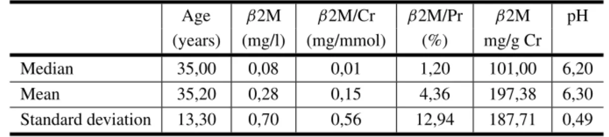
in 35 subjects (23 male) of whom 25 presented id-iopathic calcium stone disease (3 with mild renal insufficiency), 6 presented nephrocalcinosis and 4 were asymptomatic offspring of stone and nephro-calcinosis patients. The mean and median of results are shown in Table II. The urine pH varied from 5.53
to 7.60 (median 6.20). The results ofβ2M expressed
as a creatinine ratio, disclosed six patients as hav-ing abnormal low molecular weight proteinuria: the transplanted one and 5 cases with multiple urolog-ical manipulations for relief of stone obstructions; the total proteinuria was slightly increased, i.e., less than 700 mg protein/g creatinine. Overall, not
in-cluding the transplanted patient, β2M represented
less than 20% of total proteinuria (3% to 16%). In
the transplanted patient theβ2M corresponded to
more than 73% of total protein excretion. (Table II).
The normalβ2M/Cr ratio (mg/mmol) ranged
from 0.003 to 0.052; median: 0.012; men: from 0.003 to 0.052; median: 0.010, and women: from 0.006 to 0.028; median 0.0125; There was no gen-der difference, Zcalc = –1.08; p = 0.28; Mann-Whitney test.
DNA Analyses for CLCN5Mutations
The CLCN5 gene was analyzed in 26 subjects: 11 with idiopathic hypercalciuria; 6 idiopathic calcium lithiasis men with normal calciuria; 5 calcium stone disease and light to moderate renal insufficiency de-grees, 6 nephrocalcinosis (3 without renal stone). Family history of renal stone disease could be ob-tained in 23 cases and it was positive in about 70%. Some features of these patients are in Table III.
Direct DNA sequencing of the CLCN5 gene did not show any mutation even in those cases with low molecular weight proteinuria.
DISCUSSION
mor-TABLE I
Polymerase Chain Reaction (PCR) amplication conditions and primers sequences. The temperature, time of each PCR step, number of PCR cycle, and primers used to amplify the different exons of CLCN5 are shown. Denaturation (Denat); Annealing (Anneal); Extension (Ext.).
Primers Conditions No.
Denat. Time Anneal. Time Ext. Time cycles
Exon 1-2
P1:5´TGATGTGATATGGCTGCAAG3 (1-20)
P2:5´CATGTACCCTAGAACTTAAAG3´(738-718) 94◦C 45 sec 58◦C 45 s 72◦C 1 min 37
Exon 3
P3:5´GCATTACAATACTGTAGATG3´ (2784-2803)
P4:5´CAGCAATTCATTTAAGGTAG3´ (3208-3189) 94◦C 45 sec 54◦C 45 s 72◦C 1 min 37
Exon 4
P5:5´GCTTATTCTGATGACCAAAG3´ (6219-6238)
P6:5´CCATTTTAAACATAAGCGTC3´(6696-6677) 94◦C 45 sec 55◦C 45 s 72◦C 1 min 37
Exon 5
P7:5´GAATCCATGGTAAATTTCC3´(10864-10882)
P8:5´CAACTCCCTTTAATTTCC3´ (11409-11392) 94◦C 45 sec 51◦C 45 s 72◦C 1 min 37
Exon 6
P9:5´GGAACATGAAAGCTTAAAG3´ (11946-11964)
P10:5´CTATCCTTATACACCTTTG3´ (12534-12516) 94◦C 45 sec 52◦C 45 s 72◦C 1 min 37
Exon 7
P11:5´ GTAACTACTAACATTTCTTC3´ (16393-16412)
P12:5´CAATACGCAGAAACTATTTC3´ (16796-16777) 94◦C 45 sec 53◦C 45 s 72◦C 1 min 37
Exon 8
P13:5´GTACATAATGTACAATATCTG3´ (16637-16657)
P14:5´CTATCAGGATTGTATGATAC3´ (17458-17439) 94◦C 45 sec 54◦C 45 s 72◦C 1 min 37
Exon 9
P15:5´GAATCAGGGTAAGCTTCTTTG3´ (18979-18999)
P16:5´GTTTCCTCATCTGTATACCAG3´ (19553-19533) 94◦C 45 sec 60◦C 45 s 72◦C 1 min 37
Exon 10
P17:5´GTTATTTTGAGAACTCTG3´ (20437-20454)
P18:5´GGCATATTTATTGAGTTC3´ (21072-21055) 94◦C 45 sec 48◦C 45 s 72◦C 1 min 37
Exon 11
P19:5´CACGTCCATCTTCAATTTG3´ (21131-21149)
P20:5´CTCAAACACATGTTCTTTCC3´ (21590-21571) 94◦C 45 sec 55◦C 45 s 72◦C 1 min 37
Exon 12
P21:5´CAAAGACAAAGTTGAAAAG3´ (22526-22544)
P22:5´TAAATTCATTTAGAAGAGATT3´(23366-3346) 94◦C 45 sec 50◦C 45 s 72◦C 1 min 37
bidity and often requires hospitalization for relief of renal spasmatic crisis or to treat unusual complica-tions, such as acute urinary obstruction, infection or urinary sepsis.
In idiopathic calcium stone disease, the high frequency of nephrolithiasis familial history, as in this casuistic, suggest a genetic base. Although there are many possibilities of altered gene to
ac-count for idiopathic hypercalciuria (vitamin D re-ceptor gene, sodium-phosphate co-transporter gene, human homologous with the rat soluble adenylate cyclase gene, renal chloride channel gene and oth-ers), so far none has been found to be prevalent (Scheinman et al. 2000, Reed et al. 2002).
nephrolithia-TABLE II
Results ofβ2-microglobulin (β2M), total protein and pH In the first urine in the morning; n = 35 (23M/12F).
Age β2M β2M/Cr β2M/Pr β2M pH
(years) (mg/l) (mg/mmol) (%) mg/g Cr
Median 35,00 0,08 0,01 1,20 101,00 6,20
Mean 35,20 0,28 0,15 4,36 197,38 6,30
Standard deviation 13,30 0,70 0,56 12,94 187,71 0,49
TABLE III
Casuistic, ethiologic diagnoses and urinary metabolic abnormalities in selected patients for CLCN5 mutation analyses.
Gender Familial IPTH* Hypo- Hyper- Hyper- Hyper- Hyper-Antecedent (pg/ml) citraturia calciuria uricosuria phosphaturia oxaluria Idiopathic
hypercalciuric
(n=11)1 7M/4F 8/10 33 9/11 11/11 5/11 3/9 1/11
Idiophatic calcium nephrolithiasis, normal renal
funcion (n=6) 6 M 3/4 29 3/6 0/6 0/6 1/3 0/5
Idiophatic calcium nephrolithiasis and
renal falure (n=6) 3M 2/3 36 2/3 1/3 3/3 3/3 1/2
Nephrocalcinosis
(n=6)2 2M/4F 3/6 20 4/5 1/4 2/5 0/4 0/5
(X/Y) = positive/analised; (*) median; (1) one patient had iRTA; (2) including two mild renal failure, two had stones, one had tubular ectasia; intact parathormone (iPTH); Male (M), Female (F).
sis (with or without renal insufficiency) or image of nephrocalcinosis, in order to increase the odds of the group to represent CLCN5 mutation. Other cri-terion was the agreement to be subjected to blood and urine sampling for genetic analysis.
The higher percentage of idiopathic hypercal-ciuria – 52% – than previously described (Rebelo et al. 1996), is in part due to selection criterion. As it is known, absorptive hypercalciuria type II patient on low calcium diet can reduce urinary calcium excre-tion to normal range and, in this study, the diet cal-cium content was not taken into account. This was the reason to include ‘‘normal’’ calciuric idiopathic nephrolithiasis with or without nephrocalcinosis in
the search for CLCN5 mutation disease, generally described as a hypercalciuric disease.
Although clinical features of CLCN5 mutation diseases manifest mainly in affected man, female nephrolithiasis and/or nephrocalcinosis have been reported but normally the carrier woman is not symptomatic (Scheinman 1998, Reed et al. 2002).
con-trasting to idiopathic lithiasis, in men, not in women, chloride channel disease has a worse prognostic caused by progressive renal failure culminating to end stage renal failure in a young age.
None of our patients had CLCN5 gene muta-tion, supporting the well known idea that most of calcium stone formers and nephrocalcinosis patients are not fenotypes of the chloride channel disease named Dent’s disease or X-linked calcium nephro-lithiasis (Scheinman et al. 2000).
ACKNOWLEDGMENTS
The present work was supported by grants from NIH (HL 47122 and DK 32753), Fundação José Bonifácio (FUJB), Fundação Carlos Chagas Filho de Amparo à Pesquisa do Estado do Rio de Janeiro (FAPERJ), Fundação de Amparo à Pesquisa do Estado de São Paulo (FAPESP) and Conselho Nacional de Desenvolvimento Científico e Tecno-lógico (CNPQ).
RESUMO
Trinta e cinco pacientes (23 homens e 12 mulheres) com idade de 35±13 anos apresentando nefrolitíase idiopática cálcica, nefrocalcinose ou insuficiência renal leve com ne-frolitíase idiopática cálcica foram selecionados para aná-lise de proteinúria de baixo peso molecular e a ocorrên-cia de possíveis mutações no gene CLCN5. A razão en-tre aβ2-microglobulina urinária e a creatinina urinária (β2M/Cr) foi muito elevada em uma mulher transplan-tada com nefrocalcinose (>3.23 mg/mmol) e levemente elevada em cinco pacientes (>0.052 ou<1.0 mg/mmol) com manipulação urológica múltipla. Outros pacientes estudados mostraram uma razãoβ2M/Cr nos limites nor-mais (0.003-0.052 mg/mmol) sem diferença entre os sexos (p>0.05). A análise da mutação do gene do gene CLCN5 foi realizada em 26 pacientes dos 35 selecionados (11 com hipercalciúria idiopática; 6 homens com calciúria normal; 3 com leve insuficiência renal e 6 com nefrocalcinose) e não apresentou alteração em nenhum dos casos, mesmo naqueles com proteinúria de baixo peso molecular anor-mal. Conclusão: A mutação do gene do CLCN5 não é uma causa comum de calculose renal ou nefrocalcinose no grupo de pacientes brasileiros estudados.
Palavras-chave:nefrolitíase, nefrocalcinose, proteinúria de baixo peso molecular, mutação do CLCN5.
REFERENCES
Danpure CJ, Pardue PE, Fryer P, Griffiths S, Allsop J, Lumb MJ, Guttridge KM, Jennings PR, Scheinman JI and Mauer SM. 1993. En-zimological and mutational analysis of a complex primary hyperoxaluria type I phenotype involving alanine: glyoxylate aminotransferase peroxisome-to-mitochondrion mistargeting and intraperoxysomal aggregation. Am J Hum Genet 53: 417–432.
Frymoyer PA, Scheinman SJ, Dunham PB, Jones DB, Hueber P and Schroeder ET.1991. X-linked recessive nephrolithiasis with renal failure. N Eng J Med 325: 681–686.
Hoopes RR Jr, Hueber PA, Reid RJ Jr, Braden GL, Goodyer PR, Melnyk AR, Midgley JP, Moel DI, Neu AM, VanWhy SK et al.1998. CLCN5 chloride-channel mutations in six new North Amer-ican families with X-linked nephrolithiasis. Kidney Int 54: 698–705.
Igarashi T, Hayskawa H, Shiraga H, Kawato H, Yan K, Kawaguchi H, Yamanaka T, Tsuchida S and Akagi K.1995. Hypercalciuria and nephrocalcinosis in patients with idiopathic low-molecular-weight pro-teinuria in Japan: Is the disease identical to Dent’s disease in United Kingdom? Nephron 69: 242–247.
Miller DG, Tiwari R, Pathak S, Hopwood VL, Gil-bert F and Hsu TC.1998. DNA repair and muta-gen sensitivity in patients with triple primary cancers. Cancer Epidemiol Biomarkers Prev 7: 321–327.
Rebelo MAP, Leite JAG and Araújo NC.1996. Hipo-citratúria na nefrolitíase cálcica. Incidência e mor-bidade. J Bras Nefrol 18: 21–27.
Reed BY, Gitomer WL, Heller HJ, Hsu MC, Lemke M, Padalino P and Pak CY.2002. Identification and characterization of a gene with base substitutions associated with the absorptive hypercalciuria pheno-type and low spinal bone density. J Clin Endocrinol Metab 87: 1476–1485.
Scheinman SJ.1998. X-linked hypercalciuric nephro-lithiasis: Clinical syndromes and chloride channel mutations. Kidney Int 53: 3–17.
Scheinman SJ, Cox JPD, Lloyd S, Pearce SH, Sa-lenger PV, Hoopes RR, Bushinsky DA, Wrong O, Asplin JR, Langman CB et al.2000. Isolated hypercalciuria with mutation in CLCN5: Relevance to idiopathic hypercalciuria. Kidney Int 57: 232–239.
Stoller ML, Bruce JE, Bruce CA, Foroud T, Kirk-wood SC and Stambrook PJ.1999. Linkage of type II and type III cystinuria to 19q13.1: codomi-nant inheritance of two cystinuric alleles at 19q13.1 produces an extreme stone-forming phenotype. Am J Med Genet 10: 134–139.