ABCD Arq Bras Cir Dig
review Article
2017;30(2):155-160
DOI: /10.1590/0102-6720201700020017
OPEN, LAPAROSCOPIC, AND
ROBOTIC-ASSISTED HEPATECTOMY IN RESECTION OF LIVER TUMORS:
A NON-SYSTEMATIC REVIEW
Hepatectomia aberta, videolaparoscópica e assistida por robótica em ressecção de tumores hepáticos: uma revisão não sistemática
Túlio Felício da Cunha RODRIGUES1, Bianca SILVEIRA1, Flávia Pádua TAVARES1,
Gustavo Moreira MADEIRA1, Iara Proença XAVIER1, Jorge Henrique Costa RIBEIRO1,
Rayanna Mara de Oliveira Santos PEREIRA1, Sávio Lana SIQUEIRA2
From the 1Universidade Federal de Ouro
Preto, Medicina; and 2Departamento
de Cirurgia, Ginecologia, Obstetrícia e Propedêutica, Escola de Medicina, Universidade Federal de Ouro Preto
(1Federal University of Ouro Preto, Medicine;
and 2Department of Surgery, Gynecology,
Obstetrics and Propedeutics, School of Medicine, Federal University of Ouro Preto),
Ouro Preto, MG, Brazil
HEADINGS - Hepatectomy. Laparoscopy. General Surgery. Liver. Robotics.
ABSTRACT - Introduction: Several factors have made hepatectomy an increasingly safe surgery and new drugs allowed surgical treatment for patients who initially were not candidates for resection. Lesions often require resection, which can be performed by open, laparoscopic, or robotic assisted hepatectomy.Aim: Compare the surgical techniques in open, laparoscopic, and robotic assisted hepatectomy for resection of liver tumors. Methods: Literature review
based on scientific papers published on Lilacs/Pubmed/Scielo in the last 17 years regarding
the indications of these techniques for liver tumor resections and on papers comparing such techniques. Results: The comparative study shows the benefits of laparoscopic surgery
over open surgery, such as smaller incisions, less postoperative pain, shorter recovery time, smaller immune and metabolic response, and quicker restoration of oral ingestion as well as lower morbidity rates. However, the need for a specialized surgical team and the reduction in handling area still remain as disadvantages in the laparoscopic technique. It is yet not clear
whether robotic assistance presents considerable benefits over the laparoscopic technique
considering that high acquisition and maintenance costs are limiting factors. Conclusion: Despite all challenges, laparoscopic hepatectomy presents many benefits over open surgery.
The robotic assisted technique is still in evolution as many centers in the world perform hepatic resections with the platforms but only after a thorough patient selection. Thus, laparoscopy stands as the best option, unless there is some contraindication to the procedure.
RESUMO - Introdução: Nas últimas décadas, inúmeros fatores transformaram as hepatectomias em operações mais seguras. A quimioterapia, juntamente com novas drogas para o tratamento de metástases propiciaram melhores respostas, o que possibilitou a indicação cirúrgica em pacientes que inicialmente não eram candidatos a ela. Lesões hepáticas muitas vezes requerem ressecção, que pode ser realizada tanto por laparotomia, por videolaparoscopia ou assistida por plataforma robótica. Objetivo: Comparar as técnicas cirúrgicas para ressecção de tumores hepáticos. Métodos: Trata-se de revisão com base em artigos científicos publicados nos
últimos anos, sobre as indicações dessas técnicas e em artigos que comparam-nas. A pesquisa foi realizada nas bases de dados Lilacs/Pubmed/Scielo. Resultados: O estudo evidenciou vantagens da videolaparoscopia sobre a laparotomia, tais como menores incisões, redução na dor pós-operatória, menor tempo de recuperação dos doentes, dentre outras. No entanto, a necessidade de equipe especializada e a restrição na manipulação da área, consistem, ainda, em desvantagens consideráveis da técnica laparoscópica. Atualmente, ainda não está claro se o auxílio robótico demonstra vantagem substancial sobre a técnica laparoscópica, sendo o alto custo de aquisição e manutenção importante fator limitante. Conclusão: Apesar dos obstáculos
e desafios, a hepatectomia laparoscópica demonstra vantagens sobre a laparotômica. A
técnica assistida por robótica ainda está em evolução, sendo poucos os centros no mundo que a realizam nas ressecções hepáticas. Dessa forma, indica-se a laparoscopia, a menos que haja alguma contraindicação para sua realização.
Correspondence:
Túlio Felício da Cunha Rodrigues E-mail: [email protected]; [email protected] Financial source: none
Conflicts of interest: none
Received for publication: 13/10/2016 Accepted for publication: 12/12/2016
DESCRITORES - Hepatectomia. Laparoscopia. Cirurgia Geral. Fígado. Robótica.
INTRODUCTION
O
ver the last years, several factors have made hepatectomy an increasingly safe surgery. A better knowledge of liver anatomy, the development of imaging techniques, a more complete preoperative assessment of the patient and his liver function with multidisciplinary work as well as the improvement in surgical and anesthetic techniques have contributed to this28.According to Lopes-Junior et al. (2014), some strategies have been developed
Hepatic lesions can be benign or malignant and both may require resection3. Benign lesions of the liver can be
hemangiomas, adenomas, or focal nodular hyperplasia. Such lesions are generally asymptomatic, with resection being required only when they generate symptoms. However, adenomas, although benign, are at high risk of complications and, thus, must always be removed surgically. Malignant tumors of the liver may be divided into primary and secondary tumors. Among the primary ones, hepatocellular carcinoma represents 70-85% of neoplasms and requires surgical treatment26.
The National Cancer Institute (INCA) develops yearly
an estimate of cancer incidence in Brazil. The Estimate/2014
doesn’t present data regarding liver and biliary tract cancer but highlights the importance of these lesions due to their high lethality and sensitivity to preventive actions, such as immunization coverage4. Epidemiological data regarding the city of São Paulo in 2014, released by the national public health
system (SUS), showed that the incidence of primary liver cancer
was 2.07/100,000 and the mean age of patients was 54.7 years, with a male/female ratio of 3:4:1.13
As for the workup, tests such as ultrasound, computed tomography (CT), magnetic resonance imaging (MRI), angiography, and biopsy can be performed to aid in the diagnosis of liver cancer.
Surgery is the best treatment for primary liver tumors without distant metastases and metastatic liver tumors in which the primary lesion was resected or is likely to be resected in a curative way. The indication for hepatic resection depends on the clinical conditions of the patient and the expected amount of remaining liver parenchyma, which should be around 10% of the patient’s body weight. In cirrhotic patients, only the ones
with a Child A classification (early cirrhosis) are candidates for
safe liver resection5.
Another possible treatment is liver transplant, but only a small proportion of patients are good candidates for it since the criteria is based on the size and number of tumors. Currently, transplants are indicated for small tumors - three or less nodes up to 3 cm each or single nodes up to 5 cm in which there was no invasion of blood vessels or for cases where the tumor cannot be completely removed or the liver is too far compromised25.
So, the aim of this article was to compare the surgical techniques in open, laparoscopic, and robotic assisted hepatectomy for resection of liver tumors.
METHODS
This literature review is based on scientific papers published
regarding the indications for open, laparoscopic, and robotic assisted hepatectomy in resection of liver tumors and on papers comparing these techniques. The research was conducted using the databases Lilacs/Pubmed/Scielo with selection of articles published in the last 17 years related to the topic.
RESULTS
Patient preparation
A correct preoperative prepare must take into account the nature of the liver disease, its severity, and the type of operation to be performed23.
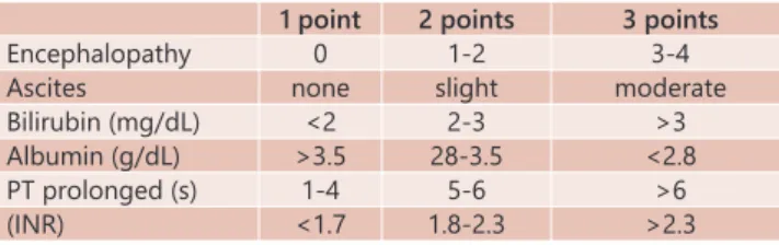
The assessment of liver function is essential and the
classification of Child-Turcotte-Pugh is the simplest form to
do that. Even small liver resections are not possible in most
patients with B or C stages of this classification23 (Table 1).
Liver resections need to be evaluated regarding residual parenchyma, especially in patients with cirrhosis or portal
hypertension. Absence of portal hypertension (defined as a
pressure gradient in the hepatic vein lower than 10 mmHg)
and a normal serum bilirubin seem to be the best predictors
of good postoperative prognosis, with a 70% five-year survival
in patients with these parameters. However, oscillations in the hepatic vein pressure gradient and an increase of bilirubin levels
were associated with a 30% five-year survival after hepatectomy, regardless of the Child-Turcotte-Pugh classification23.
TABLE 1 - Child-Turcotte-Pugh classification
1 point 2 points 3 points
Encephalopathy 0 1-2 3-4 Ascites none slight moderate Bilirubin (mg/dL) <2 2-3 >3 Albumin (g/dL) >3.5 28-3.5 <2.8 PT prolonged (s) 1-4 5-6 >6 (INR) <1.7 1.8-2.3 >2.3
Child’s A = 5-6 points; Child’s B = 7-9 points; Child’s C = 10-15 points
The presence of portal hypertension (esophageal varices, ascites, and splenomegaly with thrombocytopenia) should always be evaluated since it is a better predictor of bad prognosis than the Child-Turcotte-Pugh criteria in patients undergoing liver resections23.
The MELD (Model for End Stage Liver Disease) score, which uses INR, creatinine, and bilirubin levels, can also be used as a pre-surgical evaluation parameter. What favors the use of this score is the fact that it is continuous, being performed numerous times throughout patient care. A reduction in the score indicates that the patient’s condition has improved, being, thus, possible to recommend the surgical procedure once the MELD is below 10. When the MELD score is between 10 and 15, surgery should be performed with caution and only when strictly necessary. In people with a MELD score above 15, surgical procedures are not recommended23 (Table 2).
TABLE 2 - MELD Score
3 month mortality according to MELD score
MELD score <=9 10-19 20-29 30-39 >=40 Hospitalized pt. 4% 27% 76% 83% 100% Outpatient cirrhotic 2% 6% 50%
MELD score = 10x[0,957x log e (creatinine) + log e (bilirubin) + 1.12 x log e (INR)] + 643
The preoperative preparation can also include fasting, indwelling urinary catheter, prophylactic antibiotics (cephalothin), and two peripheral venous access, varying among cases21.
Hepatic resection by open surgery
The open operation is performed with the patient in a supine 15º Trendelenburg position with the right arm at a 90º angle. The surgical incision is made across the superior abdomen, following the curvature of the ribs. It can be bilateral subcostal for larger resections or only extended to the left for minor resections. When the mass to be addressed is very large, one can combine access between the abdomen and the chest12 (toracophrenolaparotomy, anterolateral thoracotomy
associated with laparotomy). Calne (1968) popularized the bilateral subcostal incision with midline extension. The use of retractors is essential to keep the costal railings apart,
which allows better visualization of the surgical field for
long periods of time12.
The use of intraoperative ultrasound is essential to identify the location of the nodules during surgery as well as their relationship to the blood vessels of the liver. It is also possible to identify new nodules not visualized on CT or MRI12.
of the blood flow through the inferior vena cava supra and
infrahepatically associated with clamping of the portal hilum). After vascular control, the compromised part of the liver is removed. At the end of surgery, a drain may be left near the area where the liver was cut to monitor bleeding and bile leakage12.
The extent of the resection depends on the hepatic function of the patient. If there is no cirrhosis up to two thirds of the liver can be removed surgically. When lesions are large, patients must undergo, whenever possible, major liver resection, which is related to an increased progression-free survival7. In such case, the preoperative portal embolization
of the lobe to be resected promotes hypertrophy of the remaining liver, making the resection safer and reducing the morbimortality rates14, 2, 9.
There is no consensus regarding the optimal surgical resection margins. Some studies show that segmental resections of any segment or sector where the tumor is located, including its portal pedicle, showed better results than enucleations but other papers do not demonstrate this superiority. Several studies show that resection margins larger than 1 cm are associated with higher survival rates7.
When there is macroscopic vascular invasion, resection is contraindicated, as this is known to be a poor prognostic factor associated with high recurrence and an overall survival rate of less than 10%7.
The surgery lasts 3-4 h and the patient must stay in the ICU (24 h) for monitoring of bleeding and liver function,
returning to the ward when stable. After this, the patient may remain hospitalized for as much as 10 days. After surgical removal of part of the liver (in a normal liver up to 75% of the organ can be withdrawn), the remaining tissue starts to
regenerate 48 h later and reaches a size similar to normal within 3-4 weeks. The function returns to normal in about
6-8 weeks12.
The most common clinical complications are pneumonia, deep vein thrombosis, pulmonary embolism, and liver failure. Regarding surgical complications, bleeding and bile leakage on the liver resected surface can occur. It’s important to note that liver resection has a high tumor recurrence rate of up to 50%. This may be related to metastasis of the resected tumor or to the appearance of new foci since the remaining hepatic parenchyma remains diseased. However, hepatic resection preserves the possibility of liver transplant, ablation techniques, or following resections in cases of recurrence7.
The most common contraindications for surgery are: patients with compromised cardiopulmonary function, severe malnutrition, impaired liver function, extrahepatic metastatic disease, and invasion of the portal vein bifurcation or of the hepatic veins trifurcation12.
Hepatic resection through open surgery was the treatment of choice for many years but was limited due to high rates of morbidity, mortality, and recurrence of the underlying liver disease14. Currently, liver resection can be performed in
specialized centers, with less than 5% of mortality and up to
70% of five-year survival in selected patients (asymptomatic
tumor and good liver function). However, tumor recurrence may occur in as much as 50% of cases within three years, even with proper patient selection7.
Some advantages of hepatic resection that can be cited are: a) immediate availability in specialized center; b) low risk in well-selected patients; c) precise histological evaluation; d) overall survival rates comparable to those with the intention of transplanting; e) possibility of rescue liver transplant in cases of relapse, as long as patients are monitored closely for early diagnosis of recurrences; and f) reduction of costs on the global economy of liver transplant14.
Laparoscopic hepatic resection
The technical advances in laparoscopy have revolutionized
surgical treatment for many diseases. In recent years, these advances have enabled video-assisted resection of solid organs such as the kidney and the spleen. However, some surgeries, such as liver resection, are still viewed with skepticism, since factors such as the transection of the parenchyma, the potential for intra-operative bleeding, and the risk of air embolism make the procedure a controversial theme20.
Laparoscopic hepatectomy is a difficult and laborious
procedure, requiring the availability of proper equipment and professionals with technical training in advanced laparoscopic surgery and liver surgery20.
Most of the studies indicate this technique for resecting benign hepatic tumors or treating hepatic cysts. The best candidates would be young patients with superficial or peripheral benign tumors with indication of limited parenchyma resection. It is also recommended that the method be employed in resections of the anterolateral portions of the liver, segments II, III, IV b, V, VI, or of the left portion of the caudate lobe. Within these criteria, the results are encouraging, with minimal morbidity and no complications such as bleeding or air embolism20.
The technical standardization, with use of conventional surgery technology adapted for laparoscopy is, according to Buell et al. (2005), an important aspect to facilitate the procedure. Among the new technologies available, laparoscopic ultrasound transducers should be highlighted as well as the use of hand assisted equipment, which consists of a ring set on a small skin incision of about 7 cm connected to a plastic bag, through which you can reach into the abdominal cavity without leaking gas from the pneumoperitoneum. Fong et al. (2005) reinforces that, with the use of this technology,
the procedure becomes easier and safer, the confidence in
obtaining safe margins is increased, and the removal of the specimen is facilitated27.
Bleeding is the major complication and the biggest challenge intraoperatively. Most conversions to open surgery, about 70%, happen due to intraoperative bleeding. Proper patient selection, meticulous technique, and clamping methods of the hepatic pedicle can reduce this dreaded complication. Hence, vascular control is a major concern in hepatectomies, especially in resection of tumors close to large vessels or in major resections. The clamping of the hepatic pedicle, Pringle maneuver, can be done easily by placing a shoelace around the hepatic hilum, which creates
a tourniquet to control the hepatic flow8.
Another concern is the possibility of spreading tumor cells during removal of the fragment. The specimen should always be introduced into a sturdy material bag and its removal should be done through the umbilical incision, when it has less than 3 cm, or through an appendectomy or suprapubic incision, when it is a larger piece. There is no doubt that for the treatment of benign tumors, especially liver adenomas
that affect young women, laparoscopic hepatectomy has an
important role. The whole debate should be reserved for cases of cancer in which laparoscopic surgery could increase the risk of neoplasic cells implantation, a fact that has not yet been demonstrated8.
Robotic hepatic resection
Robotics was introduced in medicine nearly two decades ago as a way of overcoming the limitations of movement of the laparoscopic instruments and providing better visualization
of the surgical field. The Da Vinci station, the most used
equipment for this type of surgery, was introduced in 2000. It has a three-dimensional view camera that allows a better sense of depth and is capable of more movements than the human hand is naturally capable of22.
In robotic assisted surgery, the patient is placed in a supine Trendeleburg position with legs apart. Commonly,
The procedure then follows three phases: portal dissection, liver mobilization, and parenchyma transection. The main surgeon sits in front of the robotic device as the assistant surgeon stands next to the patient’s right side17.
In the first phase, the liver is retracted cephalically
to expose the hilar region. The portal pedicle is dissected and its components are exposed. How the procedure will continue depends on which segment will be removed. Thus, the respective branches of the portal vein and hepatic artery are divided. The liver is mobilized after transection of the falciform and triangular ligaments, but other structures can also be sectioned depending on the procedure17.
After these steps, the capsule is incised through cauterization and the hepatic trasection begins. Two robotic arms can make the transection easier by retracting the liver and opening the surgical site. The other two arms carry a device of ultrassonic dissection and a diathermic scissor. Then a titanium clip or an endostapler is positioned to clamp the main vascular branches and biliar ducts. After the parenchyma transection is complete, the surface is inspected for any bile leakage or exudation. The specimen is then obtained through a Pfannenstiel incision inside a bag called endobag17
Robotic technique can also occur with fluorescence
guidance through application of indocyanine green that can
be injected into the bloodstream and becomes fluorescent once it gets in contact with light of a specific wavelength
near the infrared spectrum (around 820 nm) or a laser beam.
The fluorescence can be detected inside specific rooms and
chambers and then transmitted to a standard monitor that
allows the identification of anatomical structures in which the
dye is present6. This fluorescence images system associated
to robotics offers additional information to the surgeon
regarding anatomy, blood perfusion, lymphatic drainage, and functional liver reserve. The technique could become
standard in the future, considering its different diagnostic
capacities.
The robotic platform has proven to be an effective tool, particularly in the urological and gynecological field. The major drawback so far are the high costs and the difficulties
in providing on board training for surgeons. Many centers in the world have performed liver resections using the Da Vinci platform but only in highly selected patients22.
Comparison of techniques
Among laparoscopic surgeries, hepatectomies were one of the last to be carried out. The surgery was viewed with some skepticism due to concerns about bile leakage, incomplete resection, and, particularly, bleeding control, as blood loss is almost inevitable in the resection of liver tumors. Moreover, hilar dissection, liver mobilization, and parenchyma transection demand advanced technical skills and an accurate knowledge of anatomy, being, thus, potentially more dangerous than other laparoscopic procedures10.
Despite the many obstacles and challenges, laparoscopic
hepatectomy shows numerous benefits over open hepatectomy.
These are the same as in all laparoscopic surgical procedures, among which we can highlight the fact that the patient reports less pain postoperatively, has a lower incidence of ileus, less scarring, a shorter recovery time and hospital stay, and a lower rate of complications18.
A case-control study conducted by Lee et al. in 2007 helps support this view since it showed that laparoscopic hepatectomy caused less blood loss, lower morbidity rate, and less overall operative complications. In another study conducted in 2009, Zhang et al. observed 78 patients who underwent laparoscopic hepatectomies. The procedure was successful in all patients, without conversion to open procedures, and only four required blood transfusions.
Fonseca et al. (2003) and Machado et al. (2013) conducted studies with, respectively, 57 and 107 patients who underwent
laparoscopic hepatectomies. Blood transfusion was required in 29.8% and 18.7% of patients, respectively. There was no operative mortality in both studies. The rate of postoperative
complications was 57.9% in the first study and 14.9% in the
second. It is clear, therefore, that there has been an advance in the art of laparoscopic hepatectomy, as the percentage of complications and the volume of blood loss have been decreasing.
Alhomaidhi et al. (2012) conducted a comparative study through a literature review that showed that, between 1990
and 2010, 751 open hepatectomies and 4207 laparoscopic
hepatectomies were conducted. On average, it took 65 min less to perform the laparoscopic surgery. Blood loss volume was 260 ml in patients undergoing laparoscopic hepatectomy, while the loss at laparotomy was of 1290 ml on average. Postoperative morbidity was relatively low on both operations. The length of stay and mortality were also reduced.
Franken et al. (2014) conducted a comparative study of 104 patients, in which 52 underwent laparoscopic hepatectomy
(group 1) and 52 underwent open hepatectomy (group 2). They noted that the estimated blood loss average was lower in patients undergoing laparoscopy (237 ml) than in those undergoing open surgery (387 ml). The average hospital stay
was five days in group 1 and six days in group 2. Although
blood loss decreased with laparoscopic hepatectomy, the
need for blood transfusion was not significantly different and the severity of complications was not different between
the groups.
Koffron et al. (2007) conducted a study in which 300
laparoscopic liver resections were compared to open procedures. They noted that the laparoscopic approach was superior
to the open technique. The benefits were significant in the
following aspects: duration of surgery (99 vs. 182 min), blood loss (102 vs. 325 ml), need for transfusion (two out of 300 cases vs. eight out of 100 cases), hospitalization (1.9
compared to 5.4 days), overall postoperative complications
(9.3 vs. 22%), and local recurrence of malignancy (2 vs. 3%).
Wakabayashi et al. (2014) and Wang et al. (2015) also
conducted comparative studies indicating that laparoscopic hepatectomy is better than the open technique. It was noticed that the exposure of the region is better, the hospital stay is shorter, the size of the incision and the blood loss was smaller, and the cost to the hospital is lower. Despite all these advantages, it was elucidated by Wakabayashi et al. that the restriction in the handling area is a considerable disadvantage. Wang et al. also noted that the possibility
of tumor dissemination and the difficulties in maintaining
adequate margins are potential disadvantages.
According to Machado et al. (2012), in patients with a
previously identified technical difficulty for the exclusive use
of laparoscopy, hybrid techniques may be used with hand assistance or laparoscopic release followed by liver section through a small incision. The use of hand assisted techniques makes it easier to display the liver, to section the parenchyma, especially in cirrhotic livers, and also allows the surgeon the tactile sensation lost in laparoscopy. However, it is believed that a frequent use of this technique is not required, as it should be a step prior to complete conversion to laparotomy,
or an option when difficulties for the realization of a total
laparoscopic technique are expected.
The literature review shows an exponential growth in the number of and indications for laparoscopic hepatectomies. In a review of all published cases of laparoscopic hepatectomies,
held in 2009, in which 2804 cases were identified, the
mortality rate was only 0.3% and morbidity 10.5%12. Thus,
safe and effective.
Therefore, if there is indication for a hepatectomy, provided there is no contraindication for the method (intestinal obstruction, generalized peritonitis, severe cardiopulmonary disease, or severe hypovolemic shock among others reasons), laparoscopy should be the technique of choice20.
When comparing laparoscopic and robotic techniques, in accordance with Montalti et al. (2015), it’s noticeable that, despite laparoscopy’s worldwide spread since its introduction, robotics have not had the same evolution, possibly due to
the significant initial costs and the different levels of required learning. Studies show that there is no significant difference
in morbidity rate between the two techniques, although it was observed a tendency for fewer complications in the robotic group.
In a systematic review and meta-analysis, Montalti et al. (2015) states that the robotic platform is a tool with which many of the limitations of conventional liver laparoscopic
surgery could be overcome, like image amplification,
two-dimensional tremor, fulcrum effect, limited freedom of movement, and ergonomics. Furthermore, the increased dexterity activated by the endowristed movements, the
filter software for the surgeon movements, and the high definition 3-D vision provided by a stereoscopic camera
allows a constant and careful dissection of structures. All of this leads to minimal biliary leakage and overall reduction in postoperative complications22.
Despite all these advantages, robotic assisted hepatectomy has evolved slowly over the years and it is currently unable to provide useful tools to fully exploit the potential of the
movements and the vision offered by the robot, especially
when the resection space is limited.
It is important to note that to perform the robotic technique, an additional surgeon responsible for the procedure is required and the costs with the robot, the instrumentation, and the annual maintenance are high. In addition, there are few centers in the world that perform robotic liver resections and they always go through a thorough patient selection, which limits the number of procedures performed, and keeps it as non-standard, even if these centers can surpass the learning curve.
The results of the meta-analysis made by Montalti et
al. (2015) show a significant rise in bleedings during robotic assisted hepatectomies, which can be explained by the different
techniques used to execute the liver transection. The most used technique for laporoscopic hepatic transection requires
the use of a harmonic scalpel for superficial liver trasection
and Cavitron Ultasonic Surgical Aspirator for deeper cuts, to provide a more meticulous and precise parenchymal structure dissection. In turn, in robotic resection the technique is based
on crushing fixation, requiring, in most cases, the use of an intermittent occlusion of blood flow (Pringle’s Maneuver).
Another important factor regards the operation time,
which was significantly higher in robotic hepatectomy. This
may be due to the technique itself buy may also be a result of robotics being new and requiring, therefore, greater
experience and refinement.
As it is known, one of the principles of oncologic resection of malignant tumors is the maintenance of free margin to avoid incomplete resection of the tumor and possible iatrogenic spread. That is why the previously mentioned meta-analysis aimed to observe the margins
width in each technique. As a result, no significant difference
was observed, although there was a tendency for smaller margins in laparoscopic hepatectomies, which suggests a
greater difficulty in identifying the lesion through robotic
intraoperative ultrasound. This can be explained by the fact that the surgeon who performs the ultrasound is not the one responsible for the robotic hepatectomy. However, further studies are necessary for a better data analysis. The
study of Montalti et al. (2015) considers that laparoscopic hepatectomies have a reduced blood loss and a shorter operative time compared to robotic hepatectomies.
Thus, it is not clear whether robotic assistance demonstrates substantial advantage over laparoscopic techniques since both approaches are considered minimally invasive, with no
differences in safety or efficacy22.
CONCLUSION
The approach of liver tumors is considered complex as it involves factors related to the clinical condition of the patient, the function of the liver, and the stage and characteristics of malignant diseases. Some tumors develop in livers considered normal, while others arise in organs compromised by obstruction
of the biliary tract or liver diseases such as steatosis, fibrosis, or cirrhosis. Each of these factors can influence the result of
surgeries and, therefore, careful attention to all of these aspects must be paid in order to achieve good results. The laparoscopic approach is more beneficial when compared to the open technique, despite the barriers and mistrust that still remains. Laparoscopy has shorter procedure duration, shorter hospital stay, and lower incidence of complications. Furthermore, the local malignant recurrence rate is also reduced in the closed surgical procedure.When laparoscopic and robotic techniques are compared, it is clear that the robotic assistance can overcome many limitations that laparoscopic surgery presents. However,
due to the high cost and different levels of learning required,
robotic hepatectomy still hasn’t spread worldwide. It is not yet clear whether robotic assistance is an advantage over laparoscopy, since hospital stay, morbidity, and estimated blood loss are similar.
REFERENCES
1. Alhomaidhi A. Right Hepatectomy, Open vs Laparoscopy: a systematic
review. Surgical Science. 2012 Dec; 3, 580-7. doi: 10.4236/ss.2012.312115.
2. Amico, Enio Campos et al. immediate complications after 88 hepatectomies - brazilian consecutive series. ABCD, arq. bras. cir. dig., Sept 2016, vol.29,
no.3, p.180-184. ISSN 0102-6720.
3. Araujo, Raphael L. C. et al. Central hepatectomy for biliary cystadenoma: parenchyma-sparing approach for benign lesions. ABCD, arq. Bras. Cir.
Dig., Dec 2016, vol.29, no.4, p.295-296. ISSN 0102-6720.
4. Brasil. Ministério da Saúde. Câncer de fígado. Instituto Nacional do Câncer. 2016.
5. Bredt LC, Zanini JC, Fernandes F, Mierzwa TC, Rachid AF. Tratamento cirúrgico do carcinoma hepatocelular em estado inicial. Sociedade Brasileira de Cirurgia Oncológica. 2015 Oct.
6. Buell JF, Koffron AJ, Thomas MJ, Rudich S, Abecassis M, Woodle ES. Laparoscopic liver resection. J Am Coll Surg. 2005 Mar;200:472-480.
PMID:15737861
7. Coutinho AK, Hoff PMG, Costa FP, Gil RA, Sabbaga J, Marinho F, Herman
P, Bastos J, Oliveira A, Menezes M. In: Revista da Sociedade Brasileira de Oncologia Clínica. Manual de Condutas. 2011 Oct. 279-298.
8. D’Albuquerque LAC, Herman P. Hepatectomia por videolaparoscopia:
realidade? Arq. Gastroenterol. 2006 Sep. 43(3): 243-246. doi:10.1590/ S0004-28032006000300017.
9. Fernandes, Eduardo de Souza Martins et al. The largest western experience with hepatopancreatoduodenectomy: lessons learned with 35 cases. ABCD, arq. bras. cir. dig., Mar 2016, vol.29, no.1, p.17-20. ISSN 0102-6720. 10. Fong Y, Jarnagin W, Conlon KC, DeMatteo R, Dougherty E, Blumgart
LH. Hand-assisted laparoscopic liver resection: lessons from an initial
experience. Arch Surg. 2000 Jul;135(7):854-9. PubMed. PMID: 10896382
11. Franken C, Lau B, Putchakayala K, DiFronzo LA. Comparison of
short-term outcomes in laparoscopic vs open hepatectomy. JAMA Surg. 2014 Sep;149(9):941-6. doi: 10.1001/jamasurg.2014.1023.
12. Goffi SF. Técnica cirúrgica: bases anatômicas, fisiopatológicas e técnicas
de cirurgia. 3. ed. Atheneu, 2006/2007; 671-76.
13. Gomes MA, Priolli DG, Tralhão JG, Botelho MF. Carcinoma hepatocelular: epidemiologia, biologia, diagnóstico e terapias. Rev. Assoc. Med. Bras.,
2013 oct; 514-24. doi: 10.1016/j.ramb.2013.03.005.
14. Herman P. Carcinoma hepatocelular - ressecção cirúrgica. GED gastroenterol.
15. Junior AGL, Belebecha V, Jacob CE. Hepatectomy: a critical analysis on
expansion of the indications. ABCD, arq. bras. cir. dig. 2014 Jan; 47-52. doi: 10.1590/S0102-67202014000100012.
16. Lacerda CF, Bertulucci PA, Oliveira ATT. Totally laparoscopic liver resection:
New brazilian experience. 2014 Jul;27(3):191-19. doi: 10.1590/S0102-67202014000300008.
17. Lai EC, Tang CN, Li MK. Robot-assisted laparoscopic hemi-hepatectomy: technique and surgical outcomes. Int J Surg. 2012;10:11-15. PMID: 22079835 18. Laurent A, Cherqui D, Lesurtel M, Brunetti F, Tayar C, Fagniez PL. Laparoscopic liver resection for subcapsular hepatocellular carcinoma complicating chronic liver disease. Arch Surg. 2003 Jul;138(7):763-9. PMID: 12860758. 19. Lee KF, Cheung YS, Chong CN, Tsang YY, Ng WW, Ling E, Wong J, Lai
PB. Laparoscopic versus open hepatectomy for liver tumours: a case
control study. Hong Kong Med J. 2007 Dec;13:442-448. PMID:18057432
20. Machado MAC, Makdissi FF, Surjan RCT. Hepatectomia videolaparoscópica: experiência pessoal com 107 casos. Rev. Col. Bras. Cir. 2012 Dec; 39( 6):
483-488. doi: 10.1590/S0100-69912012000600007.
21. Machado MAC. Carcinoma hepatocelular. Resultados da hepatectomia parcial e análise dos fatores prognósticos. 1999.
22. Montalti R, Berardi G, Patriti A, Vivarelli M, Troisi RI. Outcomes of robotic vslaparoscopic hepatectomy: A systematic review and
meta-analysis. World Journal of Gastroenterology. 2015 Jul. 21(27):8441-8451. doi:10.3748/wjg.v21.i27.8441
23. Paes-Barbosa FC, Ferreira FG, Szutan LA. Hepatectomy preoperative
planning. Rev. col. bras. cir., 2010 oct; (5)370-75. PMID: 21181004 24. Pais-Costa SR, Araujo SLM, Lima SRM, Lima OAT, Teixeira ACP. Laparoscopic
hepatectomy: indications and results from 18 resectable cases. Einstein,
São Paulo. 2011 Sept;9(3):343-9. doi: 10.1590/S1679-45082011AO1983.
25. Pimenta JR, Massabki PS. Carcinoma Hepatocelular: um panorama clínico. Rev Bras Clin Med, 2010; 8:59-67.
26. Rao AM, Ahmed I. Laparoscopic versus open liver resection for benign and malignant hepatic lesions in adults. Cochrane database of systematic reviews. 2013 may 31; (5):CD010162. PMID: 23728700.
27. Reddy SK, Tsung A, Geller DA. Laparoscopic liver resection. World journal
of surgery. World J Surg. 2011 Jul;35(7):1478-86. PMID: 21181472
28. Salim T, Cutait R. Videolaparoscopy complications in the management of
biliary diseases. Arquivos Brasileiros de Cirurgia Digestiva. 2009 Oct;21(4), 153-157. doi: 10.1590/S0102-67202008000400001.
29. Silva M, Mattos AA, Fontes PRO, WaechterI FL, Lima LP. Avaliação da ressecção hepática em pacientes cirróticos com carcinoma hepatocelular. Arq.
Gastroenterol. 2008 Apr; 45(2) 99-105. doi: 10.1590/S0004-28032008000200002.
30. Wakabayashi G, Cherqui D, Geller DA, Han HS, Kaneko H, Buell JF. Laparoscopic hepatectomy is theoretically better than open hepatectomy: preparing for the 2nd International Consensus. doi: 10.1002/jhbp.139. 31. Wang XT, Wang HG, Duan WD, Wu CY, Chen MY, Li H, Huang X, Zhang
FB, Dong JH. Pure Laparoscopic Versus Open Liver Resection for Primary Liver Carcinoma in Elderly Patients: A Single-Center, Case-Matched
Study. Medicine, Baltimore. 2015 Oct;94(43):E1854. doi: 10.1097/ MD.0000000000001854
32. Wu YM, Hu RH, Lai HS, Lee PH. Robotic-assisted minimally invasive liver
resection. Asian J Surg. 2014 Apr.37(2):53-7. doi: 10.1016/j.asjsur.2014.01.015.
33. Zhang L, Chen YJ, Shang CZ, Zhang HW, Huang, ZJ. Total laparoscopic liver resection in 78 patients. World Journal of Gastroenterology. 2009