Effects of placebo-controlled continuous and pulsed
ultrasound treatments on carpal tunnel syndrome: a
randomized trial
Onur Armagan,IFulya Bakilan,IMerih Ozgen,IOzlem Mehmetoglu,IISetenay OnerIII
IEskisehir Osmangazi University Faculty of Medicine, Department of Physical Medicine and Rehabilitation, Eskisehir, Turkey.IIKonya State Hospital, Department of Physical Medicine and Rehabilitation, Eregli, Turkey.IIIEskisehir Osmangazi University University Faculty of Medicine, Department of Biostatistics, Eskisehir, Turkey.
OBJECTIVE: The aim of this placebo-controlled study was to evaluate the effects of pulsed and continuous ultrasound treatments combined with splint therapy on patients with mild and moderate idiopathic carpal tunnel syndrome.
METHODS:The study included 46 carpal tunnel syndrome patients who were randomly divided into 3 groups. The first group (n = 15) received a 0 W/cm2ultrasound treatment (placebo); the second group (n = 16) received a 1.0 W/cm2continuous ultrasound treatment and the third group (n = 15) received a 1.0 W/cm21:4 pulsed ultrasound treatment 5 days a week for a total of 15 sessions. All patients also wore night splints during treatment period. Pre-treatment and post-treatment Visual Analogue Scale, Symptom Severity Scale and Functional Status Scale scores, median nerve motor conduction velocity and distal latency and sensory conduction velocities of the median nerve in the 2nd finger and palm were compared. Clinicaltrials.gov: NCT02054247.
RESULTS:There were significant improvements in all groups in terms of the post-treatment Functional Status Scale score (p,0.05 for all groups), Symptom Severity Scale score (first group:p,0.05, second group:p,0.01, third group: p,0.001) and Visual Analogue Scale score (first and third groups: p,0.01, second group:
p,0.001). Sensory conduction velocities improved in the second and third groups (p,0.01). Distal latency in the 2ndfinger showed improvement only in the third group (
p,0.01) and action potential latency in the palm improved only in the second group (p,0.05).
CONCLUSION:The results of this study suggest that splinting therapy combined with placebo and pulsed or continuous ultrasound have similar effects on clinical improvement. Patients treated with continuous and pulsed ultrasound showed electrophysiological improvement; however, the results were not superior to those of the placebo.
KEYWORDS: Carpal Tunnel Syndrome; Continuous Ultrasound; Pulsed Ultrasound.
Armagan O, Bakilan F, Ozgen M, Mehmetoglu O, Oner S. Effects of placebo-controlled continuous and pulsed ultrasound treatments on carpal tunnel syndrome: a randomized trial. Clinics. 2014;69(8):524-528.
Received for publication onJanuary 24, 2014;First review completed onFebruary 21, 2014;Accepted for publication onFebruary 21, 2014 E-mail: [email protected]
Tel.:+90 222 2392979/2457
& INTRODUCTION
Carpal tunnel syndrome (CTS) is an entrapment mono-neuropathy that is commonly observed in clinical practice and is caused by the compression of the median nerve at the wrist (1). When the median nerve is compressed in the tunnel, the patient develops the signs and symptoms of
CTS. The most common symptoms of CTS include pain, paresthesia, numbness or tingling involving the fingers that are innervated by the median nerve and a weakness of thumb abduction. The symptoms are at their worst at night and often wake the patient (2).
Both conservative and surgical treatments are used to relieve the pressure on the median nerve (1). The choice between conservative and surgical treatment is determined by the severity of the symptoms and the patient’s physical limitations (3).
Conservative treatment of CTS would seem to be preferable as the initial treatment choice, particularly for mild to moderate cases (1). However, the efficacy of conservative treatment options for CTS is controversial (1). Ultrasound (US) is a widely used and accepted adjunct modality for the management of many musculoskeletal
Copyrightß2014CLINICS– This is an Open Access article distributed under
the terms of the Creative Commons Attribution Non-Commercial License (http:// creativecommons.org/licenses/by-nc/3.0/) which permits unrestricted non-commercial use, distribution, and reproduction in any medium, provided the original work is properly cited.
No potential conflict of interest was reported.
conditions (4). US converts electrical energy into an acoustic waveform, which is then converted into heat as it passes through tissues of varying resistance (5). Therapeutic US is reported to reduce edema, relieve pain and accelerate tissue repair (6-8). The exact mechanism of action of therapeutic US remains to be elucidated, although it is used to treat various musculoskeletal disorders. Analgesia that is induced by therapeutic US may be the result of increased capillary permeability and tissue metabolism, the enhance-ment of fibrous tissue extensibility and the elevation of the pain threshold by thermal mechanisms (6,9). Therapeutic US can be applied as pulsed or continuous therapy.
Only a few studies have reported the benefits of therapeutic US in CTS patients and there are conflicting results on its efficacy in the treatment of CTS (1,10,11). Recently, the effectiveness of pulsed and continuous US therapy was compared with a placebo and the authors reported satisfactory effects in patients with mild to moderate CTS (10,12,13).
Splinting is the most popular method among the conservative treatments that are available for CTS (13-15). Immobilization of the wrist in a neutral position with a splint maximizes the carpal tunnel volume and minimizes the pressure on the median nerve (1).
The aim of this study was to evaluate the effects of pulsed and continuous US treatments in combination with splint therapy on patients with mild and moderate idiopathic CTS compared to a placebo.
& METHODS
The study was carried out at the outpatient clinic of the Eskisehir Osmangazi University Faculty of Medicine, Department of Physical Medicine and Rehabilitation bet-ween October 2011 and January 2013. A total of 36 female patients with clinical and electrophysiological evidence of mild or moderate idiopathic CTS (without thenar atrophy or spontaneous activity as determined by the electrophysiolo-gical examination of the abductor pollicis brevis (APB) muscle) were included in the study. All of the patients had unilateral CTS and were right-handed.
Patients were excluded if they had secondary entrapment neuropathies, cervical radiculopathy or systemic diseases that are associated with increased CTS risk in addition to those who had undergone surgery for the syndrome, had been treated with US or had a history of steroid injections into the carpal tunnel and physical therapy within the last 3 months. Additionally, patients with either thenar atrophy or spontaneous activity (fibrillation potentials and positive sharp waves) as determined by an electrophysiological examination of the APB muscle were excluded from the study.
Study design
This study was designed as a prospective, randomized, placebo-controlled, double-blind study. All of the patients were assessed by the same physiatrist before beginning treatment and at the end of the three weeks of treatment. Before initiation and after the three weeks, all of the patients were assessed by the same physiatrist. Neither the investi-gator nor the patients were informed of the treatment assignments. The study was conducted in full compliance with the amended Declaration of Helsinki after obtaining approval from the institutional review board of Eskisehir
Osmangazi University (date/n
˚
: 18-05-2012/127). Following baseline assessments, the patients who fulfilled the inclu-sion criteria and were included in the study were randomly assigned to one of three groups using a secure system of opaque closed envelopes that were numbered from 1 to 3.Forty-six patients were randomly assigned to one of the three groups: group 1 (n = 15) received splinting and continuous US therapy; group 2 (n = 16) received splinting and pulsed US therapy and group 3 (n = 15) received splinting and a ‘sham’ (placebo) US therapy.
Treatment protocol
Custom-made neutral volar splints were given to all of the patients that were included in the study. They were instructed to wear the splints at night and during the day for a total of three weeks.
Continuous, pulsed and sham US therapies were per-formed on all of the patients by the same physiotherapist. In group 1 (continuous US group), US treatments were administered to the carpal tunnel area at a frequency of 1 with an intensity of 1 W/cm2and a transducer of 5 cm2in size (Sonopuls 434; Enraf Nonius, Delft, The Netherlands), using aquasonic gel that did not contain any pharmacolo-gically active substances. The apparatus was initially standardized and the output was controlled by a simple underwater radiation balance.
In group 2 (pulsed US group), the same US equipment was set at a frequency of 1 MHz with an intensity of 1 W/ cm2and a pulsed mode duty cycle of 1:4. The duration of the applied US and the posture of the patient being treated were the same as those that were described for the continuous US group. The patients in group 3 (placebo US group) received a ‘sham’ US application wherein the US device that was described above appeared to be working but did not deliver any output. US treatment sessions were performed once a day, five days a week, for a total of three weeks.
Evaluations
All of the patients were evaluated immediately before and after the three-week treatment by a blinded investigator with respect to the parameters that are described below.
Electrophysiological evaluations
the recording ring electrode that was looped around the proximal interphalangeal joint of the third digit. The onset latencies of the negative potentials were taken into consideration. For sensory testing, the sweep-speed velocity was set at 10 msec, whereas for motor testing, it was set at 30 msec; the duration of the stimulus was 0.1 msec in both studies. The voltage was increased until the action poten-tials reached maximal amplitude. The main electrophysio-logical criteria for the diagnosis of CTS were the slowing of sensory nerve conduction velocity of the median nerve in the palm-wrist segment or an absence of sensory nerve action potential of the median nerve that was accompanied by prolonged terminal motor latency (17). In our electro-physiology laboratory, if the median nerve DL was 3.9 msn or above, the median nerve sensory latency was 3 msn or above and the sensory nerve conduction velocity of the median nerve in the palm-wrist segment was 35.2 m/sn or below, the subjects were considered to have CTS.
Clinical assessment
A blinded physician who was unaware of the treatment allocations performed the clinical assessments at baseline and at the end of the treatments. Symptom Severity scores (SSSs), Functional Status scores (FSSs) and a Visual Analog Scale (VAS) were used for the clinical follow-up and evaluation of the patients.
The severity of the pain was assessed using a VAS consisting of 10-cm horizontal lines with anchor points of 0 (no pain) and 10 (maximum pain). The Symptom Severity Scale has 11 items in relation to pain, including nocturnal symptoms, numbness, tingling and weakness (18). The Functional Status Scale encompasses 8 items (difficulty in writing, buttoning clothes, opening jars, holding a book, gripping a telephone handle, performing household chores, carrying grocery bags, bathing and dressing). Each item in these scales has five ordinal response categories,
ranging from 1 (no symptoms or no difficulty) to 5 (severe symptoms).
Statistical analyses
Statistical analyses were performed using SPSS 18.0 for Windows. The normality of the distribution of the data was confirmed by the Kolmogorov-Smirnov test. The results were analyzed by a one-way ANOVA with post-hoc Tukey HSD tests and t-tests. The results were reported as the mean¡SD. Power calculations were evaluated using a
one-way ANOVA and the power of the study was 0.70 (70%). The significance level was set atp,0.05.
& RESULTS
Table 1 shows the demographic characteristics of the study groups. No significant differences were observed between the groups. There were significant improvements in all groups for all clinical parameters. There were no significant differences between the groups (Table 2).
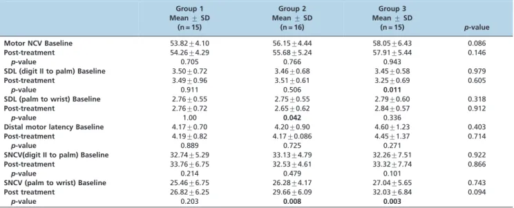
Table 3 shows a comparison of the electrophysiological measurements that were obtained before and after the treatments. The sensory conduction velocity of the median nerve showed improvement in the second and third groups, the sensory DL of the median nerve (digit II to palm) improved only in the third group, and the sensory DL of the median nerve (palm to wrist) improved only in the second group. There were no significant differences between the groups.
& DISCUSSION
CTS is one of the most common hand disorders. The highest incidence is among middle-aged and elderly women (19). Although several treatment modalities are routinely used, there is no consensus regarding the best way to manage CTS. The aim of this placebo-controlled study was to evaluate
Table 1 -Demographic characteristics of the patients.
Group 1 (n = 15) Mean¡SD
Group 2 (n = 16) Mean¡SD
Group 3 (n = 15)
Mean¡SD p-value
Age (years) 45.20¡2.98 43.31¡2.79 44.53¡2.38 0.883
Duration of symptoms (months) 13.67¡3.52 12.00¡3.29 11.87¡2.70 0.908
Table 2 -Comparison of the clinical parameters at baseline and at the end of treatment (post-treatment).
Group 1 Mean¡SD
(n = 15)
Group 2 Mean¡SD
(n = 16)
Group 3 Mean¡SD
(n = 15) p-value
FSS Baseline 21.33¡7.37 24.00¡5.58 19.00¡0.85 0.019
Post-treatment 18.80¡7.34 19.31¡9.42 14.20¡4.52 0.125
p-value 0.036 0.041 0.003
SSS Baseline 26.60¡8.11 29.75¡7.71 25.93¡4.46 0.274
Post-treatment 23.06¡8.13 22.06¡8.73 19.66¡4.60 0.442
p-value 0.047 0.002 0.001
VAS Baseline 5.40¡2.32 5.56¡1.75 5.20¡1.26 0.859
Post-treatment 4.40¡2.32 2.68¡1.92 3.53¡1.95 0.083
p-value 0.006 0.000 0.003
FSS:Functional Status Scale.
SSS:Symptom Severity Scale.
the effects of pulsed and continuous US treatments in combination with splinting therapy on various clinical and electrophysiological parameters in patients with CTS.
Splinting is the most popular method among the conservative treatment modalities that are available for CTS (14,15). The efficacy of wrist splinting therapy has been variably demonstrated in several studies (15,20,21). Premoselli et al. reported that splinting therapy improved symptoms and electrophysiological parameters in CTS patients (21).
In our study, a night rest splint was used by all of the participants and clinical improvement was observed in all of the groups. Placebo US can lead to symptom relief by producing a local massage effect; however, this sympto-matic improvement may be, in fact, related to the splinting therapy, as has been observed in recent studies investi-gating the beneficial effects of splinting therapy in CTS. Electrophysiological improvement was found in the con-tinuous and pulsed US groups but not in the placebo group. US is used extensively in musculoskeletal disorders. A few studies have found US therapy to be effective for CTS (10,12,13). However, a consensus has yet to be reached regarding the optimal therapeutic US parameters (intensity, frequency of sound waves, duration, pulse, etc.) (1,10,13,22). Therapeutic US can be applied in a pulsed or continuous manner. Pulsed US has been recommended for acute pain and inflammation, whereas continuous US has been recommended for the treatment of restricted movement (23). Pulsed US produces non-thermal effects and is used to aid in the reduction of inflammation, whereas continuous US generates thermal effects (6).
The aim of this study was to compare the effects of pulsed and continuous US treatments on patients with CTS. In the placebo-controlled study that was conducted by Ebenbichler et al., the US method was similar to that which was used in our study with the exception of the therapy time (1 MHz, 1.0 W/cm2in pulsed mode 1:4 for 15 min per session). This group revealed the clinical and electrophy-siological improvement of CTS symptoms (10). In another
study comparing the efficiency of US and laser therapy, US (1 MHz, 1.0 W/cm2 in pulsed mode 1:4 for 15 min per session) was found to be more effective than laser therapy for CTS in terms of electrophysiological parameters (22). US therapy in combination with splinting therapy was compared with ketoprofen phonophoresis in a placebo-controlled study and the electrophysiological and clinical parameters were reported to improve in both groups (24). Electrophysiological and clinical improvements were observed in our study in the pulsed US group. These results are in agreement with those from other studies, although there are some methodological differences.
In the group that received continuous US therapy in combination with splinting therapy, the treatment was applied for 10 min per session to the carpal tunnel area at a frequency of 1 and an intensity of 1 W/cm2. The efficacy of continuous US in CTS has been previously evaluated in only a few studies (12,25,26). A placebo-controlled study reported clinical and electrophysiological improvement using different doses of US therapy (1.5 W/cm2, 0.8 W/ cm2, 0 W/cm2). In addition, the NCV decreased slightly and DL increased in the US group (13). In a placebo-controlled study evaluating low-intensity (0.5 W/cm2) US, a signifi-cant improvement in clinical variables was observed after the treatment, although there were no differences in the electrophysiological variables (12). In another placebo-controlled study, clinical improvement was observed with US therapy (1.5 W/cm2
); however, no electrophysiological improvement was found (25).
Because the US intensities that were used differ in all of these studies, it is difficult to compare results. Furthermore, there is no consensus in the literature regarding the effective dose for US therapy. Similarly to our study, Dincer et al. compared splinting therapy alone with continuous US therapy (at an intensity of 1.0 W/cm2) combined with splinting therapy in addition to laser therapy combined with splinting therapy and found clinical and electrophy-siological improvement in the combined therapy groups (26).
Table 3 -Comparison of electrophysiological parameters at baseline and at the end of the treatment (post-treatment).
Group 1 Mean¡SD
(n = 15)
Group 2 Mean¡SD
(n = 16)
Group 3 Mean¡SD
(n = 15) p-value
Motor NCV Baseline 53.82¡4.10 56.15¡4.44 58.05¡6.43 0.086
Post-treatment 54.26¡4.29 55.68¡5.24 57.91¡5.44 0.146
p-value 0.705 0.766 0.943
SDL (digit II to palm) Baseline 3.50¡0.72 3.46¡0.68 3.45¡0.58 0.979
Post-treatment 3.49¡0.96 3.51¡0.61 3.25¡0.69 0.605
p-value 0.911 0.506 0.011
SDL (palm to wrist) Baseline 2.76¡0.55 2.75¡0.55 2.79¡0.60 0.318
Post-treatment 2.76¡0.72 2.65¡0.62 2.84¡0.57 0.912
p-value 1.00 0.042 0.336
Distal motor latency Baseline 4.17¡0.70 4.20¡0.90 4.60¡1.23 0.403
Post-treatment 4.19¡0.82 4.17¡0.086 4.45¡1.37 0.714
p-value 0.889 0.725 0.271
SNCV(digit II to palm) Baseline 32.74¡5.29 33.13¡4.79 32.26¡7.51 0.922
Post-treatment 33.76¡6.75 32.53¡4.61 33.32¡7.74 0.866
p-value 0.214 0.479 0.101
SNCV (palm to wrist) Baseline 25.46¡6.75 26.28¡4.17 27.04¡5.65 0.743
Post treatment 26.82¡6.25 29.66¡6.09 32.03¡6.84 0.094
p-value 0.203 0.008 0.003
SNCV:Sensory nerve conduction velocity.
US is assumed to be an anti-inflammatory procedure that increases blood flow, local metabolism and tissue regeneration in target tissues in addition to reducing edema and pain and limiting nerve compression (1).
Evidence of an anti-inflammatory effect of US treatment from experiments on the stimulation of nerve regeneration and on nerve conduction supports the idea that US treatment may facilitate recovery from nerve compression (27-30).
In our study, the patients that were treated with pulsed and continuous US showed electrophysiological improve-ment; however, these results were not superior to those that were observed with the placebo. We believe that this electrophysiological improvement is related to the mechan-ism of action of the US therapy. Further, the lack of intergroup differences in the electrophysiological para-meters may be related to the small sample size.
There are no studies currently available comparing the effects of continuous and pulsed US. The relatively small number of patients and lack of data describing a long-term follow-up of the patients were the main limitations of our study.
In our study, splinting therapy in combination with pulsed or continuous US or placebo showed similar clinical results. Patients who were treated with continuous and pulsed US showed electrophysiological improvement; how-ever, the results that were obtained, were not superior to those that were reported with the placebo. There is still no existing consensus on optimal therapeutic US parameters and well-designed studies with long-term follow-up are needed.
& AUTHOR CONTRIBUTIONS
Armagan O was the study director. Bakilan F, Ozgen M and Mehmetoglu O were the assistant directors. Oner S was the co-assistant for the statistical analyses.
& REFERENCES
1. Gerritsen AA, de Krom MC, Struijs MA, Scholten RJ, de Vet HC, Bouter LM. Conservative treatment options for carpal tunnel syndrome: a systematic review of randomised controlled trials. J Neurol. 2002; 249(3):272-80.
2. Simovic D and Weinberg DH. Carpal tunnel syndrome. Archives of Neurology. 2000;57(5):754-55, http://dx.doi.org/10.1001/archneur.57.5. 754.
3. Patijn J, Vallejo R, Janssen M, Huygen F, Lataster A, van Kleef M, et al. Carpal tunnel syndrome Pain Pract. 2011;11(3):297-301.
4. Wong RA, Schumann B, Townsend R, Phelps CA. A survey of therapeutic ultrasound use by physical therapists who are orthopaedic certified specialists. Phys Ther. 2007;87(8):986-94, http://dx.doi.org/10. 2522/ptj.20050392.
5. Klaiman MD, Shrader JA, Danoff JV, Hicks JE, Pesce WJ, Ferland J. Phonophoresis versus ultrasound in the treatment of common muscu-loskeletal conditions. Med Sci Sports Exerc. 1998;30(9):1349-55. 6. Rutjes AW, Nu¨esch E, Sterchi R, Ju¨ni P. Therapeutic ultrasound for
osteoarthritis of the knee or hip. Cochrane Database Syst Rev. 2010;20(1):CD003132.
7. Fu SC, Shum WT, Hung LK, Wong MW, Qin L, Chan KM. Low-intensity pulsed ultrasound on tendon healing: a study of the effect of treatment duration and treatment initiation. Am J Sports Med. 2008;36(9):1742-9, http://dx.doi.org/10.1177/0363546508318193.
8. Korstjens CM, van der Rijt RH, Albers GH, Semeins CM, Klein-Nulend J. Low-intensity pulsed ultrasound affects human articular chondrocytes in vitro. Med Biol Eng Comput. 2008;46(12):1263-70, http://dx.doi.org/ 10.1007/s11517-008-0409-9.
9. Baker KG, Robertson VJ, Duck FA. A review of therapeutic ultrasound: biophysical effects. Phys Ther. 2001;81(7):1351-8.
10. Ebenbichler GR, Resch KL, Nicolakis P, Wiesinger GF, Uhl F, Ghanem AH, et al. Ultrasound treatment for treating the carpal tunnel syndrome: randomised "sham" controlled trial. BMJ. 1998;7;316(7133):731-5, http:// dx.doi.org/10.1136/bmj.316.7133.731.
11. Piazzini DB, Aprile I, Ferrara PE, Bertolini C, Tonali P, Maggi L et al. A systematic review of conservative treatment of carpal tunnel syndrome. Clin Rehabil. 2007;21(4):299-314, http://dx.doi.org/10.1177/ 0269215507077294.
12. Piravej K, Boonhong J. Effect of ultrasound thermotherapy in mild to moderate carpal tunnel syndrome. J Med Assoc Thai. 2004;87( Suppl 2): 100-6.
13. Oztas O, Turan B, Bora I, Karakaya MK. Ultrasound therapy effect in carpal tunnel syndrome. Arch. Phys Med. Rehabil. 1998;79(12):1540-4, http://dx.doi.org/10.1016/S0003-9993(98)90416-6.
14. Weiss AP, Sachar K, Gendreau M. Conservative management of carpal tunnel syndrome: a reexamination of steroid injection and splinting. J Hand Surg Am. 1994;19(3):410-5, http://dx.doi.org/10.1016/0363-5023(94)90054-X.
15. Burke DT, Burke MM, Stewart GW, Cambre´ A. Splinting for carpal tunnel syndrome: in search of the optimal angle. Arch Phys Med Rehabil. 1994;75(11):1241-4, http://dx.doi.org/10.1016/0003-9993(94)90012-4. 16. Delisa JA, McKenzie K, Baran EM. Manual of nerve conduction velocity
and somatosensory evoked potentials. Newyork; Raven Press. 1987. 17. Oh SJ. Clinical Elecromyography. Nerve Conduction Studies. ed 2.
Baltimore. Williams & Wilkins. 1993.
18. Levine DW, Simmons BP, Koris MJ, Daltroy LH, Hohl GG, Fossel AH, et al. A self-administered questionnaire for the assesment of severity of symptoms and functional status in carpal tunnel syndrome. J Bone Joint Surg Am. 1993;75(11):1585-92.
19. Becker J, Nora DB, Gomes I, Stringari FF, Seitensus R, Panosso JS, et al. An evaluation of gender. obesity. age and diabetes mellitus as risk factors for carpal tunnel syndrome. Clin Neurophysiol. 2002;113(9):1429-34, http://dx.doi.org/10.1016/S1388-2457(02)00201-8.
20. Manente G, Torrieri F, Di Blasio F, Staniscia T, Romano F, Uncini A. An innovative hand brace for carpal tunnel syndrome: a randomised controlled trial. Muscle Nerve. 2001;24(8):1020-5, http://dx.doi.org/10. 1002/mus.1105.
21. Premoselli S, Sioli P, Grossi A, Cerri C. Neutral wrist splinting in carpal tunnel syndrome: a 3 and 6 month clinical and neurophysiologic followup evaluation of nightonly Splint therapy. Eura Medicophys. 2006;42(2):121-6.
22. Bakhtiary AH, Rashidy-Pour A. Ultrasoundand laser therapy in the treatment of carpal tunnel syndrome. Aust J Physiother. 2004;50(3):147-51.
23. Sharma L. Nonpharmacologic management of osteoarthritis. Curr Opin Rheumatol. 2002;14(5):603-67, http://dx.doi.org/10.1097/00002281-200209000-00022.
24. Yildiz N, Atalay NS, Gungen GO, Sanal E, Akkaya N, Topuz O. Comparison of ultrasound and ketoprofen phonophoresis in the treatment of carpal tunnel syndrome. J Back Musculoskeletal Rehabil. 2011;24(1):39-47.
25. Ekim A, C¸ olak E. Ultrasound Treatment in Carpal Tunnel Syndrome: A Placebo Controlled Study. Turk J Phys Med Rehab. 2008;54:96-101. 26. Dincer U, Cakar E, Kiralp MZ, Kilac H, Dursun H. The effectiveness
of conservative treatments of carpal tunnel syndrome: splinting, ultrasound, and low-level laser therapies. Photomed Laser Surg. 2009;27(1):119-25, http://dx.doi.org/10.1089/pho.2008.2211.
27. Currier DP, Greathouse D, Swift T. Sensory nerve conduction: effect of ultrasound. Arch Phys Med Rehabil. 1978;59(4):181-5.
28. El Hang M, Coghlan K, Christmas P, Harvey W, Haris M. The antiinflammatory effect of dexamethasone and therapeutic ultrasound in oral surgery. Br J Oral Maxillofac Surg. 1985;23(1):17-23.
29. Hong CZ, Liu HH, Yu J. Ultrasound thermotherapy effect on the recovery of nerve conduction in experimental compression neuropathy. Arch Phys Med Rehabi. 1988;69(6):410-4.