The diagnostic value of circulating microRNAs for
middle-aged (40–60-year-old) coronary artery disease
patients
Ali Sheikh Md Sayed,IKe Xia,I,IIFei Li,IXu Deng,IUmme Salma,III Tingbo Li,IVHai Deng,I Dafeng Yang,I Zhou Haoyang,ITianLun Yang,IJun PengIV
ICentral South University, Xiangya Hospital, Department of Cardiology, Changsha, China.IIHarvard Medical School, Brigham and Women’s Hospital,
Center for Vascular Biology and Inflammation, Department of Medicine, Cardiovascular Division, Boston, U.S.A.IIICentral South University, Xiangya 3rd Hospital, Department of Gynecology and Obstetrics, Changsha, China.IVCentral South University, School of Pharmaceutical Sciences, Department of Pharmacology, Changsha, China
OBJECTIVE: Circulating microRNAs have been recognized as promising biomarkers for various diseases. The present study aimed to explore the potential roles of circulating miR-149, miR-424 and miR-765 as non-invasive biomarkers for the diagnosis of coronary artery disease in middle-aged (40–60-year-old) patients.
METHODS:Sixty-five stable coronary artery disease patients (49–57 years old), 30 unstable coronary artery disease patients (49–58 years old), and 32 non-coronary artery disease patients (49–-57 years old) who were matched for age, sex, smoking habits, hypertension and diabetes were enrolled in this study. Total RNA was isolated from plasma with TRIzol reagent. Circulating miRNA levels were measured by quantitative real-time polymerase chain reaction. RESULTS:Circulating miR-149 levels were decreased 4.49-fold in stable coronary artery disease patients (1.18±0.84)
and 5.09-fold in unstable coronary artery disease patients (1.04±0.65) compared with non-coronary artery disease
patients (5.30±2.57) (po0.001). Circulating miR-424 levels were reduced 3.6-fold in stable coronary artery disease patients (1.18 ± 0.60) and 5-fold in unstable coronary artery disease patients (0.86 ± 0.54) compared with
non-coronary artery disease patients (4.35±2.20) (po0.001). In contrast, circulating miR-765 levels were elevated 3.98-fold in stable coronary artery disease patients (6.09±2.27) and 5.33-fold in unstable coronary artery disease
patients (8.17 ± 2.77) compared with non-coronary artery disease patients (1.53 ± 0.99) (po0.001). Receiver operating characteristic curve analysis revealed that the respective areas under the curve for circulating miR-149, miR-424 and miR-765 were 0.938, 0.919 and 0.968 in stable CAD patients and 0.951, 0.960 and 0.977 in unstable coronary artery disease patients compared with non-coronary artery disease patients.
CONCLUSION:Our results suggest that circulating miR-149, miR-424 and miR-765 might be novel, non-invasive biomarkers for the diagnosis of coronary artery disease in middle-aged patients. However, future prospective trials in large patient cohorts are necessary before reaching a solid conclusion.
KEYWORDS: Circulating microRNAs; Coronary artery disease; Unstable angina; Biomarkers; Non-coronary artery disease.
Md Sayed AS, Xia K, Li F, Deng X, Salma U, Li T, et al. The diagnostic value of circulating microRNAs for middle-aged (40–60-year-old) coronary artery disease patients. Clinics. 2015;70(4):257-263
Received for publication onAugust 6, 2014;First review completed onOctober 8, 2014;Accepted for publication onJanuary 27, 2015 E-mail: [email protected]; [email protected]
’ INTRODUCTION
Coronary artery disease (CAD) remains the leading cause of sudden cardiac death (SCD) in the world and a major
cause of hospital admissions (1). The total CAD prevalence is 6.4% in US adults over 20 years of age. Among Asians over 18 years of age, the estimated incidence of CAD is 4.3%, and the prevalence of CAD will increase approximately 18% by 2030 (2). CAD is caused by the build-up of atherosclerotic plaque along the tunica intima walls of the coronary arteries of the heart, which narrows the coronary arteries and reduces blood flow to the heart. An early and correct diagnosis of CAD is important for the prevention of heart attacks and SCD. CAD is hard to diagnose without the help of the well-established invasive coronary angiogram (CAG) technique. Although ECG and ETT have been widely used, there is no specific plasma
DOI:10.6061/clinics/2015(04)07
Copyright&2015CLINICS –This is an Open Access article distributed under the terms of the Creative Commons Attribution Non-Commercial License (http:// creativecommons.org/licenses/by-nc/3.0/) which permits unrestricted non-com-mercial use, distribution, and reproduction in any medium, provided the original work is properly cited.
biomarker for the clinical diagnosis of CAD, particularly for the early diagnosis of CAD. Therefore, there is a clinical demand for specific and reliable non-invasive biomarkers for the early diagnosis of CAD.
Circulating microRNAs (miRNAs) have attracted major interest as novel biomarkers for the early diagnosis of CAD (3). miRNAs are a class of small (B22 nucleotides long),
highly specific, endogenous, single-stranded, non-coding RNAs that regulate the expression of target genes by binding to the 39untranslated region and degrading or inhibiting the translation of mRNAs (4). It is well established that miRNAs play critical roles in physiological and pathological processes in the cardiovascular system, such as endothelial dysfunc-tion, inflammadysfunc-tion, apoptosis, angiogenesis, atherosclerosis, and neointimal hyperplasia or restenosis (5–8).
miRNAs in plasma or serum, called circulating miRNAs, exhibit remarkable stability and are highly resistant to plasma RNase activity due to internalization in microvesicles and the formation of protein–miRNA complexes (9–12). Therefore, the levels of individual cardiac-enriched circulat-ing miRNAs are related to the diagnosis and prognosis of heart diseases (13). Recently, it has been demonstrated that the expression levels of circulating miRNAs are significantly altered in patients with unstable angina, acute coronary syndrome, acute myocardial infarction (AMI), heart failure, atrial fibrillation, stroke and cancer (14–21). Recently, certain circulating miRNAs, such as miR-208b, miR-499, miR-1, miR-126, miR-133, miR-1291 and miR-663b, have been recognized as novel potential biomarkers for the diagnosis of AMI (22–24). The levels of circulating 423-5p, miR-103, miR-142-3p, miR-30b and miR-342-3p were highly correlated with heart failure (25,26). Circulating miR-135a and miR-31 levels are up-regulated, whereas miR-378 and miR-147 levels are down-regulated in patients with stable CAD compared with healthy subjects (12). However, there have been no reports regarding circulating miRNAs as non-invasive biomarkers for the diagnosis of CAD in middle-aged patients. In this study, we detected the plasma levels of miR-149, miR-424 and miR-765 in middle-aged (40–60 years old) CAD patients to explore their potential role as biomarkers for the diagnosis of CAD in middle-aged patients.
’ MATERIALS AND METHODS Study subjects
Thirty-two non-CAD patients, sixty-five consecutive CAD patients and thirty unstable angina (UA) patients admitted to Xiangya Hospital, Central South University, Hunan, China, from March 2012 to December 2013 were enrolled in this study. The diagnosis of CAD was confirmed by coronary angiogram. CAD was defined as at least one major coronary artery withZ50% stenosis based on a modified form of the AHA/ACC classification of the coronary tree. CAD was defined as either stable or unstable according to ACC/AHA guidelines (27,28). The exclusion criteria for CAD patients were as follows: previous history of AMI; elevated cardiac troponin I (cTnI) or creatine kinase (CK-MB) levels; impaired left ventricular ejection fraction (LVEF) (r45%); congestive heart failure; cardiac arrhythmias and pacing; severe hepatic and renal dysfunction; renal replacement therapy; chronic inflammatory and malignant disease; age over 60 years; and any major operation within the previous month. The non-CAD patients were matched for age, sex, smoking habits, hypertension and diabetes, and CAD and other
diseases of interest were excluded based on the following criteria: 12-lead ECG, ETT and echocardiogram reports within normal limits; no history of stroke; no history of acute or chronic hepatic or renal disease; and no hospitaliza-tions for at least 1 month prior to participation. The study was approved by the Local Medical Ethics Committee of Xiangya Hospital, Central South University, Hunan, China, and the protocol was performed according to the principles of the Declaration of Helsinki (2008) of the World Medical Association. All the participants provided written informed consent at the time of enrollment.
Sample collection
Peripheral 5 ml blood samples were obtained in K2-EDTA-coated tubes from the antecubital veins of CAD and non-CAD patients and were processed within 30 min of collection using two-step centrifugation. Samples were first centrifuged at 1,500 gfor 15 min at 4˚C. The supernatant was collected
and then centrifuged again at 14,000 gfor 15 min at 4˚C to
obtain pure plasma. Finally, the plasma was transferred to RNase-free tubes and stored at -80˚C.
RNA isolation from plasma
Total RNA was extracted using a TRIzol-based miRNA isolation protocol (Invitrogen, Carlsbad, CA, USA) (1). We added 750ml of TRIzol reagent to 250ml of plasma, and the resulting solution was mixed well and incubated at room temperature (RT) for 5 min prior to the addition of 200ml of chloroform and a 3-min incubation at RT. The three phases (aqueous, inter, and organic) were obtained by centrifugation at 4˚C and 12,000 rpm for 15 min. (2). The upper aqueous phase was collected, mixed with 500 ml of 100% isopropanol and incubated at -20˚C overnight; then, the samples were centri-fuged at 4˚C and 13,000 rpm for 15 min to enable RNA precipitation. RNA samples were cleaned twice with 80% ethanol (500ml) and centrifuged again at 4˚C and 7500 rpm for 10 min (3). The supernatants were removed, and the precipitates were dried at RT for 5 min. Subsequently, the RNA samples were diluted in 30ml of RNase-free water and incubated at 4˚C for 8 hours. Finally, the RNA concentrations of 2ml aliquots were measured with a NanoDrop ND-1000 spectrophotometer (NanoDrop Technologies, Inc. Wilmington, USA), and the RNA samples were stored at 80˚C for future use.
defined as 2 DCt, whereDCt = (Ct miRNA of sample x Ct miR-156a of sample x). The samples with qRT-PCR Ct values greater than 40 were considered not expressed. To reduce the number of false positives, we only measured miRNAs whose expression in CAD and unstable patients differed from the healthy controls by more than 2-fold on average.
Biochemical and clinical assays
Fasting blood sugar (FBS), triglycerides (TG), total cholesterol (TC), low-density lipoprotein cholesterol (LDL-C), high-density lipoprotein cholesterol (HDL-C), aspartate aminotransferase (AST), alanine aminotransferase (ALT), and high-sensitivity C-reactive protein (hs-CRP) were measured using an automatic analyzer (Hitach75, Tokyo, Japan). Clinical history, family history, drug history, physical examination, serial 12-lead ECG and echocardiogram reports were recorded.
Statistical analysis
Data were analyzed with SPSS software (version 20.0, SPSS, Chicago, IL) and reported as the mean ± standard deviation (SD). Differences among groups were compared using Student’s t-test and one-way ANOVA; for categorical variables, Fischer's exact test or the Chi-Square (w2) test was used, and for non-Gaussian distributions, rank transforma-tion and the Wilcoxon test were used. The miRNA expression data were used to generate graphs with Graph-Pad Prism version 6 for Windows (GraphGraph-Pad Software,
San Diego, CA, USA) and were presented as the mean ± standard error (SEM). ROC analysis was used to assess the diagnostic accuracy of each circulating miRNA for all the groups. The area under the ROC curve (AUC) was considered a diagnostic index, and the best cut-off point was obtained based on the highest sensitivity and specificity values. All the tests were 2-sided, and differences with
po0.05 were considered to be statistically significant.
’ RESULTS
Clinical participant information
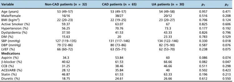
We enrolled a total of 95 CAD patients, including 65 with stable CAD (49–57 years old) and 30 with unstable CAD (49–58 years old), and 32 non-CAD patients (49–57 years old) who were matched for age, sex, hypertension and diabetes. Body mass index, diabetes mellitus, systolic blood pressure, diastolic blood pressure, LVEF, FBS, TC, TG, HDL-C, LDL-C, hs-CRP, AST, ALT, creatinine, smoking habits, hypertension and treatment records were collected. Clinical and laboratory characteristics of the participants are presented in Tables 1 and 2.
The expression levels of circulating miRNAs in CAD versus non-CAD patients
We analyzed the plasma expression levels of miR-149, miR-424 and miR-765 in stable CAD, unstable CAD and non-CAD patients. The results are presented in Figure 1. The
Table 1- Clinical data from the study subjects.
Variable Non-CAD patients (n = 32) CAD patients (n = 65) UA patients (n = 30) p1 p2
Age (years) 53 (49–57) 53 (49–57) 54 (49–58) 0.957 0.471
Male/Female 16/16 38/27 20/12 0.516 0.208
BMI (kg/m2) 22 (20–23) 22 (19–25) 23 (20–27) 0.706 0.124
Active Smoker (%) 59.37 63.07 67 0.825 0.606
Hypertension (%) 56.25 70.76 73.3 0.177 0.192
Dyslipidemia (%) 37.50 41.53 43.33 0.826 0.796
DM (%) 15.63 20 23.33 0.783 0.529
SBP (mmHg) 127 (119–135) 131 (117–146) 134 (122–146) 0.330 0.018
DBP (mmHg) 79 (72–86) 80 (73–86) 82 (75–90) 0.587 0.076
LVEF (%) 66 (60–72) 63 (55–71) 62 (53–70) 0.238 0.075
Medications
Aspirin (%) 34.3 53.84 60 0.086 0.074
b-blocker (%) 40.62 61.53 66.66 0.082 0.047
CCB (%) 31.25 38.46 46.66 0.511 0.298
ACEI/ARB (%) 28.12 35.84 40 0.502 0.423
Statin (%) 46.87 61.53 63.33 0.196 0.213
Diuretic (%) 18.75 24.61 26.66 0.612 0.550
The data are presented as the median and range. Abbreviations: UA, unstable angina; BMI, body mass index; DM, diabetes mellitus; SBP, systolic blood pressure; DBP, diastolic blood pressure; LVEF, left ventricular ejection fraction; CCB, calcium channel blocker; ACEI, angiotensin-converting enzyme inhibitor; and ARB, angiotensin receptor blocker.p1-value (non-CAD patients vs. CAD patients),p2-value (non-CAD patients vs. UA patients).
Table 2- Laboratory data from the study subjects.
Variable Non-CAD patients (n = 32) CAD patients (n = 65) UA patients (n = 30) p1 p2
FBS (mmol/L) 4.9 (4.3–5.4) 5.1 (4.2–6.1) 5.2 (3.9–6.7) 0.234 0.490
TG (mmol/L) 1.5 (0.5–2.5) 1.6 (0.6–2.7) 1.8 (0.8–2.7) 0.454 0.126
TC (mmol/L) 4.4 (2.9–5.9) 4.9 (3.3–6.5) 5.3 (3.7-7) 0.177 0.038
HDL-C (mmol/L) 0.9 (0.5–1.4) 0.9 (0.4–1.4) 0.9 (0.5–1.3) 0.595 0.746
LDL-C (mmol/L) 2.7 (1.5–3.8) 2.9 (1.4–4.4) 3.2 (2.1–4.3) 0.872 0.024
hs-CRP (mg/L) 1.4 (0.5–2.7) 19.8 (9.9–29.5) 21.3 (15–27.5) o0.001 o0.001
AST (U/L) 15.3 (6.9–24) 17.7 (8.3–27.1) 19.8 (12.6–27) 0.294 0.019
ALT (U/L) 17.1 (9.1–25) 19.1 (10.7–27.4) 21.5 (13.4–29.6) 0.244 0.042
Cr (umol/L) 71.5 (52.4–90) 74.8 (53.4–96.1) 79.8 (58.6–101.1) 0.602 0.049
levels of plasma miR-149 in stable CAD and unstable CAD patients were 4.49-fold (1.18± 0.84) and 5.09-fold (1.04± 0.65) lower, respectively, than those in non-CAD patients (5.30 ± 2.57) (po0.001). Circulating miR-424 levels were down-regulated 3.6-fold (1.18±0.60) in stable CAD patients and 5-fold (0.86±0.54) in unstable CAD patients compared with those in non-CAD patients (4.35±2.20) (po0.001). In contrast, the plasma concentrations of miR-765 were elevated 3.98-fold (6.09±2.27) in stable CAD patients and 5.33-fold (8.17±2.77) in unstable CAD patients compared with those in non-CAD patients (1.53 ± 0.99). The plasma levels of miR-149 and miR-424 were not significantly different between stable and unstable CAD patients, whereas the plasma levels of miR-765 were significantly different between stable and unstable CAD patients (po0.001).
Diagnostic role of plasma miRNAs in middle-aged CAD patients
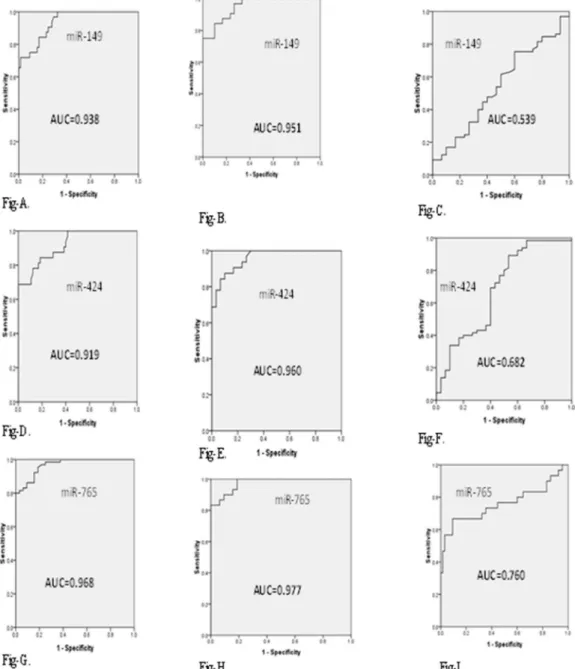
To examine the potential of miRNAs (miR-149, miR-424, and miR-765) as biomarkers for middle-aged CAD patients, ROC analysis was performed on all 127 subjects. The ROC curves of plasma miR-149 revealed a strong discrimination between non-CAD patients, stable CAD patients and unstable CAD patients, with AUCs of 0.938, 0.951 and 0.539, respectively (Figure 2A, 2B, and 2C). The ROC curves of plasma miR-424 revealed significant differences between non-CAD patients, CAD patients and unstable CAD patients, with AUCs of 0.919,
0.960 and 0.682, respectively (Figure 2D, 2E, and 2F). Furthermore, the ROC curves of plasma miR-765 also accurately differentiated stable and unstable CAD patients from non-CAD patients, with AUCs of 0.968, 0.977 and 0.760, respectively (Figure 2G, 2H, and 2I). These results indicated that plasma miR-149, miR-424 and miR-765 might be valuable biomarkers for middle-aged CAD patients. The AUCs, max-imum cut-off points, sensitivities, specificities, 95% CIs (con-fidence intervals) andp-values of the circulatory miRNAs (miR-149, miR-424, and miR-765) are briefly summarized in Table 3.
’ DISCUSSION
CAD is the most important cause of heart attacks and resulted in 3.6 million deaths in the year of 1990 in developing countries; this number will approximately increase to 7.8 million deaths by the year 2020 (29). The main cause of CAD is atherosclerosis, which is a hyperlipi-demia-induced chronic inflammatory disease of the arterial wall that occurs predominantly at sites of disturbed laminar blood flow due to endothelial dysfunction. Emerging evidence suggests that alterations in flow conditions regulate the expression of miRNAs in endothelial cells both in vitro and in vivo (30). The administration of miR-126-5p rescued EC proliferation at predilection sites and limited atherosclerosis by suppressing the Notch1 inhibitor delta-like 1 homolog (Dlk1), introducing a potential therapeutic approach (31). miR-33 has
Figure 1 -Plasma expression levels of miR-149, miR-424 and miR-765 in non-CAD patients, stable CAD patients and unstable CAD
patients.
Table 3- Receiver operator characteristic curve (ROC) analysis of miRNA ratios for predicting coronary artery disease.
miRNA AUC Cut-off point Sensitivity Specificity 95% CI p-value miR-149
Non-CAD vs. CAD 0.938 1.67 71.8% 95.3% 0.894 to 0.983 o0.0001
Non-CAD vs. UA 0.951 1.71 75% 96.6% 0.905 to 0.997 o0.0001
CAD vs. UA 0.539 1.15 75% 40% 0.415 to 0.663 0.540
miR-424
Non-CAD vs. CAD 0.919 1.61 68.7% 92.3% 0.863 to 0.975 o0.0001
Non-CAD vs. UA 0.960 1.77 84.3% 93.3% 0.920 to 1.000 o0.0001
CAD vs. UA 0.682 1.35 89.2% 46.6% 0.559 to 0.805 0.005
miR-765
Non-CAD vs. CAD 0.968 1.75 81.5% 93.7% 0.939 to 996 o0.0001
Non-CAD vs. UA 0.977 1.80 83.3% 96.8% 0.949 to 1.005 o0.0001
CAD vs. UA 0.760 1.57 66.5% 90.7% 0.635 to 0.885 o0.0001
been discovered as a key regulator of the initiation and progression of atherosclerosis by controlling lipid metabolism, the inflammatory response and cell cycle progression and proliferation (32). miR-1 prevents high-cholesterol-induced endothelial dysfunction through myosin light chain kinase (MLCK) expression and extracellular signal-regulated kinase (ERK) phosphorylation (33). Monocytes migrate from the circulation into the intima by binding to activated endothelial cells and later differentiate into macrophages. miR-424 promotes monocyte/macrophage differentiation and is intimately involved in the initiation and progression of atherosclerotic lesions and the development of associated complications. Ezetimibe (a lipid-lowering agent) decreased the expression of miR-424 by 75% and decreased atherosclerotic lesions (34).
In addition, several miRNAs are directly and indirectly involved in the pathophysiological mechanisms of athero-sclerosis, such as 19a, 21, 23b, 92a, miR-143, miR-145, miR-155, miR-663 and miR-133a (30,35). Although enormous progress has been made in the diagnosis, treatment and prognosis of CAD, there remains a clinical need for a novel diagnostic biomarker and new therapeutic interventions to decrease the incidence of CAD. Biomarkers play an essential role in the diagnosis of CAD. However, many of these biomarkers have shortcomings, such as reduced sensitivity, low specificity or unsuitability for early diagnosis. However, several recent studies have suggested that circulating miRNAs are excellent promising biomarkers for the early diagnosis of AMI (18,23–25,36).
Figure 2 -Diagnostic accuracy of circulating miR-149, miR-424 and miR-765 were analyzed by ROC curve.A)ROC curve of miR-149,
non-CAD patients vs. non-CAD. B) ROC curve of miR-149, non-CAD patients vs. unstable CAD patients. C) ROC curve of miR-149,
CAD vs. unstable CAD patients.D)ROC curve of miR-424, non-CAD patients vs. CAD.E)ROC curve of miR-424, non-CAD patients vs.
unstable CAD patients.F)ROC curve of miR-424, CAD vs. unstable CAD patients.G)ROC curve of miR-765, non-CAD patients vs. CAD.H)
Circulating levels of miR-133a, miR-208a, miR-1, miR-122, miR-133b, miR-337-5p, miR-433, and miR-485-3p were significantly up-regulated, whereas 126, 17, miR-92a, miR-145, miR-155, and miR-199a levels were markedly down-regulated in CAD (14,37).
Circulating miRNA levels in middle-aged CAD patients have not yet been investigated. We specifically focused on middle-aged people for the following reasons: one, they are the most active contributors to the development of society; and two, although the invasive CAG procedure is the standard method for diagnosing CAD, it has many complications. The present work demonstrated that circulating miRNAs might be clinically feasible biomarkers for diagnosing middle-aged CAD patients. We discovered that plasma miR-149 and miR-424 levels were significantly down-regulated, whereas plasma miR-765 levels were up-regulated in stable and unstable CAD patients compared with non-CAD patients. In addition, we measured the diagnostic value of plasma miR-149, miR-424 and miR-765 levels using ROC curves. The AUC values for miR-149, miR-424 and miR-765 were significantly higher in stable CAD patients (0.938, 0.919 and 0.968, respectively) and unstable CAD patients (0.951, 0.960 and 0.977, respectively) compared with non-CAD patients. These results strongly indicated that plasma miR-149, miR-424 and miR-765 might be valuable novel non-invasive biomarkers for the early diagnosis of middle-aged CAD patients.
Recent studies have revealed that 5,10-methylenetetrahy-drofolate reductase (MTHFR) plays a critical role in homo-cysteine metabolism. The abnormal function of MTHFR increases plasma homocysteine levels to enhance endothelial dysfunction and atherosclerosis. The human MTHFR rs4846049 (T-allele) polymorphism was significantly asso-ciated with an increased risk of CAD and dyslipidemia via modification of the binding of miR-149 in a Chinese Han population (38). Furthermore, themiR-149T4C polymorph-ism was significantly associated with ischemic stroke and silent brain infarction (SBI) risk in a Korean population (39). TNF-aplays an essential role in the initiation and progression of endothelial dysfunction. The transfection of miR-149 mimics counteracted the TNF-a-induced expression of metal-loproteinase-9 (MMP-9), inducible nitric oxide synthase (iNOS), and interleukin-6 (IL-6) and regulated TNF-a-induced endothelial dysfunction (40). miR-149 expression was sig-nificantly down-regulated in infarcted mouse hearts three days after MI and in cardiac tissue from the border zone of the infarcted region in patients receiving a cardiac transplant (41). In our study, circulating miR-424 levels were significantly decreased in CAD patients, which is consistent with studies from Fichtlscherer et al (14). Moreover, plasma miR-424 levels were significantly down-regulated in acute ischemic stroke patients as well as in mouse plasma and brain tissue 4 hours after ischemia. In addition, overexpressing miR-424 mimics decreased the ischemic brain injury by suppressing microglia activation via the translational depression of key activators of the G1/S transition, including CDC25A, CCND1, and CDK6 (42). miR-424 levels were also signifi-cantly decreased in pulmonary arterial hypertension (PAH) due to the dysregulation of the apelin (APLN) and fibroblast growth factor 2 (FGF2) signaling pathways (43). The specific target of miR-424 in cardiac cells is currently unknown and awaits elucidation in future studies.
Redell JB et al. (44) reported that plasma miR-765 levels were markedly increased in traumatic brain injury (TBI) patients compared with healthy volunteers. ROC curve
analysis indicated that miR-765 was a good biomarker, especially for severe TBI. However, the exact mechanisms for the altered expression of miR-765 in CAD remain unknown. Using target scan (www.targetscan.org), hsa-miR-765 was predicted to potentially be involved in CAD by regulating several important target genes, such as HIF3A (hypoxia inducible factor-3,asubunit), NADPH oxidase, low density lipoprotein receptor-related protein 4 (LRP4), and low density lipoprotein receptor-related protein 6 (LRP6).
To ensure that the study populations were comparable, subjects with similar clinical features, such as age, gender, TC, TG, HDL-C, LDL-C, systolic blood pressure, diastolic blood pressure, AST, ALT, creatinine, LVEF, diabetes mellitus, smoking, hypertension and medication history, were carefully considered. Statistical analyses revealed that these factors did not affect circulating miR-149, miR-424 and miR-765 levels. Importantly, three plasma miRNAs (miR-149, miR-424 and miR-765) displayed a direct relationship in stroke patients, but we excluded stroke patients in our study to minimize possible bias. To decrease potential errors resulting from qRT-PCR assays, we evaluated three potential internal controls (U6, miR-cel-39, and miR-156a) in a pre-analysis (n = 20). We ultimately chose miR-156a for standard normalization because it was more stable and reliable (36). We analyzed each sample in triplicate, and miRNAs with Ct valuesZ40 were not included in our study, which assured that our results were constant and reproducible. To the best of our knowledge, the present study is the first to demonstrate that plasma miR-149, miR-424 and miR-765 might be non-invasive novel biomarkers for middle-aged CAD patients. However, there are several limitations that need to be acknowledged and addressed regarding the present study. First, this work was performed in a single center with a small cohort. Therefore, a multi-center study with a larger cohort is necessary to verify the findings of this present study. Second, the specificity of the changes in the three measured miRNAs in middle-aged CAD patients remains unclear because we did not measure these miRNAs in other age groups of CAD patients or in other clinical settings. Third, we did not include healthy subjects as normal controls.
In conclusion, our findings suggest that circulating miR-149, miR-424 and miR-765 might be non-invasive biomarkers for the diagnosis of CAD in middle-aged patients. However, the sensitivity and specificity of these miRNAs need to be further examined in a larger clinical cohort.
’ ACKNOWLEDGMENTS
This work was supported by the National Nature Science Foundation of China (No. 81370320 to Tian-Lun Yang and No. 81373409 to Jun Peng), the Hunan Provincial Natural Science Foundation of China (No. 13JJ2008 to Peng J.) and the Doctoral Fund of Ministry of Education of China (No. 20120162110056 to Peng J.)
’ AUTHOR CONTRIBUTIONS
Sayed AS and Xia K designed the research, performed the experiments and wrote the manuscript. Deng X, Deng H, Yangda F, Li F, Haoyang Z, Li T, and Salma U collected patient samples and assembled the data. Yang T and Peng J supervised the data analysis and revised the manuscript. The work presented in this paper was performed in collaboration with all the authors.
’ REFERENCES
disease in sudden cardiac death from coronary artery disease. Cardiovasc Res. 2014;103(Suppl 1):S10.
2. Go AS, Mozaffarian D, Roger VL, Benjamin EJ, Berry JD, Blaha MJ, et al. Heart disease and stroke statistics--2014 update: a report from the American Heart Association. Circulation. 2014;129(3):e28-292, http://dx. doi.org/10.1161/01.cir.0000441139.02102.80.
3. Gupta SK, Bang C, Thum T. Circulating microRNAs as biomarkers and potential paracrine mediators of cardiovascular disease. Circ Cardiovasc Genet. 2010;3(5):484-8, http://dx.doi.org/10.1161/CIRCGENETICS.110.958363. 4. Diehl P, Fricke A, Sander L, Stamm J, Bassler N, Htun N, et al.
Micro-particles: major transport vehicles for distinct microRNAs in circulation. Cardiovasc Res. 2012;93(4):633-44, http://dx.doi.org/10.1093/cvr/cvs007. 5. Peng Y, Song L, Zhao M, Harmelink C, Debenedittis P, Cui X, et al. Critical roles of miRNA-mediated regulation of TGFbsignalling during mouse cardiogenesis. Cardiovasc Res. 2014;103(2):258-67, http://dx.doi.org/ 10.1093/cvr/cvu126.
6. Small EM, Olson EN. Pervasive roles of microRNAs in cardiovascular biol-ogy. Nature. 2011;469(7330):336-42, http://dx.doi.org/10.1038/nature09783. 7. Thum T, Catalucci D, Bauersachs J. MicroRNAs: novel regulators in car-diac development and disease. Cardiovasc Res. 2008;79(4):562-70, http:// dx.doi.org/10.1093/cvr/cvn137.
8. Dangwal S, Bang C, Thum T. Novel techniques and targets in cardio-vascular microRNA research. Cardiovasc Res. 2012;93(4):545-54, http:// dx.doi.org/10.1093/cvr/cvr297.
9. Mar-Aguilar F, Rodríguez-Padilla C, Reséndez-Pérez D. Use of serum-circulating miRNA profiling for the identification of breast cancer bio-markers. Methods Mol Biol. 2014;1165:71-80, http://dx.doi.org/10.1007/ 978-1-4939-0856-1.
10. Gidlöf O, Andersson P, van der Pals J, Götberg M, Erlinge D. Cardiospecific microRNA plasma levels correlate with troponin and cardiac function in patients with ST elevation myocardial infarction, are selectively dependent on renal elimination, and can be detected in urine samples. Cardiology. 2011;118(4):217-26, http://dx.doi.org/10.1159/000328869.
11. Arroyo JD, Chevillet JR, Kroh EM, Ruf IK, Pritchard CC, Gibson DF, et al. Argonaute2 complexes carry a population of circulating microRNAs independent of vesicles in human plasma. Proc Natl Acad Sci U S A. 2011;108(12):5003-8, http://dx.doi.org/10.1073/pnas.1019055108. 12. Sayed AS, Xia K, Salma U, Yang T, Peng J. Diagnosis, Prognosis and
Therapeutic Role of Circulating miRNAs in Cardiovascular Diseases. Heart Lung Circ. 2014;23(6):503-10, http://dx.doi.org/10.1016/j.hlc.2014.01.001. 13. Small EM, Frost RJ, Olson EN. MicroRNAs add a new dimension to
cardiovascular disease. Circulation. 2010;121(8):1022-32, http://dx.doi. org/10.1161/CIRCULATIONAHA.109.889048.
14. Fichtlscherer S, De Rosa S, Fox H, Schwietz T, Fischer A, Liebetrau C, et al. Circulating microRNAs in patients with coronary artery disease. Circ Res. 2010;107(5):677-84, http://dx.doi.org/10.1161/CIRCRESAHA.109.215566. 15. Zeller T, Keller T, Ojeda F, Reichlin T, Twerenbold R, Tzikas S, et al.
Assessment of microRNAs in patients with unstable angina pectoris. Eur Heart J. 2014;35(31):2106-14, http://dx.doi.org/10.1093/eurheartj/ehu151. 16. Widera C, Gupta SK, Lorenzen JM, Bang C, Bauersachs J, Bethmann K, et al. Diagnostic and prognostic impact of six circulating microRNAs in acute coronary syndrome. J Mol Cell Cardiol. 2011;51(5):872-5, http://dx. doi.org/10.1016/j.yjmcc.2011.07.011.
17. Wang GK, Zhu JQ, Zhang JT, Li Q, Li Y, He J, et al. Circulating microRNA: a novel potential biomarker for early diagnosis of acute myocardial infarction in humans. Eur Heart J. 2010;31(6):659-66, http://dx.doi.org/ 10.1093/eurheartj/ehq013.
18. Goren Y, Kushnir M, Zafrir B, Tabak S, Lewis BS, Amir O. Serum levels of microRNAs in patients with heart failure. Eur J Heart Fail. 2012;14(2):147-54, http://dx.doi.org/10.1093/eurjhf/hfr155.
19. McManus DD, Lin H, Tanriverdi K, Quercio M, Yin X, Larson MG, et al. Relations between circulating microRNAs and atrial fibrillation: data from the Framingham Offspring Study. Heart Rhythm. 2014;11(4):663-9, http://dx.doi.org/10.1016/j.hrthm.2014.01.018.
20. Tsai PC, Liao YC, Wang YS, Lin HF, Lin RT, Juo SH. Serum microRNA-21 and microRNA-221 as Potential Biomarkers for Cerebrovascular Disease J Vasc Res. 2013;50(4):346-54, http://dx.doi.org/10.1159/000351767. 21. Waters PS, Dwyer RM, Brougham C, Glynn CL, Wall D, Hyland P, et al.
Impact of tumour epithelial subtype on circulating microRNAs in breast cancer patients. PLoS One. 2014;9(3):e90605, http://dx.doi.org/10.1371/ journal.pone.0090605.
22. Corsten MF, Dennert R, Jochems S, Kuznetsova T, Devaux Y, Hofstra L, et al. Circulating microRNA-208b and microRNA-499 reflectmyocardial damage in cardiovascular disease. Circ Cardiovasc Genet. 2010;3(6):499-506, http:// dx.doi.org/10.1161/CIRCGENETICS.110.957415.
23. Long G, Wang F, Duan Q, Chen F, Yang S, Gong W, et al. Human circu-lating microRNA-1 and microRNA-126 as potential novel indicators for acute myocardial infarction. Int J Biol Sci. 2012;8(6):811-8, http://dx.doi. org/10.7150/ijbs.4439.
24. Peng L, Chun-guang Q, Bei-fang L, Xue-zhi D, Zi-hao W, Yun-fu L, et al. Clinical impact of circulating miR-133, miR-1291 and miR-663b in plasma of patients with acute myocardial infarction. Diagn Pathol. 2014;1(9):89, http://dx.doi.org/10.1186/1746-1596-9-89.
25. Tijsen AJ, Creemers EE, Moerland PD, de Windt LJ, van der Wal AC, Kok WE, et al. MiR423-5p as a circulating biomarker for heart failure. Circ Res. 2010;106(6):1035-9, http://dx.doi.org/10.1161/CIRCRESAHA.110.218297. 26. Ellis KL, Cameron VA, Troughton RW, Frampton CM, Ellmers LJ, Richards
AM. Circulating microRNAs as candidate markers to distinguish heart fail-ure in breathless patients. Eur J Heart Fail. 2013;15(10):1138-47, http://dx. doi.org/10.1093/eurjhf/hft078.
27. Fihn SD, Gardin JM, Abrams J, Berra K, Blankenship JC, Dallas AP, et al. 2012 ACCF/AHA/ACP/AATS/PCNA/SCAI/STS Guideline for the diagnosis and management of patients with stable ischemic heart disease: a report of the American College of Cardiology Foundation/ American Heart Association Task Force on Practice Guidelines, and the American College of Physicians, American Association for Thoracic Surgery, Preventive Cardi-ovascular Nurses Association, Society for CardiCardi-ovascular Angiography and Interventions, and Society of Thoracic Surgeons. J Am Coll Cardiol. 2012;60 (24):e44-??e164, http://dx.doi.org/10.1016/j.jacc.2012.07.013.
28. Braunwald E, Antman EM, Beasley JW, Califf RM, Cheitlin MD, Hochman JS, et al. ACC/AHA guideline update for the management of patients with unstable angina and non-ST-segment elevation myocardial infarction--2002: summary article: a report of the American College of Cardiology/ American Heart Association Task Force on Practice Guidelines (Committee on the Management of Patients With Unstable Angina). Circulation. 2002;106 (14):1893-??1900, http://dx.doi.org/10.1161/01.CIR.0000037106.76139.53. 29. Okrainec K, Banerjee DK, Eisenberg MJ. Coronary artery disease in the
developing world. Am Heart J. 2004;148(1):7-15, http://dx.doi.org/ 10.1016/j.ahj.2003.11.027.
30. Kumar S, Kim CW, Simmons RD, Jo H. Role of Flow-Sensitive microRNAs in Endothelial Dysfunction and Atherosclerosis: Mechanosensitive Athero-miRs. Arterioscler Thromb Vasc Biol. 2014;34(10):2206-16, http:// dx.doi.org/10.1161/ATVBAHA.114.303425.
31. Schober A, Nazari-Jahantigh M, Wei Y, Bidzhekov K, Gremse F, Grommes J, et al. MicroRNA-126-5p promotes endothelial proliferation and limits atherosclerosis by suppressing Dlk1. Nat Med. 2014;20(4):368-76, http:// dx.doi.org/10.1038/nm.3487.
32. Chen WJ, Zhang M, Zhao GJ, Fu Y, Zhang DW, Zhu HB, et al. MicroRNA-33 in atherosclerosis etiology and pathophysiology. Atherosclerosis. 2013;227(2):201-8, http://dx.doi.org/10.1016/j.atherosclerosis.2012.11.025. 33. Wang H, Zhu HQ, Wang F, Zhou Q, Gui SY, Wang Y. MicroRNA-1
pre-vents high-fat diet-induced endothelial permeability in apoE knock-out mice. Mol Cell Biochem. 2013;378(1–2):153-9.
34. Muñoz-Pacheco P, Ortega-Hernández A, Miana M, Cachofeiro V, Fernández-Cruz A, Gómez-Garre D. Ezetimibe inhibits PMA-induced monocyte/macrophage differentiation by altering microRNA expression: a novel anti-atherosclerotic mechanism. Pharmacol Res. 2012;66(6):536-43.
35. Gao S, Wassler M, Zhang L, Li Y, Wang J, Zhang Y, et al. MicroRNA-133a regulates insulin-like growth factor-1 receptor expression and vascular smooth muscle cell proliferation in murine atherosclerosis. Atherosclerosis. 2014;232(1):171-9, http://dx.doi.org/10.1016/j.atherosclerosis.2013.11.029. 36. Hsu A, Chen SJ, Chang YS, Chen HC, Chu PH. Systemic approach to
identify serum microRNAs as potential biomarkers for acute myocardial infarction. Biomed Res Int. 2014;418628.
37. D’Alessandra Y, Carena MC, Spazzafumo L, Martinelli F, Bassetti B, Devanna P, et al. Diagnostic potential of plasmatic MicroRNA signatures in stable and unstable angina. PLoS One. 2013;8(11):e80345, http://dx. doi.org/10.1371/journal.pone.0080345.
38. Wu C, Gong Y, Sun A, Zhang Y, Zhang C, Zhang W, et al. The human MTHFR rs4846049 polymorphism increases coronary heart disease risk through modifying miRNA binding. Nutr Metab Cardiovasc Dis. 2013; 23(7):693-8, http://dx.doi.org/10.1016/j.numecd.2012.02.009.
39. Jeon YJ, Kim OJ, Kim SY, Oh SH, Oh D, Kim OJ, et al. Association of the miR-146a, miR-149, miR-196a2, and miR-499 polymorphisms with ischemic stroke and silent brain infarction risk. Arterioscler Thromb Vasc Biol. 2013;33(2):420-30, http://dx.doi.org/10.1161/ATVBAHA.112.300251. 40. Palmieri D, Capponi S, Geroldi A, Mura M, Mandich P, Palombo D.
TNFa induces the expression of genes associated with endothelial dysfunction through p38MAPK-mediated down-regulation of miR-149. Biochem Biophys Res Commun. 2014;443(1):246-51, http://dx.doi.org/ 10.1016/j.bbrc.2013.11.092.
41. Van Rooij E, Sutherland LB, Thatcher JE, DiMaio JM, Naseem RH, Mar-shall WS, et al. Dysregulation of microRNAs after myocardial infarction reveals a role of miR-29 in cardiac fibrosis. Proc Natl Acad Sci U S A. 2008;105(35):13027-32, http://dx.doi.org/10.1073/pnas.0805038105. 42. Zhao H, Wang J, Gao L, Wang R, Liu X, Gao Z, et al. MiRNA-424 protects
against permanent focal cerebral ischemia injury in mice involving sup-pressing microglia activation. Stroke. 2013;44(6):1706-13, http://dx.doi. org/10.1161/STROKEAHA.111.000504.
43. Kim J, Kang Y, Kojima Y, Lighthouse JK, Hu X, Aldred MA. An endo-thelial apelin-FGF link mediated by miR-424 and miR-503 is disrupted in pulmonary arterial hypertension. Nat Med. 2013;19(1):74-82.