Original Article
Artigo Original
Camila Rosa de Oliveira1 Karina Carlesso Pagliarin2 Luara de Freitas Calvette3 Gigiane Gindri3 Irani Iracema de Lima Argimon3 Rochele Paz Fonseca3
Keywords
Stroke Right-hemisphere Depression
Cognition Neuropsychological Tests
Descritores
Acidente Vascular Cerebral Hemisfério Direito Depressão Cognição Testes Neuropsicológicos
Correspondence address: Camila Rosa de Oliveira
Rua Luís de Camões, 255/309, Porto Alegre (RS), Brasil, CEP: 90620-150. E-mail: [email protected]
Received: 01/19/2015
Accepted: 03/31/2015
Study carried out at the Pontifícia Universidade Católica do Rio Grande do Sul – PUC/RS – Porto Alegre (RS), Brazil.
(1) Graduate Program in Biomedical Gerontology, Pontifícia Universidade Católica do Rio Grande do Sul – PUC/RS – Porto Alegre (RS), Brazil.
(2) Graduate Program in Human Communication Disorders, Universidade Federal de Santa Maria – UFSM – Santa Maria (RS), Brazil.
(3) Graduate Program in Psicology, Pontifícia Universidade Católica do Rio Grande do Sul – PUC/RS – Porto Alegre (RS), Brazil.
Conlict of interests: nothing to declare.
Depressive signs and cognitive performance in
patients with a right hemisphere stroke
Sinais depressivos e desempenho cognitivo em
pacientes com lesão de hemisfério direito
ABSTRACT
Purpose: This study investigated the inluence of suggestive signs of depression (SSD) in right-hemisphere brain-damaged (RHD) patients following a stroke on their cognitive performance measured by a brief neuropsychological assessment battery. Methods: Forty-two adults with RHD after a single episode of stroke and 84 matched controls participated in this study. They were assessed by means of the Geriatric Depression Scale and by Brief Neuropsychological Assessment Battery NEUPSILIN. Results: Almost half of the patients showed SSD. The RHD group with SSD (RHD+) showed poorer performance in at least one task among all evaluated cognitive domains (concentrated attention, visual perception, working memory, episodic verbal memory and semantic memory, auditory and written language, constructional praxia and verbal luency). Conclusion: The association of depression and RHD seems to enhance the occurrence and the severity of cognitive déicits. A brief neuropsychological assessment can be useful to identify cognitive impairment caused by this neuropsychiatric disorder.
RESUMO
INTRODUCTION
Depression is considered to be one of the most common neuropsychiatric consequences following a stroke, reaching approximately a third of the patients. Besides generating a negative impact on functional rehabilitation, it represents one of the most prominent causes to increase mortality in this population(1). Depression has been detected in between 20
and 60% of stroke patients, this rate ranging in accordance with the evaluation criteria established (by means of scales or semi-structured interviews, for instance), as well as to the population investigated (such as ischemic and/or hemor-rhagic, chronic and/or acute stroke)(2) and lesion location.
Patients with lesions involving left-hemisphere prefrontal or basal ganglia structures had a higher frequency of depressive disorder (75%) than other left-hemisphere lesions (8%) or those with right-hemisphere lesions (29%)(3). However, studies
investigating the relationship between injury localization and depression are contradictory(4,5). Therefore, further
investiga-tions are necessary, specially considering the cognitive issues in right-hemisphere.
Despite the high incidence of depressive symptoms associ-ated with the stroke framework, it seems that literature does not have enough studies with converging evidences concerning the frequency and the relationship between this psychiatric disease and stroke aspects, such as the lesion side and associated cogni-tive impairment(6). More speciically, when comparing what has
been found about cognitive impairment following a stroke in isolation(7), as well as cognitive changes following depression
cases(8), little is known about the relationship between
depres-sion, unilateral right-hemisphere stroke and cognitive deicits derived by the association of these aspects. Thus, there is an important lack of knowledge on the comprehension of cogni-tive impairment associated with stroke and depression versus hemispheric specializations.
Although the left-hemisphere has been related to higher incidence of depression, the right-hemisphere seems to be more related to emotions in general, being pointed in the literature as the main neurobiological correlate to facial emotions and prosodic processing(9). Therefore, vascular
le-sions in this hemisphere may impair the patient’s mental and emotional state, as well as negatively affecting motivation, comprehension and prosodic emotional, metaphoric and humor expression(10).
Some few existing studies(6,11-13) propose to discuss the
relationship between cognitive dysfunctions and post-stroke depression, but not followed by the association’s investiga-tion of the neurocognitive deicits of this comorbidity with the affected hemisphere. Among those studies, the research conducted by Alexopoulos(11), for instance, emphasizes that
lesions in the frontolimbic and frontoestriatic networks are associated withwith executive functions deicits; however, the contributions of each hemisphere are not considered in this process. According to Sneed and Culang-Reinlieb(12),
post-stroke depression is also associated withwith executive dysfunction, although independently of lesion type. Brodaty et al.(13) identiied that apathy following stroke is linked to
low performance in tasks measuring concentrated attention, working memory and processing speed. Marazziti et al.(6),
on their hand, observed the incidence of attention, working memory and executive functions impairment, including cog-nitive inhibition, planning and problem solution, suggesting a strong relationship between low performance in cognitive tasks and positive signs of depression associated with stroke, not necessarily unilateral, though.
As for the characterization of cognitive processing in this neuropsychiatric framework of post-stroke depression, besides the lack of studies about several cognitive functions, some limitations may be pointed out regarding assessment methods. The researches had, in general, administrated screenings such as the Mini Mental State Exam – MMSE(14),
some tasks of expanded batteries(15) or entire expanded
bat-teries(13). Therefore, seems to be an even larger gap in the
literature on the relationship between stroke, suggestive signs of depression (SSD) and hemispheric specialization with the processing of several cognitive functions measured by brief neuropsychological batteries.
Taking into account the importance of brief batteries to the neuropsychological examination in contexts where initial diagnosis is necessary in a shorter time(16), associated with the
scarceness of empirical evidence relating unilateral stroke, post-stroke depression and cognitive impairment following this comorbidity, the present article aims to investigate the incidence of SSD in a sample of right-hemisphere brain-damaged (RHD) adults and verify the inluence of these signs and symptoms in the processing of several cognitive functions — temporo-spacial orientation, attention, perception, memory, language, arithmetical skills, praxias and executive functions.
METHOD
Participants
In this study a total sample of 126 adults participated, divided in two groups: clinical (with two subgroups) and a control group.
RHD clinical group
RHD patients with SSD (RHD+), between 5 and 15 points in 15 Item Geriatric Depression Scale (GDS-15), and with no SSD (RHD-), between 0 and 4 points.
Control Group
This group was composed by 84 neurologically preserved participants, paired with brain-damaged patients according to age criteria, years of formal education, reading and writing habits and gender. The control group formation should observe the same inclusion criteria of brain-damaged patients, including absence of the diagnosis of a cerebrovascular disease, no signs of cognitive impairment measurement by the MMSE (adapted by Chaves and Izquierdo(17)) and non-occurrence of SSD. Procedures and instruments
It is important to state that all ethical procedures have been respected, with the guarantee of voluntary participation in the study, and under the approval of a Research Committee on Ethics of a higher education institution (protocol number 10/05134). The evaluations were developed in individual sessions with approximated duration of one hour and a half. Participants answered to sociodemographic characterization and general health conditions questionnaire.
In order to verify the presence of SSD, participants were administered GDS-15 (adapted by Almeida and Almeida(18),
in order to obtain same measure in adult and elderly. The validity of the GDS-15 for other age groups, like younger and adults, has already been reported in the literature(19). The
cognitive abilities were assessed by the Instrument of Brief Neuropsychological Assessment (NEUPSLIN)(20), which is an
instrument with a brief administration time aiming at verify-ing preserved and impaired abilities in components of eight different cognitive functions, giving support to a neuropsycho-logical diagnosis. It includes the following tasks, all of them administered in this study: Concentrated attention (inverted counting and digit repetition); Visual perception (veriication of similarities and differences of lines, hemineglect, faces perception and recognition); Working memory (ascendant ordering of digits and auditory span of words in sentences); Episodic-semantic verbal memory (delayed and immediate re-call and word list recognition); Long-term semantic memory; Short-term visual memory (igures); Prospective memory;
Arithmetic abilities (simple addition, subtraction, multipli-cation and division calculation); Oral language (objects and igures’ naming, words and non-words’ repetition, automatic language, inference comprehension and processing); Written language (reading aloud of words and non-words, written comprehension, spontaneous writing, copying, words and non-words dictation); Apraxia (ideomotor, constructive and relexive), and Executive functions (simple problems’ resolu-tion and orthographic verbal luency – letter F).
Data analysis
Initially an analysis of frequency was performed to verify the incidence of SSD and its intensity in RHD patients as well as to subdivide the RHD+ and RHD- clinical groups. The dependent variables presented a normal distribution in the Kolmogorov-Smirnov Test (p>0.05). In order to com-pare the performance of RHD+, RHD- and the Clinical Group, a One-way ANOVA analysis was used, with a Bonferroni post hoc test. The incidence of hemineglect between RHD+ and RHD- groups was compared through a χ2 analysis.
RESULTS
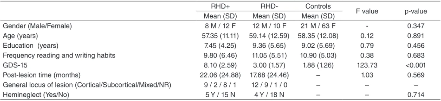
The groups’ sociodemographic and clinical characteristics are depicted in Table 1. Regarding the characterization vari-ables, the groups only present signiicant differences in the GDS-15 score (RHD+ > RHD- > Controls, p≤0.001, indicating that pairing was correctly done. Moreover, a predominance of cortical lesion was observed in the sample.
The incidence of SSD in RHD patients was of 47.62%. As for the symptoms intensity in the RHD+ group through the GDS-15 scores, 40% of the cases presented a mild SSD level, 35% moderate and 25% severe cases. Means and standard deviations of NEUPSILIN tasks are described in Table 2.
The post hoc analysis identiied a signiicant lower performance of the RHD+ group as compared to the Control Group’s performance in at least one of the subtests of all cognitive domains assessed. Regarding the tasks of Concentrated attention, Visual perception, Working memory, Auditory and Written language, in which differences were observed between RHD+ and RHD- groups, the RHD+ also presented lower scores.
RHD+ RHD- Controls
F value p-value Mean (SD) Mean (SD) Mean (SD)
Gender (Male/Female) 8 M / 12 F 12 M / 10 F 21 M / 63 F - 0.347 Age (years) 57.35 (11.11) 59.14 (12.59) 58.35 (12.08) 0.12 0.891 Education (years) 7.45 (4.25) 9.36 (5.65) 9.02 (5.69) 0.79 0.456 Frequency reading and writing habits 9.80 (6.46) 11.05 (5.51) 10.90 (5.03) 0.38 0.683
GDS-15 8.10 (2.59) 3.00 (1.57) 1.88 (1.26) 123.73 <0.001
Post-lesion time (months) 22.06 (24.88) 17.68 (24.46) – 1.03 0.569 General locus of lesion (Cortical/Subcortical/Mixed/NR) 9 / 2 / 8 / 1 12 / 9 / 1 / 0 – – –
Hemineglect (Yes/No) 5 Y / 15 N 4 Y / 18 N – – 0.714
Table 1. Sociodemographic and Clinical Characteristics
df (degrees of freedom) = 2
DISCUSSION
The SSD incidence in RHD patients in this study sample (47.62%) was compatible with indexes levels of depressive symptoms presented in the literature, suggesting that it is deinitively a framework commonly found following a stroke. According to the study developed by Kouwenhoven et al.(21),
for instance, the index of depressive signs varied from 5 to 54% of the sample. Haq et al.(22) observed depression signs in
28% of their patients. In another study, a varying level of 23
to 60% of patients’ post-stroke depression was observed(2).
This variability may be related to the variety of instruments utilized to assess depressive symptoms as well as the inclu-sion of patients with language dificulties, which limits the comprehension of depression scales and inventories (in reha-bilitation, ambulatories or hospitals). Although some studies suggested increased frequency of depression related to the left-hemisphere, mainly associated with left anterior lesions, little focus has been given to the cognitive consequences of SSD in patients with right-hemisphere lesion(5,8). Thus, there *p≤0.05; **p≤0.01; ***p≤0.001; df (degrees of freedom) = 2
Caption: SD = standard deviation; ADO = Ascendant Digit Ordering; ASWS = Auditory Span of Words in Sentences
Table 2. Groups’ Performances in NEUPSILIN Tasks
Variables RHD+ RHD- Controls F value p-value Post hoc
Mean (SD) Mean (SD) Mean (SD) Attention
Inverted counting 16.7 (6.39) 18.86 (4.25) 17.87 (5.22) 0.88 0.416
Inverted counting (time) 42.68 (26.92) 28.53 (15.06) 26.11 (14.43) 7.61 0.001 RHD+ > RHD-* RHD+ > Controls*** Digit repetition 2.6 (1.57) 2.45 (1.82) 3.17 (1.93) 1.71 0.185
Perception
Line verification 4.65 (1.53) 5.24 (1.04) 5.26 (1.04) 2.42 0.094
Visual neglect 0.85 (0.37) 0.86 (0.35) 1 (0) 6.86 0.001 RHD+ < RHD-** RHD- < Controls* Face perception 2.1 (0.64) 2.05 (0.72) 2.39 (0.64) 3.41 0.036
Face recognition 1.55 (0.51) 1.82 (0.39) 1.88 (0.33) 6.39 0.002 RHD+ < Controls** Memory
Working memory (ADO) 3.95 (2.7) 6.27 (2.47) 6.38 (2.37) 8.21 0 RHD+ < RHD-** RHD+ < Controls*** Working memory (ASWS) 9.55 (5.65) 11.91 (4.94) 13.5 (6.13) 3.84 0.024 RHBD+ < Controls* Verbal memory (immediate recall) 3.65 (1.5) 3.91 (1.27) 4.64 (1.37) 5.65 0.005 RHD+ < Controls** Verbal memory (delayed speech) 0.8 (1.32) 1.14 (1.39) 1.99 (1.89) 4.98 0.008 RHD+ < Controls* Verbal memory (recognition) 10.35 (1.5) 11.45 (1.57) 12.45 (2.39) 8.44 0 RHD+ < Controls*** Long-termsemanticmemory 4.1 (0.91) 4.55 (0.91) 4.67 (0.63) 4.84 0.009 RHD+ < Controls** Short-term visual memory 2.35 (0.93) 2.55 (0.74) 2.64 (0.61) 1.47 0.234
Prospective memory 1.25 (0.72) 1.05 (0.84) 1.39 (0.78) 1.8 0.17
Arithmetic abilities 4.8 (2.93) 6 (2.54) 6.79 (2.04) 6.37 0.002 RHD+ < Controls** Language
Oral naming 3.9 (0.45) 4 (0) 3.99 (0.11) 1.8 0.169 Repetition 9.55 (0.69) 9.59 (0.67) 9.7 (0.72) 0.5 0.611
Automatic language 1.8 (0.41) 2 (0) 1.94 (0.24) 3.54 0.032 RHD+ < RHD-* Oral comprehension 2.7 (0.57) 2.73 (0.55) 2.8 (0.51) 0.36 0.695
Inference processing 2.3 (0.73) 2.5 (0.6) 2.36 (0.77) 0.44 0.643
Reading aloud 9.75 (3.26) 11.55 (0.67) 11.46 (0.91) 11.1 0 RHD+ < RHD-*** RHD+ < Controls*** Written comprehension 2.7 (0.57) 2.5 (0.67) 2.82 (0.42) 3.79 0.025 RHD- < Controls* Spontaneous writing 1.05 (0.76) 1.41 (0.73) 1.56 (0.72) 4.02 0.02 RHD+ < Controls* Copied writing 1.5 (0.69) 1.68 (0.57) 1.82 (0.42) 3.64 0.029 RHD+ < Controls* Dictated writing 8.65 (3.08) 10.23 (1.34) 10.04 (2.27) 3.32 0.039 RHD+ < Controls* Praxias
Ideomotor 2.95 (0.22) 3 (0) 2.95 (0.21) 0.54 0.582
Constructive 8.65 (3.7) 10.59 (3.19) 11.2 (2.99) 5.35 0.006 RHD+ < Controls** Reflexive 1.8 (1.15) 2.09 (1.11) 2.15 (0.92) 1.03 0.362
Executive functions
Problem resolution 1.5 (0.61) 1.64 (0.49) 1.64 (0.53) 0.59 0.558
is still a prominent lack of epidemiologic data on depres-sion unilateral following post-stroke.Considering the aim to verify whether there is an inluence of post-RHD depression in eight cognitive functions, in a general perspective RHD+ adults presented cognitive changes in at least one subtest in all neuropsychological domains assessed. These indings corroborate the literature which investigates the relationship between post-stroke depression and cognitive performance, even when they do not consider the isolated contribution of each cerebral hemisphere in the execution of the tasks ad-opted(12). Despite of the fact that this has not been the main aim
of Barker-Collo’s(23) study, for instance, RHD patients with a
diagnosis of depression presented lower performance in late recall in semantic-verbal memory.
In adults with post-stroke depression deicits are observed in the domains of memory, visual-perception, language, ex-ecutive functions and attention(23). In a study conducted by
Nys et al.(24), in which patients were evaluated six months after
the stroke, changes were observed in abstract reasoning and in verbal memory. Verhoeven et al.(25) assessed patients with
depression following both hemorrhagic or ischemic CVA with a battery of neuropsychological tests which included the Token Test, Boston Naming Test, Trail Making Test, Rey Auditory Verbal Learning Test, Doors Test, Benton Facial Recognition Test, Judgment of Line Orientation Test, and Letter Cancelation Task. The authors found changes in lin-guistic and visuo-perceptive abilities.
Speciic late-onset depression is known for generating cog-nitive impairment which in general encompasses changes in attention, episodic, working and prospective memory, besides deicits in executive components, such as processing speed(26).
Performance in RHD+ patients was lower as compared to other groups in attention and working memory tasks, conirming this proile associated with depression. However, the battery administered in this study contains few executive functions tasks and presents reduced punctuation variability, which could have led it to not to be suficient to discriminate RHD+ and RHD- patients(27). It is possible that tasks of problem
resolu-tion may have been too simple and that other paradigms of verbal luency could have been more sensitive to discriminate it, such as free and semantic verbal luency, as well as luency tasks with higher duration time, which demand more search-ing cognitive strategies(28). Regarding the differences found in
written and auditory language, initially unexpected for being more associated with LH specialization, a plausible hypothesis is that they occurred due to comorbidity with an attentional deicit framework. This is the case because tasks used present a high accuracy level in the normative sample, being few errors enough to deicit occurrence.
An important inding in the literature refers to sensory hemineglect as a determining factor to depressive humor in stroke patients(25). In the sample of the present study, although
both groups include patients with hemiplegia, the distribu-tion of the occurrence of this syndrome did not differentiate between them, which seems not to directly justify the differ-ences in cognitive performance found between RHD+ and RHD- groups.
Although the performance in the brief neuropsychological battery has discriminated two groups (RHD+ and RHD-) in different cognitive functions, it is important to highlight that the current study presents some limitations, such as assessing SSD with a single scale. Such limitation is justiiable since that GDS-15 presents good sensitivity and speciicity when com-pared to clinical interviews based on the DSM-IV criteria(29).
Moreover, the cognitive performance was measured with a brief neuropsychological exam which did not encompass all cognitive functions, such as cognitive lexibility. However, it is noticeable that the majority of the studies which consider the post-stroke depressive signs adopt evaluations with the MMSE and the Barthel Index to assess altered cognitive functions(30).
However, the use of screenings instruments seems to be insuf-icient to assess the cognitive functioning of these patients in a broader way. Such deiciencies are frequent and may cause a signiicant impact both to the patients and to their relatives. Furthermore, participants with left-hemisphere damage were not included in this study in order to control the effect of the local brain injury as well as other variables have not been investigated that could inluence cognitive performance in addition to the occurrence of SSD (speciic injury localization and socioeconomic status, for example). Age and education are related to cognitive performance, and may have an interaction effect with depressive symptoms. However, the groups of this study had no signiicant differences in these variables.
In this way, as a follow up study, we suggest a complemen-tary neuropsychological battery with speciic tasks, validated in the literature to examine executive functions, both with formal and ecological tasks. It is important to state that, despite the limitations cited above, the brief neuropsychological battery adopted seems to be relevant applicability to differentiate RHD+ and RHD- patients, demonstrating its contribution to evaluations with a limited time in clinical routines of cerebro-vascular diseases and neuropsychiatric frameworks services. Moreover, the inclusion and comparison to other clinical samples in future studies, such as LHD patients, may promote a better understanding of the impact of SSD in the cogni-tive performance in relation to hemispheric specializations. Other aspects that should be investigated are the relationship between functional capacity and social support received and the impact on the severity of depression and cognitive functioning in post-stroke patients.
*CRO, KCP, LFC and GG contributed to data collection, data analysis and writing; IILA contributed to writing; RPF contributed to data analysis and writing.
REFERENCES
1. Rajashekaran P, Pai K, Thunga R, Unnikrishnan B. Post-stroke depression and lesion location: A hospital based cross-sectional study. Indian J Psychiatry. 2013;55(4):343-8.
3. Morris PL, Robinson RG, Raphael B, Hopwood MJ. Lesion location and poststroke depression. J Neuropsychiatry Clin Neurosci. 1996; 8(4):399-403.
4. Alajbegovic A, Djelilovic-Vranic J, Nakicevic A, Todorovic L, Tiric-Campara M. Post stroke depression. Med Arch. 2014;68(1):47-50. 5. Wei N, Yong W, Li X, Zhou Y, Deng M, Zhu H, Jin H. Post-stroke
depression and lesion location: a systematic review. J Neurol. 2015; 262(1):81-90.
6. Marazziti D, Consoli G, Picchetti M, Carlini M, Faravelli L. Cognitive impairment in major depression. Eur J Pharmacol. 2010;626(1):83-6. 7. Haan EH, Nys GM, Van Zandvoort MJ. Cognitive function following
stroke and vascular cognitive impairment. Curr Opin Neurol. 2006;19(6):559-64.
8. Terroni LMN, Amaro E, Iosifescu DV, Tinone G, Sato JR, Leite, et al. Stroke lesion in cortical neural circuits and post-stroke incidence of major depressive episode: A 4-month prospective study. World J Bio Psychiatry. 2011;12:539-48.
9. Tamietto M, Corazzini LL, Gelder B, Geminiani G. Functional asymmetry and interhemispheric cooperation in the perception of emotions from facial expressions. Exp Brain Res. 2006;171:389-404. 10. Devinsky O. Right cerebral hemisphere dominance for a sense of
corporeal and emotional self. Epilepsy Behav. 2000;1:60-73.
11. Alexopoulos GS. The vascular depression hypothesis: 10 years later. Biol Psychiatry. 2006;60:1304-5.
12. Sneed JR, Culang-Reinlieb ME. The vascular depression hypothesis: An update. Am J of Geriatr Psychiatry. 2011;19:99-103.
13. Brodaty H, Sachdev PS, Withall A, Altendorf A, Valenzuela MJ, Lorentz L. Frequency and clinical, neuropsychological and neuroimaging correlates of apathy following stroke – the Sydney Stroke Study. Psychol Med. 2005;35:1707-16.
14. Srivastava A, Taly AB, Gupta A, Murali T. Post-stroke depression: Prevalence and relationship with disability in chronic stroke survivors. Ann Indian Acad Neurol. 2010;13:123-7.
15. Paradiso S, Anderson BM, Boles Ponto LL, Tranel D, Robinson RG. Altered neural activity and emotions following right middle cerebral artery stroke. J Stroke Cerebrovasc Dis. 2011;20:94-104.
16. Vitiello APP, Ciríaco, JGM, Takahashi DY, Nitrini R, Caramelli P. Brief cognitive evaluation of patients attended in a general neurological outpatient clinic. Arq Neuropsiquiatri. 2007; 65:299-303.
17. Chaves MLF, Izquierdo I. (1992). Differential diagnosis between dementia and depression: A study of eficiency increment. Acta Neurol Scand. 1992;85:378-82.
18. Almeida OP, Almeida SA. Reliability of the Brazilian version of the abbreviated form of Geriatric Depression Scale (GDS) short form. Arq Neuropsiquiatr. 1999;57:421-6.
19. Ferraro FR, Chelminsk, I. Preliminary normative data on the Geriatric Depression Scale-Short Form (GDS-SF) in a young adult sample. J Clin Psychol. 1996;52(4):443-7.
20. Fonseca RP, Salles JF, Parente, MAMP. Development and content validity of the Brazilian Brief Neuropsychological Assessment Battery NEUPSILIN. Psych and Neuroscience, 2008;1:55-62.
21. Kouwenhoven SE, Kirkevold M, Engedal K, Kim HS. Depression in acute stroke: prevalence, dominant symptoms and associated factors. A systematic literature review. Disabil Rehabil. 2011;33(7):539-56. 22. Haq SU, Symeon C, Agius M, Brady R. Screening for depression in post
stroke patients. Psychiatr Danub. 2010;22 Suppl 1:S33-5.
23. Barker-Collo SL. Depression and anxiety 3 months post stroke: prevalence and correlates. Arch Clin Neuropsychol. 2007;22:519-31. 24. Nys GMS, Zandvoort MJE, Worp HB, Haan EHF, Kort PLM, Jansen BP,
et al. Early cognitive impairment predicts long-term depressive symptoms and quality of life after stroke. J Neurol Sci. 2006; 247:149-56. 25. Verhoeven CLM, Post MWM, Schiemanck SK, van Zandvoort MJE,
Vrancken PH, van Heugten CM. Is cognitive functioning 1 year poststroke related to quality of life domain? J Stroke Cerebrovasc Dis. 2010;20:450-8. 26. Laks J, Engelhardt E. Peculiarities of geriatric psychiatry: a focus on
aging and depression. CNS Neurosci Ther. 2010;16(6):374-9.
27. Joanette Y, Ansaldo AI, Kahlaoui K, Côté H, Abusamra V, Ferreres A, et al. The impact of lesions in the right hemisphere on linguistic skills: theoretical and clinical perspectives. Rev Neurol. 2008;46(8):481-8. 28. Beausoleil N, Fortin R, Le Blanc B, Joanette Y. Unconstrained oral
naming performance in right- and left-hemisphere-damaged individuals: When education overrides the lesion. Aphasiology. 2003;17:143-58. 29. Castelo MS, Coelho-Filho JM, Carvalho AF, Lima JWO, Noleto JCS,
Ribeiro KG, Siqueira-Neto JI. Validity of the Brazilian version of Geriatric Depression Scale (GDS) among primary care patients.Int Psychogeriatr. 2010;22:109-13.