Cognitive performance
after ischaemic stroke
Maria Gabriela R. Ferreira1, Carla Heloísa C. Moro2, Selma C. Franco3
ABSTRACT. Cognitive impairment after stroke affects the patient recovery process. Therefore, the identification of factors associated with cognitive outcomes is important since it allows risk profiles of stroke survivors to be determined. Objective: To assess cognitive outcome of stroke outpatients and investigate associations among clinical and demographic variables, vascular risk factors, depression symptoms and functional ability; and to describe the neuropsychological profile of these patients. Methods: A cross-sectional design study was conducted. Subjects who suffered a first-ever ischaemic stroke 6 to 10 months prior to data collection underwent neuropsychological assessment and screening for depressive symptoms and functional ability. The outcome “cognitive performance” was analyzed considering two groups: “cognitive impairment” and “no cognitive impairment”. Results: There was a statistically significant association between cognitive impairment and female gender, age, stroke severity and functional ability. Regarding neuropsychological profile, the cognitive impairment group exhibited more generalized deficits in attention, visuospatial organization, verbal functions and verbal memory domains compared to the community control group. Conclusion: The occurrence of cognitive impairment among patients was high, especially in women, older participants, individuals with more severe stroke, and greater impairment in functional ability. Multiple cognitive domains are affected and this may hamper recovery and negatively impact independence and quality of life after stroke.
Key words: stroke, cognition, depression, disabled persons, neuropsychology.
DESEMPENHO COGNITIVO APÓS ACIDENTE VASCULAR CEREBRAL ISQUÊMICO
RESUMO. O prejuízo na cognição após evento de Acidente Vascular Cerebral Isquêmico (AVCI) afeta a recuperação dos pacientes. Dessa forma, a identificação de fatores associados ao desfecho cognitivo torna-se relevante ao permitir traçar perfis de risco para os pacientes acometidos. Objetivo: Avaliar o desfecho cognitivo dos pacientes vitimados por AVCI em seguimento ambulatorial, verificar associação com variáveis clínicas e demográficas dos pacientes, sintomas de depressão e capacidade funcional, e descrever seu perfil neuropsicológico. Métodos: Foi realizado um estudo transversal, cuja amostra foi composta por 45 pacientes que apresentaram primeiro evento de AVCI nos 6 a 10 meses anteriores à coleta de dados. Os sujeitos foram entrevistados e submetidos à avaliação neuropsicológica e da capacidade funcional e à verificação da presença de sintomas de depressão. O desfecho “desempenho cognitivo” foi analisado considerando dois grupos: “com prejuízo cognitivo” e “sem prejuízo cognitivo”. Resultados: Identificou-se associação estatisticamente significativa entre prejuízo cognitivo e sexo feminino, idade, gravidade do AVC e capacidade funcional. Considerando o perfil neuropsicológico, o grupo de sujeitos que apresentou prejuízo cognitivo exibiu déficits mais generalizados, nos domínios de atenção, organização visuoespacial, funções verbais e memória verbal, quando seu desempenho foi comparado aos controles da comunidade. Conclusão: A ocorrência de prejuízo cognitivo entre os pacientes é elevada, especialmente em mulheres, pessoas com maior idade, naqueles que sofreram AVC mais graves e naqueles que mostraram maior prejuízo na capacidade funcional. Diversos domínios cognitivos são afetados. Isto pode dificultar a recuperação, a independência e a qualidade de vida após o AVC. Palavras-chave: acidente vascular cerebral, cognição, depressão, pessoas com deficiência, neuropsicologia.
This study was conducted at the Department of Medicine of the Regional University of Joinville, Joinville, Santa Catarina, Brazil.
1Psychologist, Specialist in Neuropsychology, Masters in Health and the Environment from the Regional University of Joinville, Professor at the Departments of Psychology and Medicine of the Regional University of Joinville (UNIVILLE), Joinville SC, Brazil. 2Neurologist, MD, Professor at the Department of Medicine of the Regional University of Joinville (UNIVILLE), Joinville SC, Brazil. 3Public Health Specialist, MD, Professor at the Department of Medicine of the Regional University of Joinville (Univille), Joinville SC, Brazil. PhD in Child and Adolescent Health from Unicamp and post-doctorate from Rovira i Virgili University, Tarragona, Spain.
Maria Gabriela Ramos Ferreira. Rua Orestes Guimarães, 240 – 89201-266 Joinville SC – Brasil. E-mail: [email protected]
Disclosure: The authors report no conflicts of interest.
Received December 24, 2014. Accepted in final form March 18, 2015.
INTRODUCTION
T
he cognitive sequelae caused by stroke can lead to serious deicits, such as disability, impairment and reduced quality of life.1 he brain is able toreor-ganize, resulting in cognitive improvement in the irst few months after stroke.2 However, some patients can
evolve without improvement or even deteriorate cogni-tively in the long term,2-4 going on to develop vascular
dementia.
Identifying the factors associated with cognitive out-come and the occurrence of dementia is important in the follow-up of stroke survivors in as far as this allows a risk proile to be established for each patient. In this line of investigation, Del Ser et al. (2005) identiied age, previous cognitive decline, polypharmacy and hypo-tension on admission as risk factors for progression of cognitive impairment in patients sufering stroke.5 Nys
et al. (2005) found diabetes mellitus to be associated as a factor for worse cognitive recovery six months after stroke, representing a risk factor for vascular dementia.2
he possible clinical outcomes post-stroke include: death, survival, impairment, disablement and deicits.6
Against this background, long-term and large-scale population-based follow-up studies assessing the neu-ropsychological outcomes and their prognostic value are needed,6 as well as functional outcomes, exploring the
intricate relationship between physical functioning (im-pairment), activity (disability) and participation (deicit and quality of life). Based on such studies, long-term outcomes can be estimated allowing valuable informa-tion to be provided to stroke survivors and their family members, and also to healthcare systems for treatment planning. he identiication of predictive factors can contribute toward developing preventive strategies and rehabilitation.7
Almost half of stroke survivors present neuropsy-chological deicits, but few studies have investigated neuropsychological sequelae as a stroke outcome.7
Bark-er-Collo et al. (2006), in a review article on the impact of neuropsychological deicits in the functional outcome of stroke, found scant adequate data available on the neu-ropsychological proile associated with diferent stroke subtypes.6 Moreover, some of the studies available have
major methodological limitations, failing to perform comprehensive neuropsychological assessment or ex-hibiting selection bias.7
he magnitude of stroke, an event afecting a large number of people worldwide every year, makes it impor-tant for patients and their families to be aware of their future possibilities. his information encompasses not only the potential beneits to the patient of
rehabilita-tion in physical, cognitive and psychological terms, but also the need to reorganize their lives (personal, social and occupational). Knowledge yielded by studies on mo-tor, cognitive and functional outcome can help teams of professionals who deal with stroke to guide patients and their family members.6
In this context, the objectives of this study were to assess the cognitive outcome of irst-ever stroke pa-tients undergoing outpatient treatment; to ascertain the association of outcome with clinical and demo-graphic variables, depression symptoms and functional ability; and to describe their neuropsychological proile.
METHODS
Casuistic. he present cross-sectional design study was performed in 45 patients followed at a Neurology Out-patient Clinic specialized in managing stroke Out-patients after discharge from a public hospital with a Stroke Unit.
Ethics. All patients agreeing to take part in the study signed the Informed Consent Form. None of the pa-tients meeting the inclusion criteria refused to partici-pate in the study. he study project was approved by the Research Ethics committee of the Regional University of Joinville under process no. 110/09.
Subject selection. A total of 45 patients undergoing out-patient treatment, all of whom had sufered irst-ever single strokes 6-10 months prior, were studied between July 2009 and 2010.
At the service of origin, a local referral hospital for treating Stroke, patients arriving at the emergency room presenting stroke are admitted to the Stroke Unit (U-AVC) which has 21 beds, without the application of any exclusion criteria such as age, severity or type of stroke. he criteria for stroke diagnosis are based on the presence of acute focal deicit, conirmed by Com-puted Tomography disclosing lesion associated with the deicit presented (NINDS criteria).8 he classiication of
Stroke subtype is performed by a neurologist after cor-relating clinical data (BAMFORD classiication),9 the
usual laboratory routine for investigating cerebrovascu-lar disease, topographic assessment of imaging exams (cranial computed tomography and/or MRI) and func-tional exams (echo Doppler of the carotids and vertebral arteries, transcranial Doppler, and echocardiogram). he investigation of possible subcortical involvement is performed by imaging analysis. he physiopathologic diagnosis of stroke subtype was protocoled according to the TOAST study.10,11 All stroke patients have access to
ac-tivator (rt-PA), via intravenous and endovenous routes. In the city, the rate of thrombolysis incidence in 2010 was 9.4 per 100,000 inhabitants.12 Having excluded
ath-erothrombotic cases, investigation of Atrial Fibrillation, including paroxysmal, takes place using electrocardio-gram (repeated when required) and cardiac holter for at least 24 hours.
Entry to the hospital takes place normally via the Emergency Ambulance Service (SAMU) which has treated patients since 2005.12 he population of the city
is a target of educational and prevention campaigns for stroke which allows symptoms to be recognized quickly and assistance sought. Patients discharged from the U-AVC of the service are selected for speciic outpatient follow-up, involving a neurologist specialized in neuro-vascular medicine, based on the following criteria: pref-erence given to younger patients, with rare stroke eti-ologies (thrombophilia, non-atherosclerotic vasculitis); individuals with diiculties accessing other outpatient follow-up services in the community, thereby ensuring adherence to treatment and access to rehabilitation in the event of further disablement following discharge; when patient is submitted to endarterectomy of the internal carotid or thrombolysis with rt-PA. Remaining patients discharged from the U-AVC of the service are re-ferred to general neurology outpatient units in the city.
he inclusion criteria adopted in this study were: pa-tients who had a irst-ever ischemic stroke within the 6-10 months leading up to data collection, were under-going outpatient follow-up, and individuals that agreed to take part. he exclusion criteria adopted were: pa-tients presenting serious psychiatric illness and/or de-mentia, serious language comprehension or production (aphasia) deicits or that refused to take part.
None of the patients were in use of antidepressive medication and/or submitted to cognitive rehabilita-tion during the months between hospital discharge and data collection.
Procedures – Neuropsychological tests. All study partici-pants were submitted to a neuropsychological assess-ment protocol, taking a mean time of two hours, carried out on the day they were seen at the neurological outpa-tient unit. he protocol included collection of sociode-mographic data, application of neuropsychological test, functional ability questionnaire and depression inven-tory. Sociodemographic and clinical data were collected using an identiication form illed out by the patient or a family member, as well as by drawing information from the medical chart.
Given that the sample size was only 45 patients and
the cross-sectional study was not part of a larger study, where cognitive screening was the only feasible ap-proach for detection of cognitive and follow-up (13), and in view of the possibility of employing a control group, it was decided to carry out a comprehensive neuropsy-chological evaluation covering all cognitive domains to detect more subtle cognitive impairments. his was the rationale for not using screening tests such as the Mini-Mental State Examination14 or the Montreal Cognitive
Assessment (MoCA).15-17 Lam et al. (2013) suggested
care when interpreting an individual’s cognitive status based on the MoCA alone, which is a screening test de-signed to provide an initial indication of the need for more in-depth neuropsychological assessment.18
Instruments. he following cognitive domains were as-sessed in the neuropsychological evaluation: reasoning, episodic declarative memory (verbal and visuospatial), attention, neglect, verbal functioning, visuospatial or-ganization and executive functions. he tests used in each domain have been described elsewhere19 and are
listed in Appendix 1.
For the assessment of functional ability, the Pfefer Functional Activities Questionnaire (PFAQ) was used, allowing identiication of functional compromise based on the presence of diiculty in performing one or more instrumental activities of daily living (food shopping, money management, transport use, preparing meals, using the telephone, taking medications, carry out light and heavy domestic tasks) or a sum of items totaling ive or greater (≥5).20 he reason for using the PFAQ
was its ability to determine the level of independence of the patient for everyday activities in performing tasks that demand recruitment of cognitive resources. here-fore, data collection was not performed using the modi-ied Rankin Scale, which measures functional ability in terms of motor skills as opposed to a cognitive aspects.21
he presence of depression symptoms was deter-mined using Beck’s Depression Inventory (BDI).22,23 For
stroke survivors, the scale adopts a cut-of of 10 points to indicate possible depression.24
Statistical analysis. he general characteristics of the ca-suistic were expressed as mean and standard deviation for the quantitative variables: age, years of schooling, months since stroke, stroke severity (NIHSS on admis-sion), the functional activities questionnaire and Beck’s Depression Inventory. Absolute and relative frequency were used to express the qualitative variables.
neuropsychological instrument were transformed into z scores, based on the means and standard deviation obtained with 54 individuals from the community who comprised the control group. Control subjects, matched for age and schooling, were submitted to the same neu-ropsychological assessment administered to the pa-tients. Cognitive impairment was deined as a z score of ≤ 1.50 standard deviations, indicative of neuropsycho-logical deicit.25 Eight cognitive domains were then
es-tablished: Reasoning, Verbal Memory, Visual Memory, Attention, Hemineglect, Verbal Functions, Visuospatial Organization and Executive Functions. An index was es-tablished for each cognitive domain, based on mean of z scores for the neuropsychological tests related to the domain. Overall cognitive performance was obtained based on a mean value, calculated as the sum of the means of each domain divided by the number of domains
After stratiication of participants according to cog-nitive performance (impairment or no impairment), their respective sociodemographic and clinical data, as well as patient performance on the neuropsychological evaluation, depression inventory and functional ability questionnaire, were compared using Student’s t-test for
quantitative variables and the Chi-Square test for quali-tative variables. Subsequently, logistic regression was performed with “cognitive performance” as the depen-dent variable, and sociodemographic and clinical char-acteristics, functional ability and mood as independent variables. Variables with p<0.10 were included in the logistic regression model. Strength of association was measured using odds ratio (OR) and 95% conidence in-tervals (95% CI).
he level of signiicance adopted was 5% and SPSS version 16.0 software26 was employed for statistical
analyses.
RESULTS
According to the Joinville Stroke Databank Registry, there were 672 cases of stroke at the service of origin between July 2009 and October 2010. However, spe-ciic data could only be accessed for the period spanning from October 2009 to October 2010. Over this period, a total of 625 cases of stroke were registered (353 cases among men – 56.48%). In general, the age group most predominantly afected was 60-69 years, accounting for 166 cases (26.56%). he majority of patients had studied up the fourth grade of Primary Education: 234 cases (37.44%), followed by 214 cases with up to three years of formal education (34.24%). Regarding stroke severity, as measured by the NIHSS on admission, most cases (275; 44%) attained a score of between 0 and 4.
In terms of etiological classiication (TOAST) at hospi-tal discharge, this was classiied as undetermined in 179 (28.64%), cardioembolic in 152 cases (24.32%), and la-cunar in 144 cases (23.04%). With regard to stroke sub-type, there were 265 cases (42.4%) of partial anterior circulation syndromes (PACS).
Mean age in the general group of patients participat-ing in this study was 60 years and 73% of participants were male. Mean schooling was 5 years. Among the study subjects, 71.1% were married and 57.8% retired. he most frequent vascular risk factors were arterial hyper-tension (68.9%), smoking (48.9%), followed by diabetes mellitus type II (20%) and alcohol use (20%). With the exception of alcohol use, the other vascular risks identi-ied in this study were the same as those observed in a population-based study conducted in the city in 2005 and 2006.27 None of the patients presented atrial
ibril-lation, a vascular risk factor strongly associated with cognitive impairment,28 probably because this study
in-volved a younger population. Mean time since stroke was 7.5 months. Stroke severity, as measured by the NIHSS, averaged 7.17 points. In relation to physiopathological diagnosis (TOAST), the majority of cases were athero-thrombotic stroke (29 cases, 64.5%). Most patients had supratentorial lesions (40 cases, 88.9%). Lesions to the right hemisphere were detected in 24 cases (53.4%) and to the left hemisphere in 17 cases (37.8%). Based on Bamford’s criteria, most strokes were classiied as PACS (22 cases, 48.9%) followed by 10 cases of POCS (22.2%). Comparison of the characteristics of the two groups (cognitive impairment and no cognitive impairment) revealed that the cognitively impaired group had higher mean age (p=0.002), greater stroke severity (p=0.005) and compromised functional ability (p=0.004). School-ing level was greater in the group without cognitive im-pairment (p=0.042).
No statistically signiicant diference was found be-tween the two patient groups for time since stroke or depression symptoms. Mean time since stroke event was 7.56 months for the general group of patients. Mean score attained on the BDI, also in the general group, was 7.13 points.
Sociodemographic variables, including socioeco-nomic level,29 marital status and retirement had no
migraine suferer, alcohol use, tobacco use, Chronic Cardiac Insuiciency, Angina, Acute Myocardial Infarc-tion, Intermittent ClaudicaInfarc-tion, Peripheral Occlusive Arterial Disease, having undergone endarterectomy or otherwise, and use of birth control pills, also showed no statistically signiicant association with cognitive impairment.
However, having female gender (p=0.002), lower
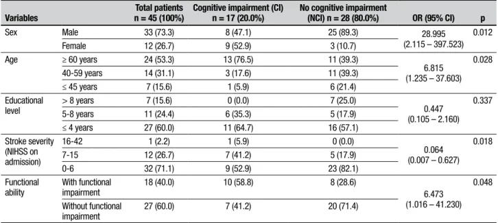
schooling (p=0.042), greater severity stroke (p=0.005) and impaired functional ability (p=0.045) were the char-acteristics most associated with cognitive impairment. For the logistic regression, categorical variables with p<0.10 were included in the model. he variables found to be associated with cognitive performance were sex (OR=28.995), age (OR=6.815), stroke severity (OR=0.064) and functional ability (OR=6.473).
Table 1. Comparison of patients exhibiting cognitive impairment and no cognitive impairment by sociodemographic, clinical, functional and mood
char-acteristics of sample (categorical and quantitative variables).
Variables n = 45 (100%)Total patients Cognitive impairment (CI) n = 17 (37.8%) No cognitive impairment (NCI) n = 28 (62.2%) p
Sex Male 33 (73.3) 8 (47.1) 25 (89.3)
0.002
Female 12 (26.7) 9 (52.9) 3 (10.7)
Age, years Mean (SD) 59.96 (13.17) 67.47 (12.54) 55.39 (11.51) 0.002 ≥ 60 years 24 (53.3) 13 (76.5) 11 (39.3)
0.050 40-59 years 14 (31.1) 3 (17.6) 11 (39.3)
18-45 years 7 (15.6) 1 (5.9) 6 (21.4)
Educational Level, years Mean (SD) 5.02 (3.56) 3.65 (2.74) 5.86 (3.79) 0.042 > 8 years 7 (15.6) 0 (0.0) 7 (25.0)
0.059 5-8 years 11 (24.4) 6 (35.3) 5 (17.9)
≤ 4 years 27 (60.0) 11 (64.7) 16 (57.1) Stroke Severity
(NIHSS on admission)
Mean (SD) 6.02 (4.28) 8.24 (5.15) 4.68 (3.03) 0.005
0-6 32 (71.1) 9 (52.9) 23 (82.1)
0.079
7-15 12 (26.7) 7 (41.2) 5 (17.9)
16-42 1 (2.2) 1 (5.9) 0 (0.0)
Functional Ability (Performance on Activities of Daily Living Questionnaire)
Mean (SD) 5.69 (7.47) 9.65 (8.91) 3.29 (5.29) 0.004 With impairment 18 (40.0) 10 (58.8) 8 (28.6)
0.045 Without impairment 27 (60.0) 7 (41.2) 20 (71.4)
Table 2. Final multiple logistic regression model best explaining association between cognitive impairment and variables.
Variables n = 45 (100%)Total patients Cognitive impairment (CI) n = 17 (20.0%) No cognitive impairment (NCI) n = 28 (80.0%) OR (95% CI) p
Sex Male 33 (73.3) 8 (47.1) 25 (89.3) 28.995
(2.115 – 397.523) 0.012 Female 12 (26.7) 9 (52.9) 3 (10.7)
Age ≥ 60 years 24 (53.3) 13 (76.5) 11 (39.3)
6.815 (1.235 – 37.603)
0.028 40-59 years 14 (31.1) 3 (17.6) 11 (39.3)
≤ 45 years 7 (15.6) 1 (5.9) 6 (21.4) Educational
level
> 8 years 7 (15.6) 0 (0.0) 7 (25.0)
0.447 (0.105 – 2.160)
0.337 5-8 years 11 (24.4) 6 (35.3) 5 (17.9)
≤ 4 years 27 (60.0) 11 (64.7) 16 (57.1) Stroke severity
(NIHSS on admission)
16-42 1 (2.2) 1 (5.9) 0 (0.0)
0.064 (0.007 – 0.627)
0.018
7-15 12 (26.7) 7 (41.2) 5 (17.9)
0-6 32 (71.1) 9 (52.9) 23 (82.1)
Functional ability
With functional impairment
18 (40.0) 10 (58.8) 8 (28.6)
6.473 (1.016 – 41.230)
0.048
Without functional impairment
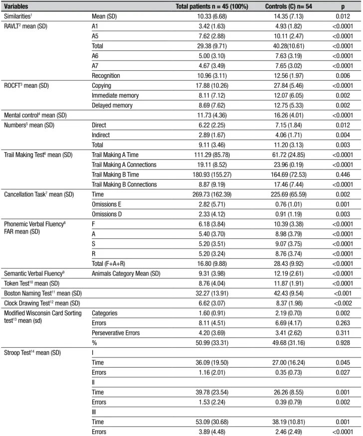
Table 3. Neuropsychological characteristics of sample and control group (quantitative variables).
Variables Total patients n = 45 (100%) Controls (C) n= 54 p
Similarities1 Mean (SD) 10.33 (6.68) 14.35 (7.13) 0.012
RAVLT2 mean (SD) A1 3.42 (1.63) 4.93 (1.82) <0.0001
A5 7.62 (2.88) 10.11 (2.47) <0.0001
Total 29.38 (9.71) 40.28(10.61) <0.0001
A6 5.00 (3.10) 7.63 (3.19) <0.0001
A7 4.67 (3.49) 7.65 (3.02) <0.0001
Recognition 10.96 (3.11) 12.56 (1.97) 0.006 ROCFT3 mean (SD) Copying 17.88 (10.26) 27.84 (5.46) <0.0001
Immediate memory 8.11 (7.12) 12.07 (6.05) 0.002 Delayed memory 8.69 (7.62) 12.75 (5.33) 0.002 Mental control4 mean (SD) 11.73 (4.36) 16.26 (4.01) <0.0001
Numbers5 mean (SD) Direct 6.22 (2.25) 7.15 (1.84) 0.012
Indirect 2.89 (1.67) 4.06 (1.71) 0.004
Total 9.11 (3.46) 11.20 (3.13) 0.003
Trail Making Test6 mean (SD) Trail Making A Time 111.29 (85.78) 61.72 (24.85) <0.0001
Trail Making A Connections 19.11 (8.52) 23.96 (0.19) <0.0001 Trail Making B Time 180.93 (155.27) 164.69 (72.53) 0.446 Trail Making B Connections 8.87 (9.19) 17.46 (7.44) <0.0001 Cancellation Task7 mean (SD) Time 269.73 (162.39) 225.69 (65.59) 0.002
Omissions E 2.82 (5.71) 0.76 (1.01) 0.001
Omissions D 2.33 (4.12) 0.91 (1.19) 0.003 Phonemic Verbal Fluency8
FAR mean (SD)
F 6.18 (3.84) 10.39 (3.38) <0.0001
A 5.40 (3.70) 8.98 (3.79) <0.0001
S 5.20 (3.51) 9.07 (3.75) <0.0001
R 5.20 (3.24) 8.76 (3.74) <0.0001
Total (F+A+R) 16.80 (9.88) 28.43 (9.92) <0.0001 Semantic Verbal Fluency9 Animals Category Mean (SD) 9.31 (3.98) 12.19 (2.61) <0.0001
Token Test10 mean (SD) 8.76 (4.04) 11.87 (1.91) <0.0001
Boston Naming Test11 mean (SD) 32.27 (13.91) 42.43 (9.54) <0.001
Clock Drawing Test12 mean (SD) 6.62 (3.07) 8.37 (1.98) <0.002
Modified Wisconsin Card Sorting test13 mean (sd)
Categories 1.60 (0.91) 2.19 (0.70) 0.002
Errors 8.11 (4.51) 6.69 (4.17) 0.263
Perseverative Errors 4.20 (3.69) 3.41 (2.62) 0.311
% 50.99 (33.31) 49.68 (31.16) 0.928
Stroop Test14 mean (SD) I
Time 36.09 (19.50) 27.00 (16.24) 0.045
Errors 1.16 (2.01) 0.35 (0.73) 0.027
II
Time 39.78 (23.54) 26.26 (8.55) 0.001
Errors 1.53 (2.24) 0.39 (0.79) 0.002
III
Time 53.09 (30.68) 38.19 (10.81) 0.001
Errors 3.89 (4.48) 2.46 (2.49) <0.0001
1Reasoning – verbal abstraction: Similarities subtest (WAIS-III subtest). 2Rey Auditory Verbal Learning Test. 3Rey-Osterrieth Complex Figure Test. 4Mental Control (WMS-III subtest). 5Numbers
(WAIS-III subtest). 6Trail Making Test - form A (time). 7Cancellation task with unstructured letters (time), cancellation task with unstructured letters (number of omissions to left),cancellation task with
unstructured letters (number of omissions to right). 8Phonemic Verbal Fluency Test (COWA).9 Semantic Verbal Fluency Test (animals category). 10Token Test. 11Boston Naming Test. 12Clock Drawing
Subjects in the general group showed worse perfor-mance on neuropsychological tests compared to the 54 healthy controls from the community, except on the Trail Making B Test, for the time parameter (p=0.495).
With regard to the neuropsychological proile, the general group of subjects exhibited greater impairment in verbal memory, attention, verbal functions, hemine-glect and visuospatial organization. he most afected domains in the group with cognitive impairment were: attention, visuospatial organization, verbal functions and verbal memory.
DISCUSSION
he aim of this study was to assess cognitive outcome and identify risk factors for impaired cognitive perfor-mance in a group of irst-ever stroke survivors followed at the specialized out-patient unit of the Uniied Health System (SUS); and to describe the neuropsychologi-cal proile of the group presenting with cognitive im-pairment. he rate of cognitive impairment found six months post-stroke was 37.8%, consistent with igures reported in the literature. Nys et al. (2005) found that
30.6% of the patients studied had cognitive impair-ment in at least one domain, six months post-stroke.2
Serrano, Domingo, Rodríguez-Garcia, Castro, and Del Ser (2007) reported 26.8% cognitive impairment 12 months after stroke.30
In a review article, Cumming, Marshall and Lazar (2013)31 reported the study of Tatemichi et al. (1994)
which detected cognitive impairment in all cognitive domains, where those most afected were attention, memory, language and orientation.32 he authors also
cited a comprehensive European study which identiied greatest cognitive impairment in the domains of atten-tion, visuospatial abilities and verbal luency.33 his
re-view noted that stroke currently has greatest impact on attention and executive functions. he disparate results found today are due to the fact that the earlier studies cited above investigated cognitive status of hospitalized patients and healthy controls, whereas current studies tend to be population-based. Consequently, earlier stud-ies may have overestimated the frequency of cognitive deicits in patients.31 he present study retains the same
design as earlier investigations (patients treated at
spe-Table 4. Neuropsychological characteristics of sample (categorical variables).
Variables Total Patients*n = 45 (100%) Cognitive impairment (CI) n = 17 (37.8%) No cognitive impairment (NCI) n = 28 (62.2%)
Reasoning1 With changes 5 (11.1) 4 (23.5) 1 (3.6)
Without changes 40 (88.9) 13 (76.5) 27 (96.4) Verbal memory2 With changes 20 (44.4) 13 (76.5) 7 (25.0)
Without changes 25 (55.6) 4 (23.5) 21 (75.0) Visual memory3 With changes 15 (33.3) 12 (70.6) 3 (10.7)
Without changes 30 (66.7) 5 (29.4) 25 (89.3) Attention4 With changes 20 (44.4) 15 (88.2) 5 (17.9)
Without changes 25 (55.6) 2 (11.8) 23 (82.1) Hemineglect5 With changes 17 (37.8) 10 (58.8) 7 (25.0)
Without changes 28 (62.2) 7 (41.2) 21 (75.0) Verbal functions6 With changes 20 (44.4) 14 (82.4) 6 (21.4)
Without changes 25 (55.6) 3 (17.6) 22 (78.6) Visuospatial organization7 With changes 17 (37.8) 15 (88.2) 2 (7.1)
Without changes 28 (62.2) 2 (11.8) 26 (92.9) Executive Functions8 With changes 7 (15.6) 7 (41.2) 0 (0.0)
Without changes 38 (84.4) 10 (58.8) 28 (100.0)
1Reasoning – verbal abstraction: Similarities subtest (WAIS-III subtest). 2Verbal Memory: delayed recall Rey Auditory Verbal Learning Test (A7 – RAVLT). 3Visual Memory: delayed recall of
Rey-Oster-rieth Complex Figure Test. 4Attention: Mental Control (WMS-III subtest), Numbers (WAIS-III subtest), Trail Making Test - form A (time). 5Hemineglect – spatial attention for determining the presence
of hemineglect: cancellation task with unstructured letters (time), cancellation task with unstructured letters (number of omissions to left), cancellation task with unstructured letters (number of omissions to right). 6Verbal Functions: Phonemic Verbal Fluency Test (COWA), Semantic Verbal Fluency Test (animals category), Token Test and Boston Naming Test.7 Visuospatial Organization: Clock
Drawing Test, .Rey-Osterrieth Complex Figure Test (copying).8Executive Functions: Modified Wisconsin Card Sorting Test (number of categories), Stroop test (time III/time I), Phonemic Verbal Fluency
ciic out-patient unit compared to control subjects from the community) and shows that the most common impairments are to verbal memory, attention, verbal functions (including verbal luency), hemineglect and visuospatial organization. hus, the pattern of cogni-tive impairments observed here was the same as those identiied in the above-cited studies.32,33
Although men predominated in the present casuis-tic, women had the most severe cognitive impairment. In fact, women had a 21-fold greater risk for cognitive impairment. Other studies2,32 have also observed this
association. Elderly women may be more vulnerable to cognitive impairment owing to vascular risk factors. he study by Millán-Callenti et al. (2009) in a population aged over 60 years found that females had a greater like-lihood of having cognitive impairment, associated with the presence of dementia, heart failure, anemia, stroke and auditory problems.34
In the present study, more advanced age was associ-ated with a 4.3 times higher risk for cognitive impair-ment. Nys et al. (2007) found advanced age to be asso-ciated with poorer cognitive recovery at 6-10 months post-stroke.35 Raz, Rodrigue, Kennedy and Acker (2007)
noted that vascular issues, such as pathological changes in the integrity of white matter and the presence of vas-cular risk factors were associated with age-related cogni-tive decline.36
Greater stroke severity was also associated with cog-nitive impairment. Possibly, serious strokes may have more sequelae that hamper recovery. Srikanth et al. (2003) showed an association between cognitive impair-ment and mild to moderate stroke severity.37 Severity
of strokes was determined using the NIHSS scale which includes a cognitive component. he study by Cumming et al. (2013) observed that this scale proved a predictor of dementia at 18 months post-stroke.31 he study of
Kauranen et al. showed that the NIHSS score at hospital discharge was strongly correlated with cognitive impair-ment. In the same study, 41% of patients with very low NIHSS scores ≤ 4, and all those with scores ≥4, present-ed cognitive impairment.38
Our study showed that the presence of neuropsy-chological impairment afects the functional abilities of the patient, particularly instrumental activities of daily living. he ARCOS study (2004) found 7% survivors 20 years after stroke, 1/5 of whom required assistance in daily living activities, particularly women and older in-dividuals.39 In a review, Feigin et al. (2008) airmed that
functional recovery of stroke survivors on basic daily living activities is possible in the presence of neuropsy-chological deicits, but this does not mean they have no
impairments in instrumental activities of daily living, which demand a higher level of physical functioning.7
Cognitive impairment can therefore represent an ob-stacle to independent life post-stroke.40
Given that stroke generally results in psychological stress and limitation in activities involving multiple do-mains of functioning, the study of stroke should include assessment of multiple functions, thereby allowing the impact of stroke to be accurately quantiied and under-stood as a whole.6 hus, investigating cognitive
tioning, presence of depression symptoms and func-tional ability after stroke is vital for the implementation of interventions in neuropsychological rehabilitation aimed at social reintegration of the patient. However, the speciic characteristics of individuals who may sufer greater cognitive impairment following stroke have only recently been elucidated and remain largely inconclu-sive.41 he results of studies on neuropsychological
out-come and those predicting cognitive recovery in strokes represent useful information for assessing the cost-beneit of neuropsychological intervention programs.2,6
Determining predictors of long-term outcome of stroke survivors can allow the identiication of which patients can beneit from certain treatment and rehabilitation strategies, and enable the provision of more consistent information to patients and their family members on the patient’s potential for recovery and chances of long-term survival.7
Finally, the patients in the present study who had cognitive decline were predominantly of female gender, older, with greater stroke severity and lower functional ability. Based on these indings, the diiculties faced by these patients upon returning to community living can be inferred. his knowledge could allow family members and caregivers to be better informed concerning the cog-nitive sequelae and the resultant restrictions in indepen-dence they impose, given that the cognitive impairments are not always evident. Such information can therefore support the neuropsychological interventions need-ed, helping patients to achieve greater independence, reintegrate socially and enhance their quality of life. he main limitations of the present study include the small sample size, the non-inclusion of the Barthel Index for measuring functionality and possible overes-timation of cognitive deicits due to the study design. he implications of the study are the inclusion of neu-ropsychology into out-patient inter-disciplinary teams, whose role will be investigating fundamental aspects in the recovery of stroke and contributing toward im-proved quality of life of patients.
stroke survivors should be conducted, investigating whether the occurrence of a single stroke signals de-mentia onset, which in turn is associated with a loss of functional ability.
Author contribution. Maria Gabriela R. Ferreira contribuiu com redação e revisão crítica do texto. Carla Heloísa C. Moro contribuiu com redação e revisão crítica do texto. Selma C. Franco contribuiu com redação e revisão críti-ca do texto. Todos os autores aprovaram a versão inal do manuscrito e declaram serem responsáveis por to-dos os aspectos do trabalho, garantindo sua precisão e integridade.
Financial support. his study was funded by the Research Support Fund (Fundo de Apoio à Pesquisa/ FAP) of the Regional University of Joinville – UNIVILLE.
Financial support. he present study is derived from a
research project entitled “Depression after Ischemic Stroke: implications for quality of life and inluence of cognitive functioning”. he data collected in the cited research project formed the basis of the Masters dis-sertation “Cognition, depression and functioning after thrombolysis in stroke patients”; Regional Univeris-ity of Joinville, 2011, 165 pages. he abovementioned research project was inancially suppored by the Re-search Support Fund /FAP of the same institution.
Acknowledgements. We extend our thanks to the Re-search Support Fund of UNIVILLE, the Departments of Psychology and Medicine, the HR sector of UNIVILLE and to the Center for Neurological Research (Neuro-logical Medicine, Joinville – SC) for their support and assistance in organizing and conducting this study. We also would like to thank the Psychology graduates who contributed to the organization and data collection of the study.
REFERENCES
1. Barker-Collo S, Feigin VL, Parag V, Lawes CMM, Senior H. Auckland Stroke Outcomes Study. Part 2: Cognition and functional outcomes 5 years poststroke. Neurology 2010;75:1608-1616.
2. Nys GMS, van Zandvoort MJE, de Kort PLM, et al. The prognostic value of domain-specific cognitive abilities in acute first-ever stroke. Neurology 2005;64:821-827.
3. Sachdev PS, Chen X, Brodaty H, Thompson C, Altendorf A, Wen W. The determinants and longitudinal course of post-stroke mild cognitive impairment. J Int Neuropsychol Soc 2009;15:915-923
4. Feign VL, Barker-Collo S, et al., and for the ASTRO Study Group. Auck-land Stroke Outcomes Study: Part 1: Gender, stroke types, ethnicity, and functional outcomes 5 years poststroke. Neurology 2010;75:1597-1607. 5. Del Ser T, Barba R, Morin M, et al. Evolution of Cognitive
Impair-ment after Stroke and Risk Factors for Delayed Progression. Stroke 2005;36:2670-2675.
6. Barker-Collo S, Feigin VL. The impact of neuropsychological deficits on functional stroke outcomes. Neuropsychol Rev 2006;16:53-64. 7. Feigin V, Barker-Collo S, McNaughton H, Brown P, Kerse N. Long-term
neuropsychological and functional outcomes in stroke survivors: current evidence and perspectives for new research. Int J Stroke 2008;3:33-40. 8. The National Institute of Neurological Disorders and Stroke rt-PA Stroke Study Group. Tissue plasminogen activator for acute ischemic stroke. New Engl J Med 1995;333:1581-1587.
9. Bamford J, Sandercock P, Dennis M, Burn J, Warlow C. Classification and natural history of clinically identifiable subtypes of cerebral infarc-tion. Lancet 1991;22;337(8756):1521-1526.
10. Adams HP, Bendixen BH, Kapelle LJ, Biller J, Love BB, Gordon DL, Marsh EE. 3rd. Classification of Subtype of Acute Ischemic Stroke
Defi-nitions for Use in a Multicenter Clinical Trial. TOAST. Trial of Org 10172 in Acute Stroke Treatment. Stroke 1993;24:35-41.
11. Amarenco P, Bogousslavsky J, Caplan LR, Donnan GA, Hennerici MG. Classification of Stroke Subtypes. Cerebrovasc Dis 2009;27:493-501. 12. Moro, CHC, Gonçalves, ARR, Longo, ALL, Fonseca, PG, Harger, R,
Gomes, DB, Ramos, MC, Estevam, ALG, Fissmer, CS, Garcia, AC, Nagel, V, Cabral, NL. Trends of the Incidence of Ischemic Stroke Throm-bolysis over Seven Years and One-Year Outcome: A Population-Based Study in Joinville, Brazil. Cerebrovac Dis 2013; 3:156-166.
13. Mellon L, Brewer L, Hall P, Horgan F, Williams D, Hickey A and on behalf of the ASPIRE-S study group. Cognitive impairment six months after ischaemic stroke: a profile from the ASPIRE-S study. BMC Neurology 2015;15-31.
14. Nys GMS, van Zandvoort MJE, de Kort PLM, Jansen BPW, Kappelle
LJ, de Haan EHF. Restrictions of the Mini-Mental State Examination in Acute Stroke. Arch Clin Neuropsychol 2005;5:623-629.
15. Mai LM, Oczkowski W, Mackenzie G, Shuster A, Wasielesky L, Franchetto A, Patlas M, Sahlas DJ. Screening for Cognitive Impairment in a Stroke Prevention Clinic Using the MoCA. Can J Neurol Sci 2013; 40:192-197.
16. Blackburn DJ, Bafadhel L, Randall M, Harkness KA. Cognitive Screening in the acute stroke setting. Age and Ageing 2013;42:113-116. 17. Memória CM, Yassuda MS, Nakano EY, Forlenza OV. Brief Screening
for mild cognitive impairment: validation of the Brazilian version of the Montreal cognitive assessment. Int J Geriatr Psychiatry 2013;28: 34-40.
18. Lam B, Middleton LE, Masellis M, Stuss DT, Harry RD, Kiss A, Black S. Criterion and Convergent Validity of the Montreal Cognitive Assess-ment with Screening and Standardized Neuropsychological Testing. J Am Geriatr Soc 2013; 61:2181–2185.
19. Ferreira MGR, Moro CHC, Franco SC. Cognition and Functional Capacity after Thrombolysis: Preliminary Results in a Brazilian Sample. Psychol Res 2013;3:83-94.
20. Pfeffer RI, Kurosaki TT, Harrah Jr. CH, Chance JM, Filos S. Measure-ment of Functional Activities in Older Adults in the Community. J Gerontol 1982;37:323-329.
21. Bonita R, Beaglehole R. Recovery of Motor Function after Stroke. Stroke 1998;19:1497-1500.
22. Beck AT, Ward CH, Mendelson M, Mock J, Erbaugh J. An inventory for measuring depression. Arch Gen Psychiatry 1961;4:561-571. 23. Cunha JA. Manual da versão em português das Escalas Beck. São
Paulo: Casa do Psicólogo; 2001.
24. Berg A, Lönnqvist J, Palomäki H, Kaste M. Assessment of Depression After Stroke: A Comparison of Different Screening Instruments. Stroke 2009;40:523-529.
25. Schoenberg MR, Dawson KA, Duff K, Patton D, Scott JG, Adams RL. Test performance and classification statistics for the Rey Auditory Verbal Learning Test in selected clinical samples. Arch Clin Neuropsy-chol 2006;21:693-703.
26. Statistical Package for the Social Sciences (SPSS for Windows). Version 16.0. [Computer program]. Chicago: SPSS Inc.;2007.
27. Cabral NL, Gonçalves ARR, Longo ALL, Eluf-Neto J. Incidence of stroke subtypes, prognosis and prevalence of risk factors in Joinville, Brazil: A two-year, community-based study. J Neurol Neurosurg Psychi-atry 2009;80:755-761.
Relationship between poststroke cognition, baseline factors, and func-tional outcome: data from: “efficacy of nitric oxide in stroke” trial. J Stroke Cerebrovasc Dis 2014;23:1821-1829.
29. Associação Brasileira de Empresas de Pesquisa (ABEP). Critério de Classificação Econômica Brasil. São Paulo: ABA, ANEP, ABEP, 2008.
30. Serrano S, Domingo J, Rodríguez-Garcia E, Castro MD, Del Ser T. Frequency of cognitive impairment without dementia in patients with stroke: a two-year follow-up study. Stroke 2007;38:105-110. 31. Cumming TB, Marshall RS, Lazar RM. Stroke, cognitive deficits, and
rehabilitation: still an incomplete Picture. Int J Stroke 2013;8:38-45. 32. Tatemichi TK, Desmond DW, Stern Y, Paik M, Sano M, Bagiella E,
1994 citado em Cumming TB, Marshall RS, Lazar RM. Stroke, cogni-tive deficits, and rehabilitation: still an incomplete Picture. Int J Stroke 2013;8:38-45.
33. Hochstenbach, Mulder, Limbeek, Donders, Schoonderwaldt, 1998, cited in Cumming TB, Marshall RS, Lazar RM. Stroke, cognitive defi-cits, and rehabilitation: still an incomplete Picture. Int J Stroke 2013;8: 38-45.
34. Millán-Callenti JC, Tubío J, Pita-Fernández S, González-Abraldes I, Lorenzo T, Maseda A. Prevalence of cognitive impairment: effects of level of education, age, sex and associated factors. Dement Geriatr Cogn Disord 2009;28:455-460.
35. Nys GMS, van Zandvoort MJE, de Kort PLM, Jansen BPW, de Haan EHF, Kapelle LJ. Cognitive Disorders in Acute Stroke: Prevalence and Clinical Determinants. Cerebrovasc Dis 2007;23:408-416.
36. Raz N, Rodrigue KM, Kennedy KM, Acker JD. Vascular health and longitudinal changes in brain and cognition in middle-aged and older adults. Neuropsychology 2007;2:149-157.
37. Srikanth, VK, Thrift AG, Saling MM, et al. Increased risk of cogni-tive impairment 3 months after mild to moderate first-ever stroke: a community-based prospective study of Nonaphasic English-speaking survivors. Stroke 2003;34:1136-1143.
38. Kauranen T, Laari S, Turunen K, Mustanoja S, Baumann P, Poutiainen E. The cognitive burden of stroke emerges even with an intact NIH Stroke Scale Score: a cohort study. J Neurol Neurosurg Psychiatry 2014;85(3):295-299.
39. Anderson CS, Carter KN, Brownlee WJ, Hackett ML, Broad JB, Bonita R. Very long-term outcome after stroke in Auckland, New Zealand. Stroke 2004;35:1920-1924.
40. Gottesmann RF, Hillis AE. Predictors and assessment of cognitive
dysfunction resulting from ischemic stroke. Lancet Neurol 2010;9: 895-905.
41. Garrett KD, Browndyke J, Whelihan W, et al. The neuropsychological profile of vascular cognitive impairment – no dementia: comparisons to patients at risk for cerebrovascular disease and vascular dementia. Arch Clin Neuropsychol 2004;19:745-757.
42. Wechsler, D. WAIS-III: Wechsler adult intelligence scale. Manual. São Paulo: Casa do Psicólogo; 2004.
43. Malloy-Diniz LF, Lasmar VAP, Gazinelli LSR, Fuentes D, Salgado JV. The Rey auditory verbal learning test: Applicability for the Brazilian elderly population. Braz Psychiatry J 2007;29: 324-329.
44. Rey, A. Copy and memory recall complex figures test: Manual (Teste de copie d’une figure complexe). São Paulo: Casa do Psicólogo; 1999. 45. Wechsler, D. WMS-III: Weschler memory scale. San Antonio: The
Psychological Corporation; 1997.
46. Lezak MD, Howieson DB, Loring DW. Neuropsychological assessment. 4th edition. New York: Oxford University Press; 2004.
47. Oliveira-Souza R, Moll J, Passman LJ, Cunha FC, Marrocos RP. Trail making and cognitive set-shifting. Arch Neuropsychiatry 2000;58:826-829.
48. Weintraub, S. Neuropsychological assessment of mental state. In: M. M. Mesulam (Ed.), Principles of behavioral and cognitive neurology, 2nd
edition. New York: Oxford University Press; 2000:121-173.
49. Machado TH, Fichman HC, Santos EL, et al. Normative data for healthy elderly on the phonemic verbal fluency task—FAS. Dement Neuropsy-chol 2009;3:55-60.
50. Fichman HC, Fernandes CS, Nitrini R, et al. Category fluency task: Influ-ence of age and education. Dement Neuropsychol 2009;3:49-54. 51. Kaplan E, Goodglass H, Weintraub S. Boston naming test. Austin:
Pro-ed; 1983.
52. Mansur LL, Radanovic M, Araújo GC, Taquemori LY, Greco LL. Boston naming test: Performance of Brazilian population from São Paulo. Pro-Fono 2006;18:13-20.
53. Spreen O, Strauss E. Clock drawing. In: O. Spreen, & E. Strauss (Eds.), A compendium of neuropsychological tests. New York: Oxford Univer-sity Press; 1998.
54. Atalaia-Silva KC, Lourenço RA. Translation, adaptation and construct validation of the clock test among elderly in Brazil. Rev Saude Publica 2008;42:930-937.
APPENDIX 1
List of neuropsychological tests and other instruments used in the study.
SIMILARITIES
Similarities subtest (Weschler Adult Intelligence Scale – WAIS-III)42.
RAVLT
Rey Auditory Verbal Learning Test43.
ROCFT
Rey-Osterrieth Complex Figure Test44.
MENTAL CONTROL
Mental Control subtest (Weschler Memory Scale – WMS-III)45.
NUMBERS
Numbers subtest (Weschler Adult Intelligence Scale – WAIS-III)2.
TRAIL MAKING TEST Trail Making Test46,47.
CANCELLATION TASK Cancellation task, unstructured letters48.
PHONEMIC VERBAL FLUENCY
COWA Test – Controlled Word Association Test46,49.
SEMANTIC VERBAL FLUENCY
Semantic Verbal Fluency Test (animals category) Category Fluency Task46,50.
TOKEN TEST Token Test46.
BOSTON NAMING TEST Boston Naming Test51,52.
CLOCK DRAWING TEST Clock Drawing Test53,54.
MODIFIED WISCONSIN CARD SORTING TEST Modified Wisconsin Card Sorting Test46,54.
STROOP TEST Stroop Test46.
PFAQ
Pfeffer Functional Activities Questionnaire20.
BDI