BBO
Case Report
Compensatory Class III malocclusion treatment
associated with mandibular canine extractions
Skeletal Class III malocclusions are ideally treated with orthodontic-surgical approaches. However, if there are no significant soft tissue implications and the patient does not want to undergo orthognatic surgery, other treatment options may be consid-ered. The current case report describes a compensatory alternative for Class III malocclusion treatment, by means of mandibu-lar canine extractions. This treatment alternative provided facial profile and occlusal improvement, which remains stable seven years posttreatment.
Keywords: Canine extraction. Class III malocclusion. Orthodontics. Corrective.
How to cite: Janson G, Maranhão OBV. Compensatory Class III malocclusion treatment associated with mandibular canine extractions. Dental Press J Orthod. 2017 Nov-Dec;22(6):86-98.
DOI: https://doi.org/10.1590/2177-6709.22.6.086-098.bbo
Submitted: August 25, 2017 - Revised and accepted: September 21, 2017
Contact address: Guilherme Janson E-mail: [email protected] » The authors report no commercial, proprietary or financial interest in the
products or companies described in this article.
» Patients displayed in this article previously approved the use of their facial and intraoral photographs.
1 Universidade de São Paulo, Faculdade de Odontologia de Bauru, Departamento de
Ortodontia (Bauru/SP, Brasil).
DOI: https://doi.org/10.1590/2177-6709.22.6.086-098.bbo
INTRODUCTION
Class III is a complex malocclusion that involves
dental, skeletal or both structures.1,2 Treatment
usual-ly consists in a compensatory or orthodontic-surgical
approach,3 but the results are not always predictable.
In cases with great skeletal vertical and anteroposte-rior discrepancies, the orthodontic treatment associ-ated with a surgical approach might be the best
treat-ment plan.1,4 However, in some cases the patient is
more interested in less invasive interventions. In these situations, one option is compensatory treatment
with extractions, which also provides good occlusal
and acceptable esthetic results, with good stability.5-8
A compensatory approach is also indicated when the patient does not have esthetic complaints and the
an-teroposterior skeletal discrepancy is not severe.9
Usually, protocols in compensatory orthodontic treat-ment involve premolar extractions, but incisor and molar extractions are also described in the literature.8,10,11 In this case report, mandibular canine extractions were performed to improve the occlusal relationships and facial esthetics.
Guilherme Janson1, Olga Benário Vieira Maranhão1
As más oclusões esqueléticas de Classe III são idealmente tratadas com intervenções ortodôntico-cirúrgicas. Contudo, se não existirem implicações estéticas faciais e se o paciente não desejar se submeter à cirurgia ortognática, outras opções de tratamento podem ser consideradas. O presente caso clínico descreve um tratamento compensatório alternativo para a má oclusão de Classe III, com extrações de caninos inferiores. Esse tratamento alternativo propiciou melhoras no perfil facial e na oclusão, que perma-neceu estável após sete anos da sua finalização.
Janson G, Maranhão OBV BBO case report
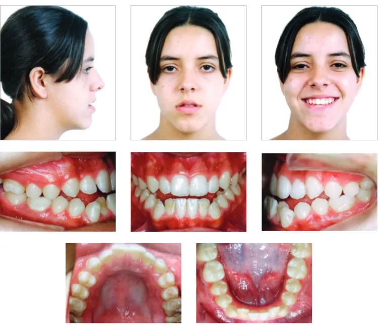
Figure 1 - Initial extra- and intraoral photographs. DIAGNOSIS
A 13-year-old female patient was referred for treat-ment by her parents ater many previous orthodontic as-sessments. The patient had a skeletal Class III malocclu-sion pattern and previous treatment plans consisted in surgical-orthodontic approaches. However, her parents did not accept a surgical treatment, and searched for a diferent opinion with the irst author. Almost all of her relatives did not present a skeletal Class III malocclusion pattern, except for her paternal grandfather who had a similar pattern.
The extraoral examination showed a skeletal Class III malocclusion pattern, vertical growth, incompetent lip seal, mouth breathing and lingual thrust during speech and swallowing. Intraorally, she presented with a
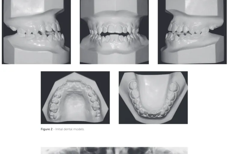
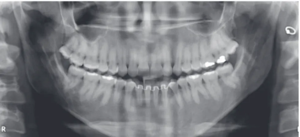
com-plete bilateral Class III malocclusion, moderate man-dibular anterior crowding, mild maxillary anterior crowding, maxillary midline deviated 1.5 mm to the let, anterior open bite, overjet of -1 mm, and tonsils hypertrophy (Figs 1 and 2). The panoramic radiograph shows that all teeth were present, with the third molars under development. No other signiicant abnormality was found (Fig 3).
Compensatory Class III malocclusion treatment associated with mandibular canine extractions
BBOcase report
Figure 3 - Initial panoramic radiograph. Figure 2 - Initial dental models.
TREATMENT PLAN
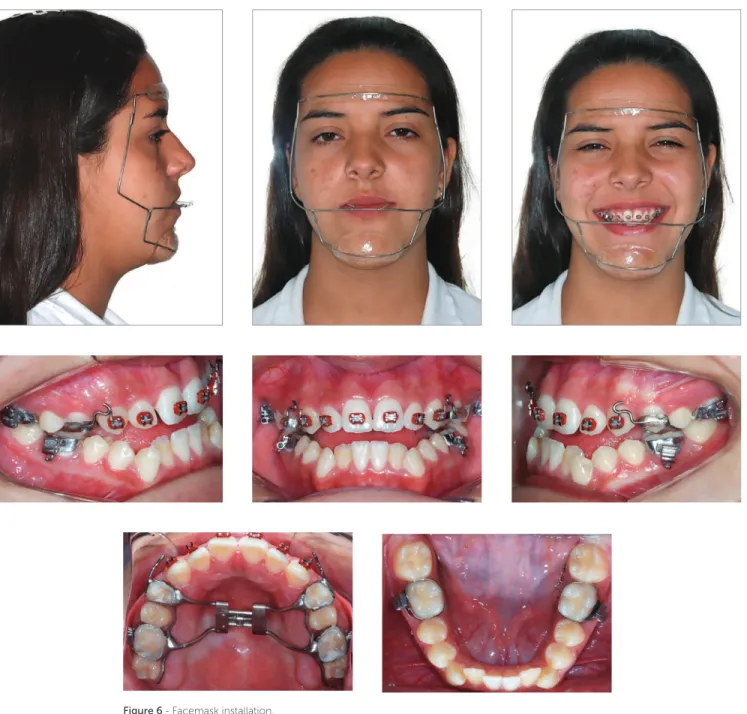
The treatment plan consisted in performing rapid maxillary expansion followed by maxillary protraction
with a facemask.12 Extraction of the mandibular canines
would be performed to correct the negative overjet. Thereater, Roth preadjusted appliances would be used, associated with Class III and anterior vertical elastics to complete correction of the anteroposterior and vertical discrepancies, respectively.
TREATMENT PROGRESS
Treatment was initiated with rapid maxillary
expan-sion, according to Liou’s protocol,13 which consists in
Janson G, Maranhão OBV BBO case report
Figure 4 - Initial cephalogram.
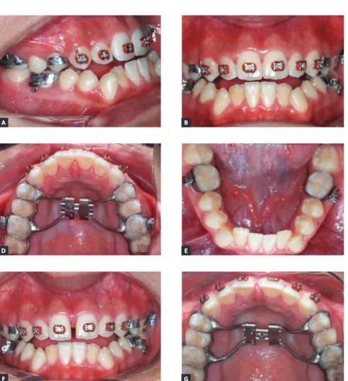
Figure 5 - Installation of Hyrax and fixed ortho-dontic appliances (A-E) and expander activation based on Liou’s protocol (F-G).13
B C
E
G F
A
Compensatory Class III malocclusion treatment associated with mandibular canine extractions
BBOcase report
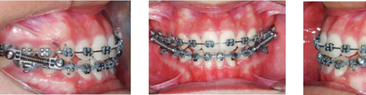
Despite the eforts, there was only small maxillary protraction, positioning the incisors in an edge to edge relationship (Fig 7). Therefore, because of the persisting Class III anteroposterior relationship and the mandibular anterior crowding, the mandibular canines were extract-ed. At this time, Roth preadjusted appliances installation was completed. Leveling and alignment proceeded with 0.014 and 0.016-in NiTi archwires, followed by 0.016, 0.018 and 0.020-inch stainless steel archwires, with a hook on the distal of the mandibular lateral incisors, to engage Class III elastics, used for 18 hours a day, with
200g of force (Figs 7 and 8). Subsequently, rectangular 0.018 x 0.025-in archwires were installed to retract the mandibular incisors and to control torque during the use of Class III CS2000 springs (DynaFlex, MO, USA) or elastics (Fig 9). A chin-cup was used during sleeping hours to redirect mandibular growth, during treatment. Ater retraction of the mandibular incisors, vertical elas-tics were used to improve interdigitation. The total treat-ment time was of 3 years and 3 months.
Janson G, Maranhão OBV BBO case report
Figure 8 - Mandibular anterior retraction and Class III elastics. Figure 7 - Intraoral photos of mandibular canine extractions.
Compensatory Class III malocclusion treatment associated with mandibular canine extractions
BBOcase report
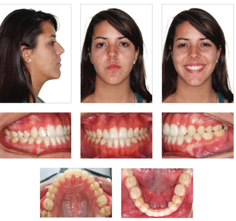
Figure 10 - Final extra- and intraoral photographs.
used 20 hours a day during 6 months; and night time use only, during the following 6 months. In the man-dibular arch, a ixed irst premolar-to-irst premolar re-tainer was bonded on each tooth and recommended to be used for 3 years. The chin-cup was recommended to be used at night, as active retention, until the end of growth, which is approximately at age 20.14,15
TREATMENT RESULTS
The facial profile improved, showing passive lip seal and improvement of the zygomatic
promi-nence (Fig 10). Consequent to crossbite and anterior open bite corrections, there was significant improve-ment of the smile esthetics (Figs 10 and 11).
Janson G, Maranhão OBV BBO case report
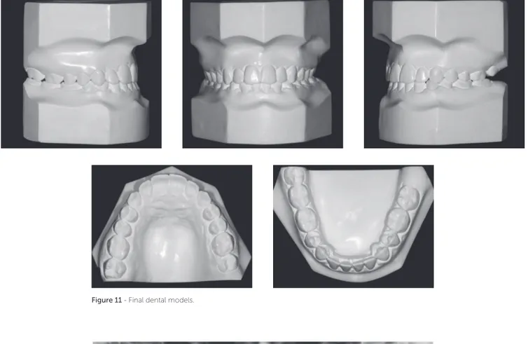
Figure 12 - Final panoramic radiograph. Figure 11 - Final dental models.
There was correction of the open and anterior cross-bites, the maxillary incisors were palatally tipped and protruded and the mandibular incisors were lingually tipped and slightly protruded (Figs 10 to 13, and Table 1). The mandibular irst premolars replaced the canines and were positioned in Class I relationship with the maxillary canines, and the molars presented Class III relationship due to extractions of the mandibular canines (Fig 12).
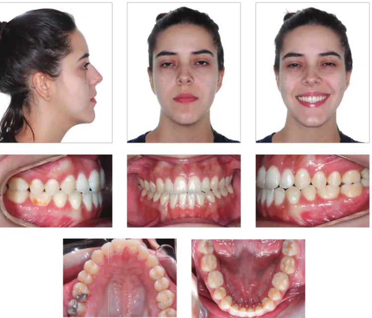
Treatment remained fairly stable 7 years posttreat-ment, with the patient presenting good facial
esthet-ics and occlusal relationships (Figs 14-17 and Table 1). The overjet and overbite are still positive and the trans-verse relationship is very satisfactory. All teeth are in contact and the third molars are present (Fig 16).
Compensatory Class III malocclusion treatment associated with mandibular canine extractions
BBOcase report
Figure 13 - Final cephalometric radiograph and superimposition of initial (black) and final (red) tracing.
Figure 14 - Seven-years posttreatment extra- and intraoral photographs. B
Janson G, Maranhão OBV BBO case report
Figure 16 - Seven-years posttreatment panoramic radiograph. Figure 15 - Seven-years posttreatment dental models.
Compensatory Class III malocclusion treatment associated with mandibular canine extractions
BBOcase report
Figure 18 - Superimposition of final (red) and long-term posttreatment (green) cephalometric tracings. B
A
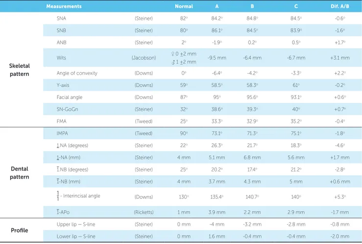
Table 1 - Cephalometric status at the initial, final and posttreatment stages.
Measurements Normal A B C Dif. A/B
Skeletal pattern
SNA (Steiner) 82o 84.2o 84.8o 84.5o -0.6o
SNB (Steiner) 80o 86.1o 84.5o 83.9o -1.6o
ANB (Steiner) 2o -1.9o 0.2o 0.5o +1.7o
Wits (Jacobson) ♀ 0 ±2 mm
♂ 1 ±2 mm -9.5 mm -6.4 mm -6.7 mm +3.1 mm
Angle of convexity (Downs) 0o -6.4o -4.2o -3.3o +2.2o
Y-axis (Downs) 59o 58.5o 58.3o 61o -0.2o
Facial angle (Downs) 87o 95o 95.6o 93.1o +0.6o
SN-GoGn (Steiner) 32o 38.6o 39.3o 40o +0.7o
FMA (Tweed) 25o 33.3o 32.9o 35.2o -0.4o
Dental pattern
IMPA (Tweed) 90o 73.1o 71.3o 75.1o -1.8o
1.NA (degrees) (Steiner) 22o 26.3o 21.7o 18.3o -4.6o
1-NA (mm) (Steiner) 4 mm 5.1 mm 6.8 mm 5.6 mm +1.7 mm
1.NB (degrees) (Steiner) 25o 20.2o 17.4o 21.2o -2.8o
1-NB (mm) (Steiner) 4 mm 3.7 mm 4.3 mm 5 mm +0.6 mm
1
1- Interincisal angle (Downs) 130o 135.4o 140.7o 140o +5.3o
1-APo (Ricketts) 1 mm 3.9 mm 2.2 mm 2.9 mm -1.7 mm
Profile
Upper lip — S-line (Steiner) 0 mm -4 mm -3.2 mm -2.8 mm -0.8 mm
Janson G, Maranhão OBV BBO case report
FINAL CONSIDERATIONS
The initial maxillary expansion was able to im-prove the transverse deficiency of the maxilla. How-ever, maxillary protraction, following Liou’s expan-sion protocol produced only slight improvement in the anteroposterior position of the maxilla (Table 1 and Fig 10). Perhaps patient’s age was a bit
ad-vanced.16,17 Besides, not every patient responds very
favorably to maxillary protraction.17
There was relative retrusion of the mandible, which was probably due to retraction of the mandibular inci-sors and also to the efects of the CS2000 spring and Class III elastics2,18-20 (Figs 8, 9 and Table 1). The asso-ciation of a slight maxillary protraction and mandibular retrusion produced improvement of the Class III an-teroposterior relationship and decreased proile
concav-ity.19 Despite the accentuated vertical growth pattern,
the orthodontic mechanics did not produce a clockwise mandibular rotation. Probably the extraction mechanics helped in the vertical control.21
The negative overjet improved due to maxillary in-cisor protrusion and mandibular inin-cisor lingual tipping during retraction (Table 1). Although the maxillary incisors were protruded, they also experienced palatal tipping. This demonstrates that there was excessive pal-atal resistant torque during Class III elastics/spring me-chanics.2,22 A positive overbite was obtained consequent to extrusion of the mandibular incisors with the use of Class III elastics and vertical anterior elastics in the in-ishing procedures.2,18,22,23
The dentoskeletal changes provided improvement of the sot tissue proile, causing slight protrusion of the upper lip and retrusion of the lower lip, which contrib-uted to establish a passive lip seal (Table 1 and Fig 10).
It was felt that a compensatory orthodontic treatment could provide satisfactory results in this patient because her facial esthetics was not signiicantly compromised and more importantly, because the patient and her par-ents did not want to undergo surgery. Perhaps an ortho-dontic-surgical approach would provide a better result. However, the patient and her parents were very satisied with the obtained results.
The option of extracting the mandibular canines was taken because it would require less anchorage reinforce-ment to retract the anterior teeth. One can visualize that the irst mandibular premolars were almost in a Class I relationship with the maxillary canines (Figs 1 and 7). Therefore, extracting the mandibular canines would only require incisor retraction and slight improvement of the anteroposterior discrepancy to obtain good ante-rior relationship. The irst mandibular premolars would then replace the mandibular canines. There are no static
or functional implications with this procedure.5
Evidently this treatment option also required great patient compliance in using the facemask and Class III elastics. The patient was not an excellent complier, but was satisfactory. This is the reason for the CS2000 spring have been used, especially in a time when the patient was already tired of using the Class III elastics. Howev-er, considering the obtained results, she performed well. Ater ixed appliances removal, she was instructed to use a chin-cup during the sleeping hours until the end of growth.24 However, she did not use it for a long time.
Despite her little compliance with posttreatment ac-tive retention, treatment has demonstrated to be very stable ater 7 years (Figs 14 to 18). Her maxillary third molars erupted, but without antagonists. If they were overerupted in the next follow-ups, they would need to be extracted.
Compensatory Class III malocclusion treatment associated with mandibular canine extractions
BBOcase report
REFERENCES
1. Stellzig-Eisenhauer A, Lux CJ, Schuster G. Treatment decision in adult patients with Class III malocclusion: orthodontic therapy or orthognathic surgery? Am J Orthod Dentofacial Orthop. 2002 July;122(1):27-37; discussion 37-8.
2. Janson G, Souza JE, Alves FA, Andrade P Jr, Nakamura A, Freitas MR, et al. Extreme dentoalveolar compensation in the treatment of Class III malocclusion. Am J Orthod Dentofacial Orthop. 2005 Dec;128(6):787-94.
3. Benyahia H, Azaroual MF, Garcia C, Hamou E, Abouqal R, Zaoui F.
Treatment of skeletal class III malocclusions: Orthognathic surgery or orthodontic camoulage? How to decide. Int Orthod. 2011 June;9(2):196-209.
4. Poletti L, Silvera AA, Ghislanzoni LTH. Dentoalveolar class III treatment using retromolar miniscrew anchorage. Prog Orthod. 2013 May;14(1):7. 5. Zimmer B, Gaida S, Dathe H. Compensation of skeletal Class III
malocclusion by isolated extraction of mandibular teeth. J Orofac Orthop. 2016 Mar;77(2):119-28.
6. Costa Pinho TM, Ustrell Torrent JM, Pinto JGC. Orthodontic camoulage
in the case of a skeletal class III malocclusion. World J Orthod. 2004 Fall;5(3):213-23.
7. Niwa K, Kushimoto K, Yamamaoto T. Mandibular irst premolar teeth
extraction in skeletal Class III malocclusion. Gifu Shika Gakkai Zasshi. 1990 June;17(1):330-8.
8. Ruellas AC, Baratieri C, Roma MB, Izquierdo AM, Boaventura L, Rodrigues
CS, et al. Angle Class III malocclusion treated with mandibular irst molar extractions. Am J Dentofacial Orthop. 2012 Sept;142(3):384-92.
9. Lima E, Brum F, Mezomo M, Pasquali CE, Farret M. Orthodontic treatment
of Class III malocclusion with lower extraction and anchorage with mini implants: case report. J World Fed Orthod. 2017 Mar;6(1):28-34. 10. Lin J, Gu Y. Lower second molar extraction in correction of severe
skeletal Class III malocclusion. Angle Orthod. 2006 Mar;76(2):217-25. 11. Canut JA. Mandibular incisor extraction: indications and long-term
evaluation. Eur J Orthod. 1996 Oct;18(5):485-9.
12. Janson G, Canto GL, Martins DR, Pinzan A, Vargas Neto J. Tratamento precoce da má oclusão de classe III com a máscara facial individualizada. Rev Dent Press Ortod Ortop Facial. 1998 Maio;3(3):41-51.
13. Liou EJ, Tsai WC. A new protocol for maxillary protraction in cleft patients: repetitive weekly protocol of alternate rapid maxillary expansions and constrictions. Cleft Palate Craniofac J. 2005 Mar;42(2):121-7.
14. Barrett AA, Baccetti T, McNamara JA. Treatment efects of the light-force chincup. Am J Orthod Dentofacial Orthop. 2010 Oct;138(4):468-76. 15. Janson G, Caldas W, Garib DG, Foncatti CF. Long-term stability of Class
III malocclusion nonextraction treatment. J World Fed Orthod. 2017 Mar;6(1):20-7.
16. Halicioglu K, Yavuz I, Ceylan I, Erdem A. Efects of facemask treatment with and without rapid maxillary expansion in young adult subjects. Angle Orthod. 2014 Sept84(5):853-61.
17. Kanekawa M, Shimizu N. Age-related changes on bone regeneration in midpalatal suture during maxillary expansion in the rat. Am J Orthod Dentofacial Orthop. 1998 Dec;114(6):646-53.
18. Lin J, Gu Y. Preliminary investigation of nonsurgical treatment of severe skeletal Class III malocclusion in the permanent dentition. Angle Orthod. 2003 Aug;73(4):401-10.
19. Janson G, Freitas MR, Araki J, Franco EJ, Barros SEC. Class III subdivision malocclusion corrected with asymmetric intermaxillary elastics. Am J Orthod Dentofacial Orthop. 2010 Aug;138(2):221-30.
20. Janson G, Souza J, Bombonatti R, Gigliotti M, Andrade Júnior P. Evaluation of dentoalveolar compensation in the treatment of Class III Malocclusion. J Interdiscipl Med Dent Sci. 2014 Oct;2(6):1-6.
21. Kocadereli Il. The efect of irst premolar extraction on vertical dimension. Am J Dentofacial Orthop. 1999 July;116(1):41-5.
22. Nakamura M, Kawanabe N, Kataoka T, Murakami T, Yamashiro T, Kamioka H. Comparative evaluation of treatment outcomes between temporary anchorage devices and Class III elastics in Class III malocclusions. Am J Orthod and Dentofacial Orthop. 2017 June;151(6):1116-24.
23. Zimmer B, Nischwitz D. Therapeutic changes in the occlusal plane inclination using intermaxillary elastics. J Orofac Orthop. 2012 Sept;73(5):377-86.
24. Janson G, Cruz KS, Henriques JFC, Freitas MR, Bombonatti R, Melucci N. Tratamento ortodôntico da Classe III, subdivisão: apresentação de um caso clínico Parte 1. Rev Dent Press Ortod Ortoped Facial. 2000 Mar;5(2):59-63.
25. Jacobson A. The “Wits” appraisal of jaw disharmony. Am J Orthod. 1975 Nov;67(2):125-38.
26. Steiner CC. Cephalometrics in clinical practice. Angle Orthod. 1959 Jan;29(1):8-29.