Cytotoxicity of alginate for orthodontic use
Matheus Melo Pithon1, Rogério Lacerda dos Santos2, Fernanda Otaviano Martins3, Maria Teresa Villela Romanos4
Objective: To evaluate the cytotoxicity of three different alginate impression materials for orthodontic use. Methods:
Three different brands of alginate were divided into three groups, namely, Group JCO (Jeltrate Chromatic Ortho), OP (Orthoprint) and CO (Cavex Orthotrace). Three control groups were also included: Group C+ (positive control), consist-ing of detergent Tween 80; Group C- (negative control), consistconsist-ing of PBS, and Group CC (cell control), consistconsist-ing of cells not exposed to any material. After manipulating the materials according to the respective manufacturer instruc-tions, samples were made with the use of silicon rings. Then the samples were immersed in Eagle’s minimum essential medium (MEM) for 2 minutes. The supernatants were then removed and brought into direct contact with L929 fibro-blasts. After exposure to the medium, the cells were incubated for 24 hours. Then 100 μl of 0.01% neutral red dye were added. The cells were incubated again for 3 hours so that the dye could be absorbed. After this 3-hour period, the cells were fixed to perform the viable cell count, using a spectrophotometer (BioTek, Winooski, Vermont, USA) at a wave-length of 492 nm. Results: Statistical differences were found when Groups CC and C- were compared with the other experimental groups. Group JCO had the highest cytotoxicity, followed by Groups OP and CO. Conclusion: Based on the results obtained in this work, it was concluded that all alginate impression materials are potentially cytotoxic.
Keywords:Cytotoxicity. Dental impression materials. Cell culture techniques.
How to cite this article: Pithon MM, Santos RL, Martins FO, Romanos MTV. Cytotoxicity of alginate for orthodontic use. Dental Press J Orthod. 2012 Nov-Dec;17(6):21.e1-5.
» The author reports no commercial, proprietary or financial interest in the products or companies described in this article.
Contact address: Matheus Melo Pithon Av. Otávio Santos, 395 – Sala 705 – Centro CEP: 45020-750 – Vitória da Conquista/BA – Brazil E-mail: [email protected]
1 Professor of Orthodontics, Southeast of Bahia State University. 2 Professor of Orthodontics, Federal University of Campina Grande. 3 MSc in Immunobiological Technology, FIOCRUZ.
4 Professor of Virology , Federal University of Rio de Janeiro.
Submitted: January 05, 2009 - Revised and accepted: August 16, 2009
Objetivo: avaliar a citotoxicidade de três diferentes alginatos de uso ortodôntico. Métodos: foram avaliados três diferen-tes alginatos divididos em três grupos, denominados grupo JCO (Jeltrate Chromatic Ortho), OP (Orthoprint) e CO (Car-rex Orthotrace). Três grupos controle também participaram: controle + (C+), constituído pelo detergente celular Tween 80; controle - (C-) PBS; e controle de célula (CC) onde as células não foram expostas a nenhum material. Após manipu-lação dos materiais, seguindo as orientações do fabricante, foram confeccionados corpos de prova utilizando-se anéis de silicone. Em seguida, esses foram imersos em meio mínimo essencial de Eagle (MEM) por 2min, onde, então, procedeu-se à remoção do sobrenadante e à colocação em contato com fibroblastos L929. Após contato com o meio, as células foram in-cubadas por mais 24h onde, então, foi adicionado o corante vermelho neutro a 0,01%. Passado esse período, foram fixadas e, então, realizada contagem de células viáveis em espectrofotômetro (BioTek, Vermont, EUA) em um comprimento de onda de 492nm. Resultados: os resultados demonstraram diferenças estatística entre os grupos CC e C- com os demais. O grupo experimental JCO mostrou-se com maior citotoxicidade, seguido pelos grupso OP e CO. Conclusões: pode-se concluir, com a realização desse trabalho, que todos os alginatos testados mostraram caráter citotóxico.
Palavras-chave: Citotoxicidade celular anticorpo-dependente. Materiais para moldagem odontológica. Técnicas de
intROduCtiOn
During the pretreatment stage, the orthodontist must collect detailed and complete documentation in order to obtain all the information required for es-tablishing a correct diagnosis and subsequent treat-ment plan.4,19
According to Monti15 impression taking is the first operation to be performed when beginning orthodon-tic treatment, because it is important to complement diagnosis. Adequate impression taking is a fundamen-tal requisite for fabricating orthodontic study models, from which data will be extracted that will help per-form treatment.4,19,22
Alginate or irreversible hydrocolloid is the most ac-cepted and used impression material in Orthodontics. Aiming to improve characteristics that are important to the orthodontist, the manufacturers have produced alginate powder with changes in the components. Many substances such as zinc, barium, cadmium, lead silicates and fluorides have been added to some com-mercial brands with the aim of improving the physical, chemical and mechanical properties, causing concern with regard to the toxicity of these materials.11
Basically, intoxication by alginate may occur by inhalation of the powder by the patient and profes-sional, accidental ingestion by the patient and ab-sorption by the oral mucosa in cases of repeated im-pression taking.2,3,24
During impression taking the alginate comes into intimate contact with the oral mucosa, which is highly vascularized and has great absorption potential, for a time interval of around 2 minutes. Therefore, the rep-etition of consecutive impression takings may cause a certain degree of toxicity to the patient, depending on the material composition.2,20
Based on this premise, the aim of the present ar-ticle was to evaluate the cytotoxicity of three different alginate brands for orthodontic use, in a cell culture experiment.
MateRial and MetHOdS Cell culture
The cell lineage used was L929 (mouse fibroblast) obtained from the American Type Culture Collection (ATCC, Rockville, MD) cultivated in Eagle’s mini-mum essential medium (MEM) (Cultilab,
Campi-nas, São Paulo, Brazil) supplemented with 2 mm of
L-glutamin (Sigma, St. Louis, Missouri, USA), 50 µg/ ml of gentamicin (Schering Plough, Kenilworth, New
Jersey, USA), 2.5 µg/ml of fungizone
(Bristol-Myers-Squibb, New York, USA), 0.25 ml sodium bicarbonate solution (Merck, Darmstadt, Germany), 10 mm of
HEPES (Sigma, St. Louis, Missouri, USA), and 10%
Fetal bovine serum (FBS) (Cultilab, Campinas, São
Paulo, Brazil) and maintained at 37 oC in an
environ-ment containing 5% of CO2.
evaluated alginates
The sample was composed of three different algi-nate brands for orthodontic use, divided into three groups: JCO (Jeltrate Chromatic Ortho, Dentsply, Petrópolis, Brazil, Lot 955069), OP(Orthoprint, Zher-mack, Rovigo, ltaly, Lot 72251) and CO (Cavex Or-thotrace, Cavex, Nederland, Lot 080910).
Composition of evaluated alginates (Provided by the manufacturer)
Diatomite, potassium alginate, calcium sulfate, magnesium oxide, iron oxide, tetrasodium pyrophos-phate, and spearmint oil.
test specimen fabrication
To fabricate the test specimens, the material was manipulated for 1 minute using a rubber bowl and plastic spatula in accordance with the manufacturer’s recommendations. After correct homogenization, the alginate was inserted in silicone rings measuring 4 mm in diameter and 4 mm height, until it was com-pletely jellified.
Controls
To verify the cell response to the extreme condi-tions, another three groups were inserted: Group CC (cell control) in which the cells were not exposed to any material; Group C+ (positive control) consisting of a detergent Tween 80 (Polyoxyethelene-20-Sorbi-tan) at 10%; Group C- (negative control), 100% PBS Solution (Phosphate-buffered saline).
Cytotoxicity test of the materials
(Cultilab, Campinas, São Paulo, Brazil). After 2 min-utes in contact with the culture medium, the superna-tants were collected for posterior evaluation.
The supernatants were placed, in triplicate, in a 96-well plate containing a confluent monolayer of L929 cells and incubated for 24 hours at 37 oC in an environment containing 5% of CO2.After incuba-tion time, the effect on cell viability was determined by means of the dye-uptake technique, described by Neyndorff et al16 (1990). After 24 hours of incubation, 100 µl of 0.01% neutral red (Sigma, St. Louis,
Mis-souri, USA), was added to culture medium in each
well of the miniplates, and these were incubated at 37 oC for 3 hours for the dye to penetrate into the live cells. After this interval, and after dispensing the dye, 100 µl of 4% formaldehyde solution (Reagen) was added to PBS (NaCl 130 mm; KCl 2 mm; Na2HPO4 2H2O 6 mm; K2HPO4 1 mm, pH 7.2) for 5 minutes, to promote cell fixation to the plates. Next, in order to extract the dye, 100 µl of 1% acetic acid solution (Vetec, Rio de Janeiro, Brazil) with 50% methanol was added (Reagen, Rio de Janeiro, Brazil). After 20 minutes the readout was taken in a spectrophotom-eter (BioTek, Winooski, Vermont, USA) at a wave-length of 492 nm.
Statistical analysis
Statistical analyses were performed with the pro-gram SPSS 13.0 (SPSS Inc., Chicago, Illinois, USA). Descriptive statistical analysis including mean and standard deviation were calculated for the groups evaluated. The values for the quantity of viable cells were submitted to the analysis of variance (ANOVA) to determine whether there was statistical differ-ence among the groups, and afterwards the Tukey test was performed.
ReSultS
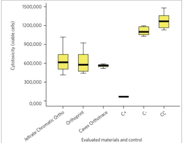
The results demonstrated statistical differences between Group C+ and all the others, and between Groups C- and CC and the experimental Groups JCO, OP and CO (p < 0.05). No statistical significance was observed between Group JCO, OP and CO, and be-tween Groups C- and CC (p > 0.05) (Table 1).
With regard to cell viability Group C+ presented the lowest cell viability followed by experimental groups CO, OP and JCO (Fig 1).
diSCuSSiOn
Alginate is the most accepted and used impression material in Orthodontics. Manufacturers produce orthodontic alginate powder containing various com-ponents with different purposes. Many substances such as zinc, barium, cadmium, lead silicates and fluo-rides have been added to some commercial brands with the aim of improving the physical, chemical and mechanical properties, causing concern with regard to the toxicity of these materials.6
According to Syndiskis et al,24 alginate is capable of affecting the ability of cells reproduction. The sub-stance may not be sufficiently toxic to kill the cells, but is toxic enough to inhibit cell growth, or on a small scale, affect normal cell function. In this context, while a single contact may not cause clinical symptoms, re-peated contacts with the material alter or affect cell vi-ability, and may result in a late toxic or allergic reaction.
Table 1 - Mean, standard deviation, percentage of viable cells and statisti-cal analysis of the groups evaluated.
Figure 1 - Diagram showing the values of cellular viability between evalu-ated groups.
M. Cell: Mean values of quantity of viable cells; SD: Standard Deviation:
Stat: Statistics: In which equal letters represent absence of statistical dif-ferences.
Groups M. Cell / S.D. % Viable Cells Stat.
JCO 644.25 ± 193.59 50.6 A
OP 617 ± 173.86 48.46 A
CO 560.87 ± 23.60 44.05 A
C+ 67 ± 2.20 5.26 B
C- 1111.5 ± 67.85 87.31 C
CC 1273.75 ± 125.71 100 C
1500,000
C
yt
ot
o
xicit
y (
viable c
ells
)
Jeltrate Chr omatic Or
tho
Cave x Or
thotr ace
Evaluated materials and control
Orthoprint
C+ C- CC
1200,000
900,000
600,000
300,000
Thus, the aim of the present study was to evaluate the cytotoxicity of three alginate brands for orthodontic use, in a cell culture experiment.
The cell cultures have been used as part of a series of tests recommended for evaluating the biologic be-havior of materials to be placed in contact with hu-man tissues.9,14,21 In this study, cytotoxicity tests were performed to evaluate the cytotoxicity of alginates. The L929 cell lineage (mouse fibroblasts) was used be-cause these cells are frequently used in various studies in which the intention was to evaluate the cytotoxicity of materials for dental use.1,8,10,13
The neutral red assay method was used to evalu-ate cell viability. The analysis procedure with neu-tral red is a cell survival/viability assay, based on the capacity of viable cells to incorporate and process the neutral red within the lysosomes. Normally, it is performed in adherent cells. Neutral red is a weak cationic dye that promptly penetrates into the cell membrane and accumulates intracellularly in the ly-sosomes (lysosomal pH < cytoplasmic pH), combin-ing with the anionic part of the lysosomal matrix.12 The changes in cell surface or sensitive lysosomal membrane led to lysosomal weakness and other changes that gradually become irreversible. These alterations that occur by the action of xenobiotics re-sult in the reduction in absorption and combination of neutral red. Thus, it is possible to distinguish vi-able, damaged or dead cells, which is the basis of the test. The quantity of dye incorporated into the cells is measured by spectrometry, and is directly propor-tional to the number of cells with intact membranes. This method was introduced to evaluate the cyto-toxicity of materials for orthodontic use by Pithon et al,17 comparing with the agar diffusion method. In the mentioned study, both methods were shown to be apt for the evaluation of cytotoxicity.
The results of the present study demonstrated the cytotoxicity of the three studied brands of algi-nates, as follows: Jeltrate (50.6% cell viability) fol-lowed by Orthoprint (48.46%) and Cavex Orthotrace (44.05%). Statistical differences were found among the experimental groups and the cell control (CC) and negative control (C-). No statistical differenc-es were observed among the experimental groups evaluated. This result may be justified with the sim-ilar constitution of these materials.
The evaluation time interval was 2 minutes, be-cause this is the interval that usually alginate stays in contact with the oral mucosa during the impression taking as recommended by the manufacturer. The test specimens remained in contact with the culture medi-um for this period. After this, the supernatant was col-lected from the culture medium, and was then placed in contact with the cells. The test specimens were not placed in direct contact with the cells, since the mechanical contact of these with the cells may harm them, as suggested by Costa.5
In order to evaluate the cell response to extreme situations, a positive control group (C+) was inserted in the study, the purpose was to generate lesions to the cells. The material used as positive control was 10% Tween, which is a non ionic surfactant, toxic to biologic membranes,18 composed of polyoxyethylene derivatives of sorbitan fatty acid esters, with the char-acteristics of stimulating the secretion of proteins in microorganism,23 in addition to altering the morphol-ogy and surface of the cell wall.7 As expected, the posi-tive control presented high toxicity, differing statisti-cally from all the other groups (p < 0.05).
The negative control group, consisted of a 100% PBS solution (Phosphate-buffered saline), recog-nized as non toxic to cells. The aim of this control was to evaluate only the physical action on cells. This procedure demonstrated low cytotoxicity, with absence of statistical significance from the cell trol group, in which no substance was placed in con-tact with the cells.
Based on the present results it should be consid-ered that success in the orthodontic clinic involves not only mastering the corrective technique to achieve an ideal dental occlusion, but also requires the application of biosafety rules and concern about the local and systemic consequences of the dental materials used for this purpose. The evaluation with regard to the possible cytotoxic effects must be verified in order to obtain safety with respect to the use of a certain material.
COnCluSiOn
1. Alcaide M, Serrano MC, Pagani R, Sánchez-Salcedo S, Nieto A, Vallet-Regí M, et al. L929 fibroblast and Saos-2 osteoblast response to hydroxyapatite-betaTCP/ agarose biomaterial. J Biomed Mater Res A. 2009;89(2):539-49.
2. Braga SRS, Braga AS, Catirse ABCEB, Vaz LG, Spadaro ACC. Potencial tóxico dos alginatos para uso odontológico. Rev Ciênc Farm Básica Apl. 2007;28(2):153-8. 3. Braga AS, Catirse ABCEB, Vaz LG, Spadaro ACC. Quantitative analysis of
potentially toxic metals in alginates for dental use. Rev Ciênc Farm Básica Apl. 2005;26(2):125-30.
4. Camargo EL, Mucha JN. Moldagem e modelagem em Ortodontia. Rev Dental Press Ortod Ortop Facial. 1999;4(3):37-50.
5. Costa CA, Edwards CA, Hanks CT. Cytotoxic effects of cleansing solutions recommended for chemical lavage of pulp exposures. Am J Dent. 2001;14(1):25-30. 6. De Freitas JF. Potential toxicants in alginate powders. Aust Dent J. 1980;25(4):224-8. 7. Domingues FC, Queiroz JA, Cabral JM, Fonseca LP. The influence of culture
conditions on mycelial structure and cellulase production by Trichoderma reesei Rut C-30. Enzyme Microb Technol. 2000;26(5-6):394-401.
8. Donadio M, Jiang J, Safavi KE, Zhu Q. Cytotoxicity evaluation of Activ GP and Resilon cones in vitro. Oral Surg Oral Med Oral Pathol Oral Radiol Endod. 2008;106(1):e76-9.
9. Estrela C. Metodologia científica: ensino e pesquisa em Odontologia. São Paulo: Artes Médicas; 2005. 15 p.
10. Feizzadeh B, Afshari JT, Rakhshandeh H, Rahimi A, Brook A, Doosti H. Cytotoxic effect of saffron stigma aqueous extract on human transitional cell carcinoma and mouse fibroblast. Urol J. 2008;5(3):161-7.
11. Freitas JF. Potential toxicants in alginate powders. Aust Dent J. 1980;25(4):224-8. 12. Griffon G, Marchal C, Merlin JL, Marchal S, Parache RM, Bey P. Radiosensitivity of multicellular tumour spheroids obtained from human ovarian cancers. Eur J Cancer. 1995;31A(1):85-91.
REFERENCES
13. Jin CY, Zhu BS, Wang XF, Lu QH. Cytotoxicity of titanium dioxide nanoparticles in mouse fibroblast cells. Chem Res Toxicol. 2008;21(9):1871-7. Epub 2008 Aug 5. 14. Jorge JH, Giampaolo ET, Pavarina AC. Cytotoxicity of the dental materials. A
literature review. Rev Odontol UNESP. 2004;33(2):65-8. 15. Monti E. Tratado de Ortodoncia. Buenos Aires: El Ateneo; 1953. 16. Neyndorff HC, Bartel DL, Tufaro F, Levy JG. Development of a model to
demonstrate photosensitizer-mediated viral inactivation in blood. Transfusion. 1990;30(6):485-90.
17. Pithon Mm, Santos RL, Ruellas ACO, Fidalgo TKS, Romanos MTV, Mendes GV. Citotoxicidade in vitro de elásticos ortodônticos: comparação entre duas metodologias. Rev Saúde Com. 2008;4(1):19-26.
18. Rege BD, Kao JP, Polli JE. Effects of nonionic surfactants on membrane transporters in Caco-2 cell monolayers. Eur J Pharm Sci. 2002;16(4-5):237-46.
19. Romano FL, Pereira Neto JS, Magnani MBBA, Nouer DF, Siqueira VCV. Moldagem ortodôntica. Rev Clín Ortod Dental Press. 2005;4(1):15-22.
20. Samuel SW, Miranda LA, Dutra CAV. Potencial tóxico dos alginatos. Rev Fac Odontol Porto Alegre. 1995;36(2):14-6.
21. Santos RL, Pithon Mm, Oliveira MV, Mendes GS, Romanos MTV, Ruellas ACO. Cytotoxicity of intraoral orthodontic elastics. Braz J Oral Sci. 2008;7(24):1520-5. 22. Strang RHW. Tratado de Ortodoncia. Rio de Janeiro: Editorial Bibliográfica; 1957. 23. Stutzenberger FJ. Interference of the detergent Tween 80 in protein assays. Anal
Biochem. 1992;207(2):249-54.