R E S E A R C H
Open Access
Molecular characterization of
Dirofilaria
spp.
circulating in Portugal
Cátia Ferreira
1†, Ana Afonso
1†, Manuela Calado
1, Isabel Maurício
1, Ana Margarida Alho
2, José Meireles
2,
Luís Madeira de Carvalho
2and Silvana Belo
1*Abstract
Background:Dirofilariosis is a potentially zoonotic parasitic disease, mainly transmitted by mosquito vectors in many parts of the world. Data concerning the canineDirofilariaspecies currently circulating in Portugal is scarce. Thereby, a large-scale study was conducted to determine theDirofilariaspp. present in Portugal, based on a molecular approach, and also to optimize a reliable and highly sensitive species-specific polymerase chain reaction (PCR) assay that could be used for the simultaneous detection and differentiation ofDirofilaria immitis,Dirofilaria repens, and other concurrent filarial species in animal reservoirs.
Methods:Blood samples were collected from three districts of Portugal (Coimbra, Santarém and Setúbal) between 2011 and 2013. Samples were tested using rapid immunomigration tests (Witness®Dirofilaria), modified Knott’s technique and acid phosphatase histochemical staining. In addition, molecular analysis was performed by
amplification of the internal transcribed spacer (ITS) region using two different PCR protocols, specific for molecular screening of canine filarial species.
Results:Of the 878 dogs sampled, 8.8% (n= 77) were positive forD. immitiscirculating antigen and 13.1% (n= 115) positive for microfilariae by the modified Knott’s technique. Of the 134 samples tested by acid phosphatase
histochemical staining, 100 (74.6%) were positive forD. immitis. Overall, 13.7% (n= 120) were positive by PCR forD. immitisby ITS2, of which 9.3% (67/720) were also positive by ITS1. ITS2 PCR was the most sensitive and specific method, capable of detecting mixedD. immitisandA. recondituminfections. Heterozygosity, in the form of double peaks, was detected by sequencing of both ITS regions. NoD. repenswas detected by any of the diagnostic methods. Conclusions:The present study confirmedD. immitisas the dominant species of the genusDirofilariainfecting Portuguese dogs, based on sequencing of ITS1 and ITS2 PCR fragments. Additionally, ITS2 PCR was the most adequate method for diagnosis and prevalence estimation.
Keywords:Dirofilaria, PCR, Internal transcribed spacer, Dog, Portugal
Background
Dirofilariosis is a potentially zoonotic filarial parasitic dis-ease, present in several parts of the world, transmitted mainly by mosquito vectors. The speciesDirofilaria immi-tisand Dirofilaria repens(Filarioidea, Onchocercidae) are widely present in the Mediterranean basin and are the causative agents of cardiopulmonary and subcutaneous dir-ofilariosis, respectively. Both nematodes are transmitted by mosquito species of the family Culicidae and can infect
domestic and wild canids and felids, causing severe patho-logical effects [1]. Dirofilaria immitis is considered the most virulent filarial species in dogs, as the long-lived adult worms reside in the right ventricle and pulmonary artery, leading to pulmonary hypertension, congestive heart failure and even death [2, 3]. Instead,D. repensadult forms live in subcutaneous tissue, where they cause dermatological problems, such as multifocal nodular and prurigo papularis dermatitis. Moreover, both species may also infect humans. Dirofilaria immitis pre-adult forms can cause pulmonary nodules and D. repens adult/pre-adult stages may induce subcutaneous and ocular lesions [4, 5]. Other less known canine filarial parasites, such as Acanthocheilonema * Correspondence:[email protected]
†
Equal contributors
1Global Health and Tropical Medicine, GHTM, Instituto de Higiene e Medicina
Tropical, IHMT, Universidade Nova de Lisboa, UNL, Lisboa, Portugal Full list of author information is available at the end of the article
dracunculoides (tick- and fly-transmitted) and Acantho-cheilonema reconditum (flea- and lice- transmitted), may also infect companion animals [6, 7]. AdultA. reconditum and A. dracunculoidesreside in the peritoneal cavity and adipose tissue of the host, and thus seem to be less virulent for canine reservoirs. Nevertheless,A. reconditumhas also been reported in humans [8].
These filarial species release circulating microfilariae (Mf ) in the blood of their definitive hosts. These Mf can be diagnosed by microscopy through specific morpho-logical identification or Mf histochemical staining [9, 10]. Other diagnostic methods are also available, such as de-tection of circulating adult female antigens (currently only for D. immitis) and molecular approaches [1, 11, 12]. Modified Knott’s and acid phosphatase histochemical staining tests of blood smears remain the most commonly used parasitological tests for Mf detection, but are labour-intensive and require expertise. Thus, the prevalence of Dirofilaria spp. can be over-estimated if other filarial species are present and misidentified [13, 14]. Molecular protocols have been developed for reliable detection and differentiation of filarial species, in particular, a species-specific PCR assay and multiplex PCR and restriction fragment length polymorphism (RFLP) assays for simul-taneous detection of different Dirofilaria spp., either in the vector or in blood [12, 14–21].
Canine dirofilariosis due to D. immitisis known to be endemic and widely distributed in Portugal, with preva-lence ranging between 0.9 and 27.3% in mainland regions to over 30% in Madeira Island [22–25]. Dirofi-laria repenswas recently detected for the first time, in a dog, presenting as mixed infection with D. immitis[26]. This is a worrying finding, as the occurrence of autoch-thonous infections in domestic animals and the numbers of notified human cases of dirofilariosis, mainly attrib-uted toD. repens, have increased substantially in several European countries in recent years [5, 27, 28].
The aim of the present study was to identify the Diro-filaria species currently circulating in Portuguese dogs through an optimised reliable and highly sensitive species-specific PCR assay for the simultaneous detec-tion and differentiadetec-tion of D. immitis, D. repens and other concurrent filariids in animal reservoirs.
Methods
Study areas and canine sampling examination
The study areas, as well as the clinical and parasitological procedures, were as previously described [25]. Briefly, canine surveys were conducted in kennels (run by local authorities or animal protection associations) in three districts of Portugal: Coimbra (northern-Centre region), Santarém (central-Centre region) and Setúbal (southern-Centre region) during three consecutive years: 2011, 2012 and 2013. Three surveys were carried out each year, in
spring (March-April), summer (July-August) and autumn (October-November). Only dogs older than 6 months of age and residing in the kennels for at least 6 months were included.
Direct and serological tests
For clinical and parasitological examination, dogs were randomly sampled in each kennel. Physical examination was performed prior to blood collection. Blood was col-lected from the cephalic vein (5 ml) and stored (2.5 ml) with either anticoagulant EDTA or in a dry tube, and later processed for parasitological, serological and mo-lecular analyses. The modified Knott’s technique (KN) and the acid phosphatase histochemical staining test (AP) were used for microscopic detection and identifica-tion of Mf in blood smears. The commercial kit WIT-NESS® Dirofilaria (WT) (Synbiotics, San Diego, CA, USA) was employed for detection ofD. immitis circulat-ing antigen in serum.
Molecular analysis
DNA isolation
DNA was extracted from whole blood using CTAB (cetyltrimethyl ammonium bromide) method, adapted from Stothard et al. [29]. Briefly, 100 μl blood with
EDTA (ethylenediamine tetraacetic acid) was incubated with 600μl CTAB buffer and 0.2 mg proteinase K
(Bio-line, London, UK) at 56 °C for 2 h, with agitation. DNA precipitation was done with 0.6 ml absolute ethanol and the pellet hydrated in 50 μl TE buffer (10 mM Tris,
1 mM EDTA, pH 7.0). DNA samples were stored at -20 °C until further use.
ForD. immitispositive control, DNA was extracted, as above, from a small macerated section of two adult worms. For D. repens positive control, DNA was extracted from infected canine blood and from a worm (kindly provided, respectively, by Prof. Eva Fok, Univer-sity of Veterinary Medicine, Budapest, Hungary, and by Prof. Claudio Genchi, University of Milan, Italy). Deio-nised water was used as a PCR negative control.
DNA amplification
region was carried out using the primers DIDR-F1 and DIDR-R1 [21], with expected amplification product sizes of 542, 484, 578 and 584 bp forD. immitis,D. repens, A. reconditum andA. dracunculoides, respectively. All PCR reactions were performed in 25 μl reaction mixtures,
containing PCR buffer (Promega, Madison, WI, USA), 6 mM MgCl2 (Promega), 10 pmol of each primer, 12 mM dNTPs (Promega), 2.5 U GoTaq® DNA polymer-ase (Promega), 10–40 ng of template DNA in deionized water. The temperature profile for both steps of the semi-nested ITS1 PCR was: 94 °C for 5 min, followed by 35 cycles of 94 °C for 30 s, 58 °C for 30 s and 72 °C for 45 s, with a final extension step at 72 °C for 10 min. Amplification of the ITS2 region had the following temperature profile: 94 °C for 2 min and 32 cycles of 30 s at 94 °C, 30 s at 60 °C and 30 s at 72 °C, with a final extension step for 7 min at 72 °C. Amplification prod-ucts were separated by electrophoresis in 2% agarose gels, stained with ethidium bromide, and visualized under UV light.
PCR analytical sensitivity was tested with serial dilu-tions (by a factor of 10) of DNA from a female adult worm of D. immitis, canine blood infected with D. repens and from dog blood samples with positive PCR (ITS1/ITS2) forD. immitis.
DNA sequencing and phylogenetic analysis
PCR amplicons were purified using a commercial kit (Qia-gen, QIAquick PCR Purification Kit, Germantown, USA) and sequenced commercially (Macrogen, Seoul, South Korea) using the PCR primers. A BLAST search was per-formed to confirm species identity of the sequenced amplicons. Homologous sequences available in GenBank/ EMBL/DDBJ databases were retrieved by BLAST and all sequences were aligned in BioEdit 7.2.5 [31]. Some se-quences exhibited regions of double peaks, and haplotypes were inferred manually to correspond to homozygous sequences in circulation (for ITS1), or using the programme PHASE [32] with 100 iterations, 100 thinning interval and 100‘burn-in’settings (for ITS2).
Phylogenetic relationships were estimated using MEGA 7.0 [33], based on an alignment of regions with no gaps. The phylogenetic trees were inferred by the Maximum Parsimony method parameter, CNI (level = 1) with initial tree by random addition (10 reps) with 1,000 bootstrap replicates and a cut-off value of 74%.
Statistical analysis
Pearson’s Chi-square and Fisher’s exact tests were used to evaluate the differences between the proportions of species-specific infected dogs detected by each PCR protocol, among different age groups (0.5–3 years, > 3–6 years, > 6 years), gender and district as compared with parasitological and serological tests. Level of agreement
was calculated using Cohen’s kappa coefficient (K). Stat-istical analysis was carried out using statStat-istical software SPSS 15.0 for Windows 10.0; a P< 0.05 was considered significant.
Results
Overall, 878 dogs (400 males and 478 females) were sam-pled from the three areas, Coimbra (n= 268), Santarém (n= 465) and Setúbal (n= 155). The dogs were 0.5 to 16 years old, with a median age of 4.5 years (IQR 2.5–7.0).
The analytical sensitivity of ITS1-PCR and ITS2-PCR were, respectively, 4.5 and 0.09 pg DNA from female adultD. immitis, 118 and 200 pg DNA fromD. repens-infected dog blood, and 250 and 2.5 pg DNA from aD. immitis-infected dog. Statistical sensitivity (i.e. the pro-portion of true positives) and specificity (i.e. the propor-tion of true negatives) of ITS2-PCR were significantly higher (McNemar test,P< 0.05) than of ITS1-PCR.
In the 720 dogs tested using both PCR targets, samples positive for ITS1-PCR were also positive for ITS2-PCR. Higher analytical sensitivity was observed for ITS2-PCR, with 12.9% of the blood samples positive forD. immitis (Table 1). Using ITS2-PCR it was possible to amplify, not only both species of Dirofilaria spp., but also A. reconditum in canine blood. Two samples that were ITS1-PCR-positive forD. immitis, were characterized by ITS2-PCR-RFLP as A. reconditum (accession number ENA: HG964682–HG964684) and were not included in the calculations. DNA from species (D. immitis and Acanthocheilonema spp.) was detected by ITS2-PCR in two samples.
The performance of the PCR with the highest analyt-ical sensitivity (ITS2) was compared with serologanalyt-ical and direct parasitological tests for all samples (Table 2). Out of the 878 samples tested,D. immitiscirculating antigen was detected in 77 (8.8%) by WT, whereas Mf were found in 115 (13.1%) stained slides by KN method. Sam-ples with inconsistent results between WT and KN (n= 19 WT-positive, KN-negative) and KN-positive blood slides (n= 115) were submitted to AP analysis (n= 134). Out of the 134 stained slides, D. immitis Mf were identified in 100 (74.6%) and A. reconditum in two (1.5%). Dirofilaria repens was not identified in blood smears through any method.
ITS2-PCR and KN presented the highest level of agreement (Cohen’s kappa coefficient), which was lower, but also statistically significant, between ITS2-PCR and WT (Table 3).
Characterization ofDirofilariaspp.
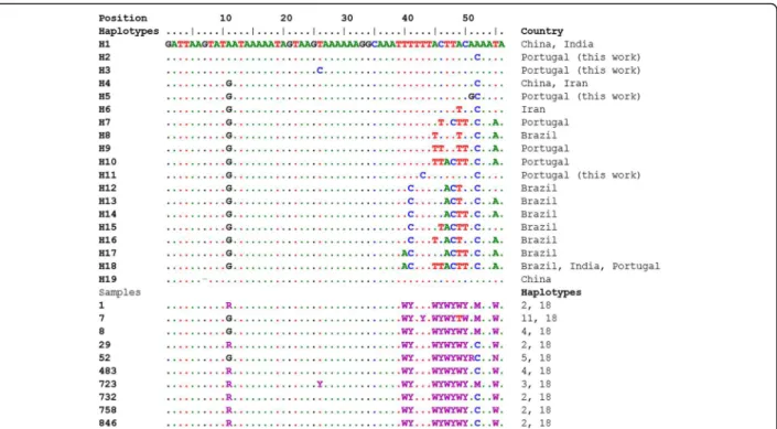
ITS region); KY014643–KY014648 (samples 483, 394, 350, 361, 488, female adult worm, respectively; ITS1) and KY644132–KY644141 (samples 1, 7, 8, 29, 52, 483, 723, 732, 758, 846, respectively; ITS2). Three out of nine ITS1 sequences analysed from PCR products obtained from ca-nine blood were found to have a string of double peaks, as did a femaleD. immitisworm (Fig. 1). The haplotypes for the ITS1 heterozygous sequences were inferred manually by assuming one sequence to be identical to the most common homozygous sequence (or haplotype) found in local samples, which in this case was H4 (Fig. 1). H4 was found in one sample from Japan, as well as the sequences AY621480.1 and AY621481.1, labelled asD. repensin the GenBank database (Fig. 1). The other inferred haplotype (H9) presented similarities with sequence EU087700, from India, but only from position 50 onwards in the alignment in Fig. 1. The region up to position 20 was more similar to other Portuguese and Japanese samples.
ITS2 sequences also presented several heterozygous sites, in particular after an A and T rich region. By com-parison with sequences obtained from a BLAST search, the ITS2 sequences obtained here were most similar to D. immitis and quite distinct from D. repens sequences present in GenBank, as revealed by phylogenetic analysis (not shown). Statistical reconstruction of haplotypes by comparison with sequences present in GenBank identi-fied 19 different haplotypes (Fig. 2). All heterozygous Portuguese samples included haplotype H18, which was present in sequences from India and Brazil (dog). Five samples had H2 as the other haplotype, which has no correspondence in the database, and two samples had haplotype H4, which was present in sequences from China (red panda) and Iran (dogs). The main difference between haplotype H18 and other haplotypes was a gap of two nucleotides in a T repeat. The other three haplo-types identified (H3, H5 and H11) were not found elsewhere in the database.
A BLAST analysis of the entire ITS region showed greatest similarities to D. immitis, with a sequence similarity that ranges from 89% to 97% with sequences available at NCBI database (JX866681.1; DQO18785.1; JX866681.1; FJ263464.1; FJ2634571; HM126606.1).
Pattern of canineD. immitisinfection related to gender and age
Based on ITS2-PCR, the prevalence of D. immitis infec-tion found in males (63/400; 15.8%) was significantly higher (P= 0.032) than in females (57/478; 11.9%). There were also significant differences (P= 0.01) in prevalence between age groups; the highest was found in dogs > 6 years of age (76/426; 17.8%), followed by the group with > 3–6 years of age (32/265; 12.1%) and the lowest in the 0.5–3 years age group (12/187; 6.4%). Similarly, sta-tistically significant differences (P= 0.016) in prevalence were found between districts: Setúbal had the highest (29/155; 18.7%), followed by Santarém (63/455; 13.8%) and Coimbra (28/268; 10.4%).
Discussion
The application of molecular analyses targeting filarial genomic DNA in blood samples proved in this work to be a highly sensitive and specific analytical tool for the diagnosis and simultaneous characterization of canine filarial infections [19, 21, 34]. In comparison with sero-logical and parasitosero-logical methods, PCR provided more reliable data for clinical and epidemiological purposes.
In the present study, the ITS2-PCR had higher analyt-ical sensitivity and specificity than the ITS1-PCR, particularly in samples with low microfilaremia (< 5 Mf per 20 μl of blood), for which ITS1 amplification failed
or gave non-specific results. In addition, even in single or mixed infection cases, species identification of the filariae in infected dogs was also more consistent for ITS2 (Table 1).
Table 1Performance of ITS1vsITS2-PCR in 720 dog samples
D. immitis A. reconditum Mixed K P
Positive (%) Negative (%) Positive (%) Positive (%)
ITS1 67 (9.3) 652 (90.6) 1 (0.1) 0 (0) 0.767 0.037
ITS2 93 (12.9) 620 (86.1) 5 (0.7) 2 (0.3)
K: level of agreement (K= 0.767,P= 0.037) between each pair of tests (positive or negative results in both tests)
Table 2Prevalence of filarial infection according to the diagnostic assays performed
Total no. of samples D. immitis Acanthocheilonemaspp. Mixed
Positive (%) Positive (%) Positive (%)
Witness 878 77 (8.8) – –
Knott 878 115 (13.1) – –
Acid phosphatase 134 100 (74.6) 2 (1.5) –
Although parasitological and serological methods are still the most frequently used techniques for the diagno-sis of canine dirofilariodiagno-sis [35], the present results showed that ITS2-PCR performs better in different as-pects (sensitivity, specificity and species identification), thus contributing to improve diagnosis and to provide a more accurate estimation of the epidemiological pattern in the country. The ITS2-PCR assay detected mostly D. immitis single infections, but also 5 (0.6%) cases of A. reconditumand 2 (0.2%) of mixed infections (D. immitis+ A. reconditum) (Table 2). ITS2-PCR was the most sensitive method, but with very similar analytical sensitivity to KN, followed by WT.
Agreement was strongest and statistically significant be-tween PCR-ITS2 and KN test, but the molecular assay has the advantage of detecting filarial DNA in co-infected ani-mals. Agreement between ITS2-PCR and AP or WT was much weaker. Serology is still useful for epidemiological surveys, as it can be faster and easier to use, allowing re-sults launching to dog owners in a short time. However, detection ofD. immitisDNA in unapparent infections can complement serology in canine surveys.
Molecular results based on ITS2-PCR also confirmed previous findings of D. immitisinfection in dogs related to sex, age, regional distribution and prevalence [25]. In fact, previous results based on WT, KN and AP tests have also shown a higher prevalence in male dogs, older than 6 years of age and from Setúbal, confirming the North-South prevalence increase trend, as reported previously based on a fast serological diagnostic kit [24].
Sequence analyses of ITS1 and ITS2 fragments identi-fied a high number of samples with at least two different alleles, which differed in sequence length, as per the inferred haplotype sequences. Although at least one of the alleles detected in each ITS region had also been found in isolates from Portugal and other regions, some samples had inferred haploid sequences that were described here for the first time. It was not possible to determine if the parasites were heterozygous or if these were cases of mixed infections in the dog. However, one adult worm presented the same heterozygous profile for ITS2, and the same ITS heterozygous patterns had been observed in the PCR product from a mosquito in Portugal, Aedes detritus (s.l.) [36]. PCR on individually isolated Mf should clarify this issue. It is of note that some ITS1 sequences in the database had been errone-ously labelled as D. repens, when, in fact, they corres-pond toD. immitis. Such observations raise the question over earlier publications of D. repens occurrence or prevalence based on this target.
Acanthocheilonemaspp. are also common filarial nem-atodes that infect dogs in Europe and, although less virulent for animals, identification of Mf of this species in blood samples by microscopy is complex and misdiag-nosis asD. immitiscan often occur. The species-specific ITS2-PCR applied in this study detected a 0.8% prevalence of A. reconditum, which is similar to the prevalence found by Menn et al. [37].
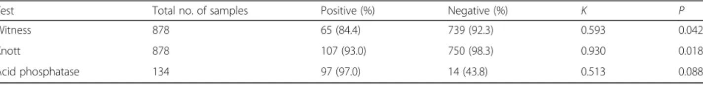
The present study showed thatD. immitisremains, so far, the dominant species of Dirofilaria genus in Table 3Agreement between ITS2-PCR in relation to direct and serological methods
Test Total no. of samples Positive (%) Negative (%) K P
Witness 878 65 (84.4) 739 (92.3) 0.593 0.042
Knott 878 107 (93.0) 750 (98.3) 0.930 0.018*
Acid phosphatase 134 97 (97.0) 14 (43.8) 0.513 0.088
K: level of agreement between each pair of tests (positive or negative results in both tests)
*P< 0.05
Portugal, as confirmed by sequencing of ITS1 and ITS2 fragments from canine blood samples. These results are consistent with the results by Ferreira et al. [36], who only detected D. immitis in mosquito vectors collected in the same time period in the same districts.
However,D. repenshas recently been identified in one dog in the Algarve [26] in southern Portugal. The Algarve has the highest number of days per year with suitable conditions for Dirofilariatransmission [38] and it is, thus, likely that it has been the point of introduc-tion of this species in Portugal. Although with very low prevalence, the presence of D. repens in the Algarve is worrying since this species has been implicated in the increasing number of reports of human dirofilariosis in Europe [28]. Such introduction was expected, as is the establishment and an increase in prevalence of this para-site species in Portugal, given the ongoing north- and eastward expansion of both Dirofilaria species that has been observed. Such expansion has been mainly attrib-uted to global warming, as well as environmental changes, which promote the expansion of mosquito vec-tors, along with the increased international mobility of infected vertebrates [27, 39–41]. Moreover, many wild
animals can also act as sylvatic reservoirs forDirofilaria spp., thus maintaining transmission of this parasite. In Portugal, the prevalence of D. immitis in red foxes, as determined by necropsy, has ranged from 3.2% in northern-Centre locations, such as Coimbra [42], to 11.8% in southern and central-Centre districts, such as Santarém and Setúbal [43]. Additionally, in a national serological survey conducted in red foxes, 8.5% were positive for D. immitiscirculating antigen, with positive animals found in northern and southern areas of Portugal [44].Dirofilaria immitis has also been reported in three Eurasian otters, Lutra lutra, in Portuguese natural freshwater habitats [45, 46] and, recently, in a collection of pinnipeds from Algarve [47].
Conclusions
In conclusion, our data strongly suggest thatD. immitis is the main etiological agent of dirofilariosis in Portugal and that PCR of the region ITS2, as applied here, could be a valuable tool for the diagnosis and screening of filarial infections in dogs, given its fast, accurate, specific detection and differentiation of Dirofilaria spp. from other concurrent blood microfilariae.
Abbreviations
AP:Acid phosphatase histochemical staining test; CTAB: Cetyltrimethyl ammonium bromide; EDTA: Ethylenediamine tetraacetic acid; ITS: Internal transcribed spacer; KN: Modified Knott’s technique; Mf: Microfilariae; PCR: Polymerase chain reaction; RFLP: Restriction fragment length polymorphism; WT: Kit WITNESS® Dirofilaria
Acknowledgements
The authors are grateful to the veterinary and auxiliary staff from municipal kennels for their collaboration; to Prof. Eva Fok (University of Veterinary Medicine, Budapest, Hungary) and Prof. Claudio Genchi (Faculty of Veterinary Medicine, University of Milan, Italy) for providingD. repens; to FCT for funds to GHTM - UID/Multi/04413/2013.
Funding
This work was sponsored by the Fundação para a Ciência e a Tecnologia (FCT), Portugal, project PTDC/SAU-SAP/113523/2009. AMA held a FCT PhD grant # SFRH/BD/85427/2012 and was partly funded by Project UID/CVT/ 00276/2013 supported by CIISA-FMV-ULisboa, FCT, Portugal. The funders had no role in study design, data collection and analysis, decision to publish, or preparation of the manuscript. This work was done under the frame of the EurNegVec COST Action TD1303.
Availability of data and materials
All data generated or analyzed during this study are included in the article and its Additional files. Sequences obtained from selected PCR amplicons were deposited in the GenBank database under accession numbers LN626257–LN626259 and LN626261 (samples 391, 623, 360 and 363; complete ITS region); KY014643–KY014648 (samples 483, 394, 350, 361, 488, female adult worm, respectively; ITS1); and KY644132–KY644141 (samples 1, 7, 8, 29, 52, 483, 723, 732, 758, 846, respectively; ITS2).
Authors’contributions
SB and LMC conceived and designed the research project. AMA, JM and LMC participated in the field work, conducted clinical examination and sample collection. AMA performed direct and serological analysis. CF, AA and MC carried out the PCR reactions. MC and IM carried out sequence analyses and alignments. CF, AA, AMA, IM and SB wrote the paper and supervised the statistical analysis. All authors contributed, read and approved the final manuscript.
Competing interests
The authors declare that they have no competing interests.
Consent for publication
Not applicable.
Ethics approval
All the clinical procedures in this study were in accordance with Portuguese (Decree-Laws no. 314/2003 and no. 113/2013) and European legislation for the protection of animals and met the International Guiding Principles for Biomedical Research Involving Animals by the Council for the International Organizations of Medical Sciences. The protocol was approved by the Committee on Ethics of Animal and Animal Welfare (CEBEA) of the Faculdade de Medicina Veterinária, Universidade de Lisboa.
Publisher’s Note
Springer Nature remains neutral with regard to jurisdictional claims in published maps and institutional affiliations.
Author details
1Global Health and Tropical Medicine, GHTM, Instituto de Higiene e Medicina
Tropical, IHMT, Universidade Nova de Lisboa, UNL, Lisboa, Portugal.2CIISA, Faculdade de Medicina Veterinária, Universidade de Lisboa (ULisboa), Lisboa, Portugal.
Received: 16 November 2016 Accepted: 9 May 2017
References
1. Simón F, Siles-Lucas M, Morchón R, González-Miguel J, Mellado I, Carretón E, et al. Human and animal dirofilariasis: the emergence of a zoonotic mosaic. Clin Microbiol Rev. 2012;25:507–44.
2. McCall JW, Genchi C, Kramer LH, Guerrero J, Venco L. Heartworm disease in animals and humans. Adv Parasitol. 2008;66:193–285.
3. Giangaspero A, Marangi M, Latrofa MS, Martinelli D, Traversa D, Otranto D, et al. Evidences of increasing risk of dirofilarioses in southern Italy. Parasitol Res. 2013;112:1357–61.
4. Pampiglione S, Canestri Trotti G, Rivasi F. Human dirofilariasis due to
Dirofilaria(Nochtiella)repens: a review of world literature. Parassitologia. 1995;37:149–93.
5. Simón F, López-Belmonte J, Marcos-Atxutegi C, Morchón R, Martín-Pacho JR. What is happening outside North America regarding human dirofilariasis? Vet Parasitol. 2005;133:181–9.
6. Lindemann BA, Evans TL, McCall JW. Clinical responses of dogs to experimentally inducedDipetalonema recondituminfection. Am J Vet Res. 1983;44:2170–2.
7. Rani PA, Coleman GT, Irwin PJ, Traub RJ.Hippobosca longipennis- a potential intermediate host of a species ofAcanthocheilonemain dogs in northern India. Parasit Vectors. 2011;4:143.
8. Huynh T, Thean J, Maini R.Dipetalonema reconditumin the human eye. Br J Ophthalmol. 2001;85:1391–2.
9. Magnis J, Lorentz S, Guardone L, Grimm F, Magi M, Naucke TJ, et al. Morphometric analyses of canine blood microfilariae isolated by the Knott's test enablesDirofilaria immitisandD. repensspecies-specific and
Acanthocheilonema(syn.Dipetalonema) genus-specific diagnosis. Parasit Vectors. 2013;6:48.
10. Chalifoux L, Hunt RD. Histochemical differentiation ofDirofilaria immitisand
Dipetalonema reconditum. J Am Vet Med Assoc. 1971;158:601–5. 11. Venco L, Genchi C, Simón F. La filariosis cardiopulmonar (Dirofilaria immitis)
en el perro. In: Simón F, Genchi C, Venco L, Montoya MN, editors. La filariosis en las especies domésticas y en el hombre. Barcelona: Merial Laboratorios; 2011. p. 19–60.
12. Casiraghi M, Bazzocchi C, Mortarino M, Ottina E, Genchi C. A simple molecular method for discriminating common filarial nematodes of dogs (Canis familiaris). Vet Parasitol. 2006;141:368–72.
13. Scoles GA, Kambhampati S. Polymerase chain reaction-based method for the detection of canine heartworm (Filarioidea: Onchocercidae) in mosquitoes (Diptera: Culicidae) and vertebrate hosts. J Med Entomol. 1995;32:864–9. 14. Mar PH, Yang IC, Chang GN, Fei AC. Specific polymerase chain reaction for
differential diagnosis ofDirofilaria immitisandDipetalonema reconditum
using primers derived from internal transcribed spacer region 2 (ITS2). Vet Parasitol. 2002;106:243–52.
15. Nuchprayoon S, Junpee A, Poovorawan Y, Scott AL. Detection and differentiation of filarial parasites by universal primers and polymerase chain reaction-restriction fragment length polymorphism analysis. Am J Trop Med Hyg. 2005;73:895–900.
16. Gioia G, Lecová L, Genchi M, Ferri E, Genchi C, Mortarino M. Highly sensitive multiplex PCR for simultaneous detection and discrimination ofDirofilaria immitis
andDirofilaria repensin canine peripheral blood. Vet Parasitol. 2010;172:160–3. 17. Gasser RB, LeGoff L, Petit G, Bain O. Rapid delineation of closely-related filarial
parasites using genetic markers in spacer rDNA. Acta Trop. 1996;62:143–50. 18. Watts KJ, Courteny CH, Reddy GR. Development of a PCR- and probe-based
test for the sensitive and specific detection of the dog heartworm,Dirofilaria immitis, in its mosquito intermediate host. Mol Cell Probes. 1999;13:425–30. 19. Latrofa MS, Weigl S, Dantas-Torres F, Annoscia G, Traversa D, Brianti E, et al.
A multiplex PCR for the simultaneous detection of species of filarioids infesting dogs. Acta Trop. 2012;122:150–4.
20. Nuchprayoon S, Junpee A, Nithiuthai S, Chungpivat S, Suvannadabba S, Poovorawan Y. Detection of filarial parasites in domestic cats by PCR-RFLP of ITS1. Vet Parasitol. 2006;140:366–72.
21. Rishniw M, Barr SC, Simpson KW, Frongillo MF, Franz M, Dominguez Alpizar JL. Discrimination between six species of canine microfilariae by a single polymerase chain reaction. Vet Parasitol. 2006;135:303–14.
23. Vieira AL, Vieira MJ, Oliveira JM, Simões AR, Diez-Baños P, Gestal J. Prevalence of canine heartworm (Dirofilaria immitis) disease in dogs of central Portugal. Parasite. 2014;21:5.
24. Cardoso L, Mendão C, Madeira de Carvalho L. Prevalence ofDirofilaria immitis,Ehrlichia canis,Borrelia burgdorferisensu lato,Anaplasmaspp. and
Leishmania infantumin apparently healthy and CVBD-suspect dogs in Portugal - a national serological study. Parasit Vectors. 2012;5:62. 25. Alho AM, Landum M, Ferreira C, Meireles J, Gonçalves L, Madeira de
Carvalho L, et al. Prevalence and seasonal variations of canine dirofilariosis in Portugal. Vet Parasitol. 2014;206:99–105.
26. Maia C, Lorentz S, Cardoso L, Otranto D, Naucke TJ. Detection ofDirofilaria repensmicrofilariae in a dog from Portugal. Parasitol Res. 2016;115:441–3. 27. Pampiglione S, Rivasi F, Gustinelli A. Dirofilarial human cases in the Old
World, attributed toDirofilaria immitis: a critical analysis. Histopathology. 2009;54:192–204.
28. Genchi C, Kramer LH, Rivasi F. Dirofilarial infections in Europe. Vector Borne Zoonotic Dis. 2011;11:1307–17.
29. Stothard JR, Hughes S, Rollinson D. Variation within the internal transcribed spacer (ITS) of ribosomal DNA genes of intermediate snail hosts within the genusBulinus(Gastropoda: Planorbidae). Acta Trop. 1996;61:19–29. 30. Nuchprayoon S, Sangprakarn S, Junpee A, Nithiuthai S, Chungpivat S,
Poovorawan Y. Differentiation ofBrugia malayiandBrugia pahangiby PCR-RFLP of ITS1 and ITS2. Southeast Asian J Trop Med Public Health. 2003;34 Suppl 2:67–73.
31. Hall TA. BioEdit: a user-friendly biological sequence alignment editor and analysis program for Windows 95/98/NT. Nucl Acids Symp Ser. 1999;41:95–8. 32. Stephens M, Scheet P. Accounting for decay of linkage disequilibrium in
haplotype inference and missing-data imputation. Am J Hum Genet. 2005; 76:449–62.
33. Kumar S, Stecher G, Tamura K. MEGA7: Molecular Evolutionary Genetics Analysis Version 7.0 for Bigger Datasets. Mol Biol Evol. 2016;33:1870–4. 34. IonicăAM, Matei IA, Mircean V, Dumitrache MO, D'Amico G, Győrke A, et al.
Current surveys on the prevalence and distribution ofDirofilariaspp. and
Acanthocheilonema recondituminfections in dogs in Romania. Parasitol Res. 2015;114:975–82.
35. American Heartworm Society. Current canine guidelines for the prevention, diagnosis and management of heartworm (Dirofilaria immitis) infection in dogs. 2014. https://heartwormsociety.org/images/pdf/2014-AHS-Canine-Guidelines.pdf. Accessed 5 Feb 2017.
36. Ferreira C, Mixão V, Novo T, Calado M, Gonçalves L, Belo S, et al. First molecular identification of mosquito vectors ofDirofilaria immitisin continental Portugal. Parasit Vectors. 2015;8:139.
37. Menn B, Lorentz S, Naucke TJ. Imported and travelling dogs as carriers of canine vector-borne pathogens in Germany. Parasit Vectors. 2010;3:34. 38. Alho AM, Nunes T, Rinaldi L, Meireles J, Belo S, Deplazes P, et al. Transmission
risk of dirofilariosis in Portugal. Parasit Vectors. 2014;7 Suppl 1:O16. 39. Genchi C, Rinaldi L, Mortarino M, Genchi M, Cringoli G. Climate and
Dirofilariainfection in Europe. Vet Parasitol. 2009;163:286–92.
40. Morchón R, Carretón E, Grandi G, González-Miguel J, Montoya-Alonso JA, Simón F, et al. Anti-Wolbachiasurface protein antibodies are present in the urine of dogs naturally infected withDirofilaria immitiswith circulating microfilariae but not in dogs with occult infections. Vector Borne Zoonotic Dis. 2012;12:17–20. 41. Fuehrer HP, Auer H, Leschnik M, Silbermayr K, Duscher G, Joachim A.
Dirofilariain humans, dogs, and vectors in Austria (1978–2014) - from imported pathogens to the endemicity ofDirofilaria repens. PLoS Negl Trop Dis. 2016;10:e0004547.
42. Eira C, Vingada J, Torres J, Miquel J. The helminth community of the red fox,
Vulpes vulpes, in Dunas de Mira (Portugal) and its effect on host condition. Wildl Biol Pract. 2006;2:26–36.
43. Carvalho-Varela M, Marcos MVM. A helmintofauna da raposa (Vulpes vulpes silacea) em Portugal. Acta Parasitol Port. 1993;1:73–9.
44. Alho AM, Cortes H, Lopes AP, Vila-Viçosa MJ, Cardoso L, Belo S, et al. Detection ofDirofilaria immitisantigen in red foxes (Vulpes vulpes) from Portugal. Parasit Vectors. 2016;10 Suppl 1:A16.
45. Torres J, Feliu C, Fernández-Morán J, Ruíz-Olmo J, Rosoux R, Santos-Reis M, et al. Helminth parasites of the Eurasian otterLutra lutrain southwest Europe. J Helminthol. 2004;78:353–9.
46. Saraiva AL, Sousa S, Silva J, Andrade S, Botelho N, Canavarro I, et al.Dirofilaria immitisin an Eurasian Otter (Lutra lutra). J Comp Pathol. 2013;148:88. 47. Alho AM, Marcelino I, Colella V, Flanagan C, Silva N, Correia JJ, et al.Dirofilaria
immitisin pinnipeds and new host record. Parasit Vectors. 2017;10:142.
• We accept pre-submission inquiries
• Our selector tool helps you to find the most relevant journal
• We provide round the clock customer support
• Convenient online submission
• Thorough peer review
• Inclusion in PubMed and all major indexing services • Maximum visibility for your research
Submit your manuscript at www.biomedcentral.com/submit