Cop
yright
© ABE&M t
odos os dir
eit
os r
eser
vados
.
Is thyroid stunning clinically
relevant? A retrospective
analysis of 208 patients
O atordoamento da tireoide é clinicamente relevante? Análise retrospectiva de 208 pacientes
Elba C. S. C. Etchebehere¹, Allan O. Santos¹, Patrícia S. Matos², Lígia V. M. Assumpção3, Maria Cecília V. L. Lima¹, Mariana C. L. Lima1, Laura S. Ward4
ABSTRACT
Objective: Current guidelines have advised against the performance of 131I-iodide diagnostic
whole body scintigraphy (dxWBS) to minimize the occurrence of stunning, and to guarantee the eficiency of radioiodine therapy (RIT). The aim of the study was to evaluate the impact of stunning on the eficacy of RIT and disease outcome. Subjects and methods: This retrospective analysis included 208 patients with differentiated thyroid cancer managed according to a same protocol and followed up for 12-159 months (mean 30 ± 69 months). Patients received RIT in do-ses ranging from 3,700 to 11,100 MBq (100 mCi to 300 mCi). Post-RIT-whole body scintigraphy images were performed 10 days after RIT in all patients. In addition, images were also perfor-med 24-48 hours after therapy in 22 patients. Outcome was classiied as no evidence of disease (NED), stable disease (SD) and progressive disease (PD). Results: Thyroid stunning occurred in 40 patients (19.2%), including 26 patients with NED and 14 patients with SD. A multivariate analysis showed no association between disease outcome and the occurrence of stunning (p = 0.3476). Conclusion: The eficacy of RIT and disease outcome do not seem to be related to thyroid stunning. Arq Bras Endocrinol Metab. 2014;58(3):292-300
Keywords
Thyroid cancer; radioiodine therapy; radionuclide scintigraphy; stunning; 131I-iodide
RESUMO
Objetivo: As diretrizes atuais alertam contra a execução da cintigraia de corpo inteiro com iodo-131 (dxWBS) para minimizar a ocorrência de atordoamento e garantir a eiciência do
tra-tamento com radioiodo (RIT). O objetivo deste estudo foi avaliar o impacto do atordoamento sobre a eicácia do RIT e desfechos da doença. Sujeitos e métodos: Esta análise retrospectiva incluiu 208 pacientes com câncer diferenciado de tireoide submetidos ao mesmo protocolo e acompanhados por 12-159 semanas (média de 30 ± 69 meses). Os pacientes receberam RIT com doses variando de 3.700 a 11.100 MBq (100 mCi a 300 mCi). As imagens da cintigraia após a RIT foram feitas 10 dias depois da RIT em todos os pacientes. Além disso, as imagens foram também obtidas após 24-48h em 22 pacientes. O desfecho foi classiicado como nenhu-ma evidência de doença (NED), doença estável (SD) e doença progressiva (PD). Resultados: O atordoamento da tireoide ocorreu em 40 pacientes (19,2%), incluindo 26 pacientes com NED e 14 pacientes com SD. A análise multivariada não mostrou associação entre o desfecho da doen-ça e a ocorrência de atordoamento (p = 0,3476). Conclusão: A eicácia da RIT e o desfecho da doença não parecem estar relacionados com o atordoamento da tireoide. Arq Bras Endocrinol Metab. 2014;58(3):292-300
Descritores
Câncer de tireoide; tratamento com radioiodo; cintigraia com radionucleotídeos; atordoamento; iodo-131
1 Division of Nuclear Medicine of the
Department of Radiology, School of Medical Sciences, Universidade Estadual de Campinas (Unicamp), Campinas, SP, Brazil
2 Department of Pathology, School
of Medical Sciences, Unicamp, Campinas, SP, Brazil
3 Division of Endocrinology of the
Department of Internal Medicine, School of Medical Sciences, Unicamp, Campinas, SP, Brazil
4 Laboratory of Molecular Cancer
Genetics, School of Medical Sciences, Unicamp, Campinas, SP, Brazil
Correspondence to:
Elba C. S. C. Etchebehere Division of Nuclear Medicine, Faculdade de Ciências Médicas, Universidade Estadual de Campinas Av. Zeferino Vaz, s/n
13081-970 – Campinas, SP, Brazil [email protected]
Cop
yright
© ABE&M t
odos os dir
eit
os r
eser
vados
.
INTRODUCTION
Differentiated thyroid cancer (DTC) may be treated by total thyroidectomy, 131I-iodide therapy (RIT),
and hormone suppression therapy (1,2). Prior to RIT, whole-body diagnostic scintigraphy with 131I-iodide
(dxWBS) may be performed for treatment planning. However, the performance of dxWBS, as well as the dose recommended for the procedure is controversial due to many descriptions regarding thyroid stunning. Thyroid stunning is deined as a reduction or absence of 131I-iodide uptake in the cervical region observed
on post-therapy whole body scintigraphy performed after a diagnostic dose of 131I-iodide (post-RIT-WBS)
(3). This phenomenon, as hypothesized, would impair the uptake of a subsequent RIT, leading to ineffec-tive treatment (4). Therefore, some institutions have abandoned dxWBS, preferring to perform RIT with empirical doses to prevent the occurrence of thyroid stunning. Other institutions have substituted diagnos-tic 131I-iodide-WBS for 123I-iodide-WBS, although the
costs of the latter are higher, and the stunning effect, due to the emission of Auger electrons by 123I-iodide,
could be more severe than with 131I-iodide (5).
Howe-ver, the true nature of this phenomenon remains uncer-tain. Moreover, there is no clear evidence that stunning actually interferes with disease outcomes.
Conversely, dxWBS has many beneits, distinguish-ing patients who may actually beneit from treatment from those who should not be treated (no cervical up-take with low thyroglobulin levels; large surgical thy-roid remnants; undetected distant metastases). The correct assessment of disease sites can be used to deter-mine and program RIT without exposing the patient to the risk of ineffective empirical doses. If the Nuclear Medicine physician decides not to perform a dxWBS prior to RIT, he may have to assume legal responsibility for performing empirical treatment doses of iodine-131I
in patients without cervical 131I-iodide uptake.
This study aimed to retrospectively evaluate the clinical impact and eficacy of RIT in patients with DTC with and without stunning.
SUBJECTS AND METHODS
This retrospective study was approved by the Resear-ch Ethics Committee of the SResear-chool of Medical Scien-ces of the Universidade Estadual de Campinas (CEP # 1079/2009).
Patients
A total of 208 patients with differentiated thyroid car-cinoma consecutively referred to our Nuclear Medicine facility for RIT from 1998 to 2012 were retrospectively reviewed.
All patients were submitted to total thyroidectomy, with or without lymph node dissection, depending on neck ultrasound and intraoperative indings, and re-ferred for RIT. Serum thyroglobulin (Tg) levels, anti-thyroglobulin antibodies (TgAb), and TSH were ob-tained before and after thyroid hormone withdrawal, immediately prior to a 131I-iodide dxWBS. All patients
were followed up with regular serum Tg, TSH and TgAb measurements; annual cervical ultrasonography; annual dxWBS; and other eventual diagnostic imag-ing modalities, such as computed tomography, 99m
Tc-sestamibi whole body scintigraphy, 201-thallium whole body scintigraphy, 18F-FDG-PET/CT (after 2008),
and bone scintigraphy for a period range of 12-159 months (mean 30 ± 69 months). Patients with high or increasing serum Tg levels (> 2 mg/dL) and/or suspi-cious whole body scans were submitted to a thorough imaging studies.
Patients were classiied according to pTNM stag-ing for DTC (1,2). Urine iodine measurements are not routinely measured in our facility.
131I-Iodide DxWBS
All patients were oriented to follow a low iodine diet for 15 days, refraining from iodide-rich substances, as well as intravenous iodide contrasts for three months prior to dxWBS. All patients received 185 MBq (5 mCi) of 131I-iodide. Images were performed after 48
hours using a dual-head scintillation camera, with high--energy parallel-hole collimators. The photopeak was centered at 364 KeV with 15% energy window level, and bed speed of 5 cm per minute.
RIT and post-RIT-WBS
Cop
yright
© ABE&M t
odos os dir
eit
os r
eser
vados
.
7,400 MBq or 200 mCi for lung metastases, and 9,250 - 11,100 MBq or 250 - 300 mCi for bone involvement. Post-therapeutic WBS (post-RIT-WBS) was per-formed on all 208 patients 10 days after discharge, with the same parameters used on the dxWBS.
A subgroup of 22 patients also underwent two post-RIT-WBS: one after 24-48 hours and another after 10 days. In this subgroup of patients, the bed speed of the 24-48 hour post-RIT-WBS was 10 cm per minute due to higher radioactivity counts.
Classiication of thyroid stunning
Two experienced nuclear medicine physicians visually assessed images from the dxWBS and post-RIT-WBS. In case of discordant indings, a third nuclear medicine physician reviewed the study. The readers were blinded to other studies and clinical outcomes. They blindly classiied the patients as: no stunning, partial stunning, or total stunning. The classiication of stunning was performed only after the irst radioiodine treatment, taking into consideration cervical uptake and eventual metastases. Subsequent treatments were not conside-red for the occurrence of stunning. Uptake conside-reduction in at least one of the foci was enough to classify the patient as having thyroid stunning.
Classiication of stunning was performed as follows: 1. No stunning: uptake in post-RIT-WBS was
equal to uptake on the dxWBS.
2. Partial stunning: uptake in 10 days post-RIT-WBS was partially reduced when compared with dxWBS (Figure 1).
3. Total stunning: uptake in 10 days post-RIT-WBS was absent when compared with dxpost-RIT-WBS (Figure 2).
Among the 22 patients who also underwent a post-RIT-WBS 24-48 hours after discharge, there were six patients presenting a partial stunning with washout pat-tern: the 24-48 hours uptake post-RIT-WBS was equal to the uptake seen on dxWBS. However, 10 days post-RIT-WBS, the uptake was partially reduced, as demon-strated in igure 3.
Disease status classiication
Patients were classiied as: no evidence of disease (NED); stable disease (SD); and progressive disease (PD). This classiication was performed one year after each RIT and at the end of the follow-up period. Thus, for example, if a patient was submitted to three RIT,
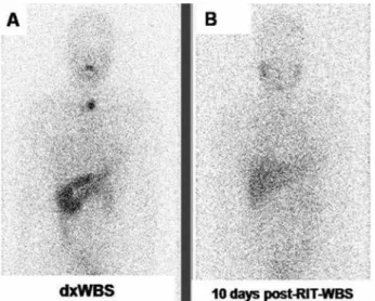
Figure 1. An example of a partial stunning. (A) 131I-iodide dxWBS shows
increased cervical uptake. (B) Post-RIT-WBS shows a partial reduction of
the cervical uptake when compared with (A).
Figure 2. An example of a total stunning. (A) 131I-iodide dxWBS shows
increased cervical uptake. (B) Post-RIT-WBS shows a no cervical uptake
when compared with (A).
he was reclassiied one year after each treatment and at the end of follow-up. This classiication was based on the results of the annual dxWBS; the suppressed and stimulated serum Tg levels; the neck ultrasound and on other eventual imaging data (18F-FDG PET/CT or 99mTc-sestamibi).
Cop
yright
© ABE&M t
odos os dir
eit
os r
eser
vados
.
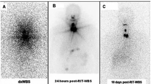
Figure 3. An example of progressive washout in a young female patient. (A) 131I-iodide dxWBS depicts markedly increased cervical uptake. (B) 24 hours
post-RIT-WBS shows basically the same cervical uptake when compared with 131I-iodide dxWBS. (C) 10 days post-RIT-WBS shows a marked reduction in
the cervical uptake.
The SD classiication was based on one or more of the following conditions: persistent cervical uptake and/or metastases on the annual dxWBS or neck ul-trasound; Tg non-stimulated levels above 1.0 ng/ mL, with stimulated Tg levels above 10.0 ng/mL (but without progressive elevation); positive stable levels of TgAb.
Patients were classiied as PD when presenting any of the following parameters: new metastases detected at annual dxWBS or neck ultrasound; new metastases de-tected in other imaging modalities; progressive increase of serum Tg levels despite RIT.
Statistical analysis
The statistical analysis was carried out using the SAS System for Windows (Statistical Analysis System, ver-sion 9.1.3, Service Pack 3. SAS Institute Inc., 2002-2003, Cary, NC, USA). The Pearson’s chi-square test was performed to evaluate the association be-tween the occurrence of stunning and gender, histo-logical subtype, aggressive variants, the presence of metastases at diagnosis, Tg levels and disease status. Nonparametric analysis was performed using either the chi-square or Fisher’s exact tests as indicated. The Mann-Whitney was performed to compare the pre-sence of stunning with cumulative doses of 131
I-iodi-de. A multiple logistic regression analysis was used to evaluate the role of the occurrence of stunning and the pTNM stage; the initial and inal serum Tg and TgAb levels; tumor histology; and the presence of
metastases at diagnosis. All tests were conducted with a 0.05 level of signiicance.
RESULTS
Patient population data
As expected, most DTC patients were female (85%), with a mean age of 43.3 ± 14.1 years, as represented in table 1. There were 178 (85.6%) papillary thyroid car-cinomas including 46 (22.1%) cases of variants conside-red aggressive (tall cell, columnar cell, diffuse sclerosing, solid/trabecular, and insular variants); and 30 (14.4%) follicular carcinomas (all of them widely invasive).
According to the pTNM staging, there were 105 (50.5%) stage I patients; 43 (20.7%) stage II; 18 (8.7%) stage III; and 42 (20.2%) stage IV patients. Among the stage IV patients, 6 were IVa and 36 were IVb. Distant metastases at diagnosis occurred in 28 (13.5%) patients.
Only one patient did not present any cervical uptake in the dxWBS. However, RIT was performed because of high serum thyroglobulin (89.8 ng/mL) levels.
Cop
yright
© ABE&M t
odos os dir
eit
os r
eser
vados
.
Thyroid stunning
There was no inter-observer variability in any of the stu-dies. Thyroid stunning occurred in 40 patients (19.2%) (Table 1). Thirty-four out of these patients (85%) were classiied as partial stunning and 6 (15%) were classiied as total stunning. There was a signiicant association be-tween stunning and gender, which was more common in females, and may be related to sample size (p = 0.05;
Pearson’s Chi-Square test). However, a signiicant in-verse relationship between stunning and age was noted (Z = - 3.74; p < 0.000001; Mann-Whitney test). Youn-ger patients tended to have more stunning effects than older patients.
There was no association between stunning and the histological classiication (p = 0.059; Pearson’s Chi-Square test). However, we observed that nonaggres-sive variants of papillary thyroid carcinomas presented
Table 1. Comparison of clinical, pathological and outcome data between patients who showed stunning or not
Mean ± SD (min – max)
Total number of patients (percentage)
Non-stunning (percentage)
Stunning (percentage)
p-value
Patients 208 168 (80.8) 40 (19.2)
Initial Tg levels 136.8 ± 638.3
(0.2 to 6,000)
194 157 (80.9) 37 (19.1) Z-score = -1.422
P < 0.155***
Final Tg levels 185.0 ± 1053.4
(0.2 to 11,480)
207 168 (81.2) 39 (18.8) Z-score = -1.309
P < 0.191***
Cumulative RIT dose 263.8 ± 219.1
(92 to 1,100)
208 168 (80.8) 40 (19.2) Z-score = -2.239
P < 0.025***
Age 43.3 ± 14.1 208 168 (80.8) 40 (19.2) Z-score = -3.740
P < 0.0001***
Gender Female 177 (85.1) 139 (78.5) 38 (21.5) 0.05*
Male 31 (14.9) 29 (93.5) 2 (6.5)
Histology Follicular 30 (14.4) 28 (93.3) 2 (6.7) 0.059*
Papillary 178 (85.6) 140 (78.7) 38 (21.3)
PTC variant Aggressive 46 (22.1) 43 (93.5) 3 (6.5) 0.013*
Non-aggressive 162 (77.9) 125 (77.2) 37 (22.8)
Initial TgAb levels Negative 176 (84.6) 145 (82.4) 31 (17.6) 0.337**
Positive 18 (8.6) 13 (72.2) 5 (27.8)
Final TgAb levels Negative 195 (93.7) 158 (81.0) 37 (19.0) 0.718**
Positive 13 (6.3) 10 (76.9) 3 (23.1)
TNM I/II 148 (71.2) 108 (73.0) 40 (23.0) < 0.0001*
III/IV 60 (28.8) 60 (100) 0 (0)
Presence of metastases at diagnosis
No 179 (86.1) 142 (79.3) 37 (20.7) 0.215*
Yes 28 (13.4) 25 (89.3) 3 (10.7)
Clinical outcome NED 121 (58.2) 95 (78.5) 26 (23.5) 0.34****
SD 64 (30.8) 50 (78.1) 14 (21.9)
PD 23 (11.1) 23 (100) 0 (0)
* Pearson’s Chi-Square Test; ** Fisher’s Exact Test; *** Mann-Whitney U Test; **** Multiple Logistic Regression Analysis.
Table 2. Initial radioiodine therapy doses in the entire patient population, according to the presence and region of metastases
Location of uptake MBq (mCi) Total number of patients (percentage)
Initial radioiodine therapy dose in MBq
Cervical uptake without lymph node metastases 3,700 (100) 116 (55.8)
Lymph node metastases 5,550 (150) 70 (33.7)
Lung metastases 7,400 – 9,250 (200 – 250) 21 (10.0)
Cop
yright
© ABE&M t
odos os dir
eit
os r
eser
vados
.
stunning more frequently then aggressive variants (p = 0.013; Pearson’s Chi-Square test). In addition, the phenomenon was more frequent in pTNM stages I and II than in stages III and IV patients (p < 0.001; Pear-son’s Chi-Square test) (Table 1).
The initial serum Tg levels (before thyroid hormone withdrawal) were of 136.8 ± 638.3 (mean ± standard deviation) and ranged from 0.2 to 6000.0. There was no association between stunning and initial serum Tg levels (Z = - 1.422; p = 0.155; Mann-Whitney test).
Final serum Tg levels were of 185.0 ± 1053.4 (mean ± standard deviation) and ranged from 0.2 to 11480.0. There was no association between stunning and inal serum Tg levels (Z = - 1.309; p = 0.191; Mann-Whit-ney test).
No associations were noted between stunning and initial serum TgAb levels as well (p = 0.337; Fisher’s exact test); follow-up TgAb levels (p = 0.718; Fisher´s exact test); disease recurrence (p = 1.000; Fisher’s exact test); or the presence of metastases at diagnosis (p = 0.215; Pearson’s chi-square test).
The RIT cumulative doses were of 263.8 ± 219.1 mCi and ranged from 92 to 1100 mCi. Stunning did not occur in patients who required more RITs (p = 0.020; nonparametric Mann-Whitney test) or higher cumulative doses (p = 0.025; nonparametric Mann-Whitney test). Thus, stunning did not occur in patients who were followed-up for a longer period of time due to PD (p = 0.002; nonparametric Mann-Whitney test) (Table 1).
Disease status data
One year after the irst RIT, patients were classiied according to the disease status, as follows: 127 NED patients (61.1%), 59 SD patients (28.4%), and 22 PD patients (10.5%). Six patients (2.9%) recurred (3 pa-tients after two years of the irst RIT and 3 papa-tients after two years of the second RIT). Therefore, patients were reclassiied as: NED – 121 patients (58.2%), SD – 64 patients (30.8%) and PD – 23 patients (11.1%).
In the NED group, 21.5% (26/121) of the patients presented stunning, while in the SD group, stunning occurred in 21.8% (14/64) of the cases. None of the PD patients presented stunning.
Multiple logistic regression analysis showed that the presence of stunning had no association with gender type nor with pTNM stages III and IV, the presence of metastasis or Tg levels. In addition, stunning occur-rence had no impact on disease outcome (p = 0.33).
DISCUSSION
There is no agreement whether to perform diagnostic whole-body scintigraphy (dxWBS) prior to radioiodine therapy (RIT) or not in patients with well-differentia-ted thyroid cancer. Numerous authors and organiza-tions have argued either to perform dxWBS prior to RIT (6-8), or not to perform it (9,10) in these patients. Although this is a controversial issue in the literature, recent studies published by well-known journals and several world-wide renowned institutions still consider not only that dxWBS is an essential exam, but also an important tool to better understand the stunning phe-nomenon. Chen and cols. (8) recommend dxWBs prior to RIT to provide the information required to prescri-be an appropriate dose of 131I-iodide for effective
treat-ment, stating the importance of unsuspected lymph no-des or distant metastases, which involve a signiicantly higher dose, besides the diagnosis of unexpected large thyroid remnants, in which a new surgery or a two-step ablation may be necessary.
Another study that supports the dxWBS prior to ablation was published by Van Nostrand and cols. (6). The authors support the performance of this exam, for it can be useful by demonstrating indings that may al-ter the patient management prior to RIT in the fol-lowing situations: absence of 131I-iodide uptake in the
thyroid bed; a signiicant number of foci of uptake in the thyroid bed/neck region; multiple foci in the neck outside the thyroid bed; distant metastases and/or breast uptake.
The European consensus (9) also questions the use of dxWBS prior to ablation, stating that this exam may be avoided without loss of information. However, at the same time, the authors comment that dxWBS could be performed by some centers in some situations, for example when there is uncertainty concerning the ex-tent of thyroidectomy, for large thyroid remnants that may require new surgery, or the use of corticosteroids to avoid radiation thyroiditis. According to the edito-rial by McDougall (11), the European consensus ac-knowledges that patient information regarding stage obtained after RIT is contrary to “good medicine” should be practiced, considering that all the diagnostic data should be obtained irst and that the treatment should be based on that information.
sur-Cop
yright
© ABE&M t
odos os dir
eit
os r
eser
vados
.
gical description at all, many times with large thyroid remnants that may beneit from a second thyroidecto-my before RIT. These large remnants may cause radia-tion thyroiditis if treated blindly (12). Besides, dxWBS provides important information considering not only the amount of thyroid remnants, but also a complete evaluation and diagnosis of unsuspected lymph node metastases, which require a signiicantly higher dose of RIT (8).
No cervical uptake (no remnants), on the other hand, may occur following total thyroidectomy. An ul-trasound to detect the presence of absence of thyroid remnants has low sensitivity and does not substitute dxWBS for this purpose (8). Patients that are found to have no cervical uptake (no remnants) on a post-therapy WBS most likely will not beneit from RIT. Therefore, if the Nuclear Medicine physician does not perform a dxWBS prior to RIT, it is the Nuclear Medi-cine physician who may have to assume possible legal responsibility for performing empirical RIT in patients without cervical 131I-iodide uptake.
Thus, as McDougall again states in his editorial (11), one of the essentials of treating patients with can-cer is to know the extent of the disease as accurately as possible. The authors’ opinion considering the legal responsibility is exactly supported by this precaution, always searching for the best evaluation of each patient.
On the contrary, larger dosages of radioiodine may be administered at the time of ablation if regional or distant metastases are detected on a pre-ablation scan (8). In addition, the use of dxWBS prior to RIT may identify unsuspected uptake in the breast due to lacta-tion or medicalacta-tion (13) and distant brain metastases (14). Patients with breast uptake should only be treated after the uptake is reduced to prevent mastitis. Brain metastases, albeit in a minority of patients, should be treated with corticosteroids or radiotherapy prior to RIT in order to prevent swelling (13,14).
However, most guidelines, including the ones by the American Thyroid Association and the National Comprehensive Cancer Network, advise against the performance of dxWBS in order to minimize the oc-currence of stunning and guarantee the eficiency of RIT (1,2,15), even though they recognize that this phenomenon is not clearly understood and does not seem to have an impact on the patient outcome (16). Thyroid stunning is deined as a reduction or absence of 131I-iodide uptake in the cervical region observed on
the post-RIT-WBS due to impairment of uptake of a
subsequent RIT, leading to ineffective treatment. This phenomenon is not known to be due to post-diagnostic scan time dependent. In fact, a few studies concluded that thyroid stunning could cause a worse clinical out-come, as it is associated with the need for more RIT sessions (4,17). On the contrary, other studies compar-ing the eficacy of radioiodine therapy between patients submitted to dxWBS prior to RIT, and patients not submitted to pretreatment scan prior to RIT failed to show differences in success rates of RIT or in patient outcomes (18-20).
The current study, although retrospective, is com-posed by a large cohort of patients submitted to the same management protocol and followed up for a considerable period of time (mean of 60 months). We clearly documented stunning in 40 (19.2%) patients. Nevertheless, no difference in outcomes occurred among them, and the rest of the 168 (80.8%) patients did not show stunning. It would be interesting to fol-low up patients for a longer period of time, considering that recurrence of thyroid cancer can occur up to 10 years after the initial treatment.
Cop
yright
© ABE&M t
odos os dir
eit
os r
eser
vados
.
the 24-48 hours following RIT, thyroid stunning could pass unnoticed in the majority of the patients.
The weaknesses in our study include the fact that it is retrospective and, therefore, we could not compare the outcome of patients who did and did not receive dxWBS prior to treatment. Also, unfortunately, only a subset of patients (22) underwent a 24-48 hours post-RIT-WBS, which we deined as “partial stunning with washout”. We do not know how many other subjects would have demonstrated this inding if 24-48 hour post-RIT-WBS were performed in all patients. How-ever, this does not lead to misclassiication of the other patients, since all patients were classiied as stunning according to the deinition in the literature. We did not perform quantitative analysis of the images to clas-sify the cervical uptake in the post-RIT-WBS as posi-tive, negaposi-tive, or with reduction compared to dxWBS. These indings were undisputable.
The lack of data of a control group (patients not receiving a dxWBS) and the impossibility to evaluate all the patients with 24-48 hours post-RIT-WBS appar-ently did not interfere with the results obtained, and this argument may be corroborated by the fact that, in the current study, none of the 23 patients (11.1%) with progressive disease had thyroid stunning. Con-versely, stunning occurred in 21.5% of patients without evidence of disease, and in 21.8% of patients with stable disease. In fact, stunning was also more likely to oc-cur in younger patients and in those with nonaggressive variants, and this may also be related to better outcome. The patient population was also not heterogeneous as all patients were submitted to an initial dose for thyroid remnants and the majority (89%) solely to this initial dose for thyroid remnants. The remaining 11% were submitted to more doses because of metastatic disease that developed later during follow-up.
Although many guidelines recommend 37 – 101 MBq of iodine-131 dose for dxWBS in part due to stunning concerns, even with our dose of 185 MBq we did not ind any difference of outcome among groups. Considering the use of 131I-iodide and its doses for
dxWBS, this is exactly the protocol still used by many institutions and in recent studies, as described by Amin and cols. (23). The option for this protocol is based on the concern that the sensitivity in the detection of foci that present 131I-iodide uptake may be reduced, and
the substitution for 123I-iodide, besides its high costs,
is controversial, for this radiopharmaceutical treatment could increase the so-called stunning effect, due to the
emission of Auger electrons by 123I-iodide (5). The
stunning effect is not well-known, as also highlighted by the American Thyroid Association and the National
Comprehensive Cancer Network (16), demanding new
scientiic studies with methodologies utilized in the routine clinical practice for better understanding of this phenomenon.
In conclusion, the occurrence of thyroid stunning did not reduce the eficiency of radioiodine therapy and did not impact negatively on patient outcomes in the group studied. Although all the limitations of the study, mainly the lack of control group and of a longer follow-up period, dxWBS may be performed safely and when necessary in DTC patients, and post-RIT-WBS after 24-48 hours, rather than after 10 days, should be considered.
Acknowledgments: we are grateful to Cleide Silva and Eduar-do Hoehne from the Department of Statistics for their statis-tical analysis of the data. The São Paulo Research Foundation (Fapesp) provided inancial support for this study (Protocol #09/548663).
Disclosure: no potential conlict of interest relevant to this article was reported.
REFERENCES
1. Cooper DS, Doherty GM, Haugen BR, Kloos RT, Lee SL, Mandel SJ, et al. Revised American Thyroid Association Management Guidelines for Patients with Thyroid Nodules and Differentiated Thyroid Cancer: The American Thyroid Association (ATA) Guideli-nes Taskforce on Thyroid Nodules and Differentiated Thyroid Can-cer. Thyroid. 2009;19:1167-214.
2. Rosário PW, Ward LS, Carvalho GA, Graf H, Maciel RB, Maciel LMZ, et al. Thyroid nodule and differentiated thyroid cancer: update on the Brazilian consensus. Arq Bras Endocrinol Metabol. 2013;57:240-64.
3. Medvedec M. Thyroid stunning in vivo and in vitro. Nucl Med Commun. 2005;26:731-5.
4. Lees W, Mansberg R, Roberts J, Towson J, Chua E, Turtle J. The clinical effects of thyroid stunning after diagnostic whole-body scanning with 185 MBq 131I. Eur J Nucl Med. 2002;29:1421-7.
5. Lundh C, Lindencrona U, Postgård P, Carlsson T, Nilsson M, Forssell-Aronsson E. Radiation-induced thyroid stunning: diffe-rential effects of 123I, 131I, 99mTc, and 211At on iodide transport
and NIS mRNA expression in cultured thyroid cells. J Nucl Med. 2009;50:1161-7.
6. Van Nostrand D, Aiken M, Atkins F, Moreau S, Garcia C, Acio E, et al. The utility of radioiodine scans prior to iodine 131 abla-tion in patients with well-differentiated thyroid cancer. Thyroid. 2009;19:849-55.
Cop
yright
© ABE&M t
odos os dir
eit
os r
eser
vados
.
8. Chen MK, Yasrebi M, Samii J, Staib LH, Doddamane I, Cheng DW. The utility of I-123 pretherapy scan in I-131 radioiodine therapy for thyroid cancer. Thyroid. 2012;22:304-9.
9. Pacini F, Schlumberger M, Dralle H, Elisei R, Smit JW, Wiersinga W. European Thyroid Cancer Taskforce. European consensus for the management of patients with differentiated thyroid carcinoma of the follicular epithelium. Eur J Endocrinol. 2006;154:787-803. 10. Schlumberger M, Pacini F, Wiersinga WM, Toft A, Smit JW,
San-chez Franco F, et al. Follow-up and management of differentiated thyroid carcinoma: a European perspective in clinical practice. Eur J Endocrinol. 2004;151:539-48.
11. McDougall IR. The case for obtaining a diagnostic whole-body scan prior to iodine 131 treatment of differentiated thyroid can-cer. Thyroid. 2009;19:811-3.
12. Samuel AM, Rajashekharrao B. Radioiodine therapy for well--differentiated thyroid cancer: a quantitative dosimetric eva-luation for thyroid remnant ablation after surgery. J Nucl Med. 1994;35:1944-50.
13. Hammami MM, Bakheet S. Radioiodine breast uptake in nonbre-astfeeding women: clinical and scintigraphic characteristics. J Nucl Med. 1996;37:26-31.
14. Datz FL. Cerebral edema following iodine-131 therapy for thyroid carcinoma metastatic to the brain. J Nucl Med. 1986;27:637-40. 15. National Comprehensive Cancer Network (NCCN) 2010, NCCN
Guidelines for Treatment of Cancer by Site, Thyroid Carcinoma. Available at: http://www. nccn.org/ professionals/physician_gls/ pdf/thyroid.pdf.
16. Morris LF, Waxman AD, Braunstein GD. Thyroid stunning. Thyroid. 2003;13:333-40.
17. Leger FA, Izembart M, Dagousset F, Barritault L, Baillet G, Cheva-lier A, et al. Decreased uptake of therapeutic doses of 131-iodi-ne after 185-MBq and 123-iodi131-iodi-ne diagnostic imaging for thyroid remnants in differentiated thyroid carcinoma. Eur J Nucl Med. 1998;25:242-6.
18. Morris LF, Waxman AD, Braunstein GD. The non-impact of thyroid stunning: remnant ablation rates in 131I-scanned and non-scanned
individuals. J Clin Endocrinol Metab. 2001;86:3507-11.
19. Rosario PWS, Barroso AL, Rezende LL, Padrão EL, Fagundes TA, Reis JS, et al. Outcome of ablation of thyroid remnants with 100 mci (3.7 Gbq) 131-iodine in patients with thyroid cancer. Ann Nucl Med. 2005;19:247-50.
20. Dam HQ, Kim SM, Lin HC, Intenzo CM. 131I therapeutic eficacy
is not inluenced by stunning after diagnostic whole-body scan-ning. Radiology. 2004;232:527-33.
21. Sisson JC, Avram AM, Lawson SA, Gauger PG, Doherty GM. The so-called stunning of thyroid tissue. J Nucl Med. 2006;47:1406-12. 22. Silberstein EB, Alavi A, Balon HR, Becker D, Charkes D, Clarke S,
et al. Society of nuclear medicine procedure guideline for the-rapy of thyroid disease with Iodine-131, version 2.0. Available at: http://interactive.snm.org/docs/ Therapy% 20of% 20Thyroid% 20Disease%20with%20Iodine-131%20v2.0.pdf.