Copyright
© ABE&M todos os dir
eitos r
eser
vados.
Simultaneous evaluation of
in vivo
glucocorticoid sensitivity and
expression of glucocorticoid
receptor alpha-isoform in
rheumatoid arthritis patients
Avaliação concomitante da sensibilidade in vivo aos
glicocorticóides e da expressão da isoforma alfa do receptor de glicocorticóide em pacientes com artrite reumatóide
Jayme F. Cobra1, Murilo R. Melo2, Claudia D. C. Faria2,
Carlos Alberto Longui2, Osmar Monte2
1 Rheumatology Discipline, Internal
Medicine, Irmandade da Santa Casa de Misericordia de Sao Paulo
2 Molecular Medicine Laboratory,
Physiology Department, Irmandade Santa Casa de Misericórdia de Sao Paulo, Faculty of Medical Sciences, Sao Paulo, SP, Brazil
Correspondence to:
Murilo R. Melo Rua Maestro Elias Lobo, 126 01433-000 São Paulo SP murilo.melo@fcmscsp.edu.br
Received in May/26/2008 Accepted in Nov/24/2008
Copyright
© ABE&M todos os dir
eitos r
eser
vados.
ABSTRACT
Objectives: To analyze glucocorticoid (GC) sensitivity using intravenous very low dose de-xamethasone suppression test (IV-VLD-DST) in patients with rheumatoid arthritis (RA) and its correlation with glucocorticoid receptor alpha-isoform (GRα) gene expression. Methods: We evaluated 20 healthy controls and 32 RA patients with Health Assessment Questionnaire (HAQ) and Disease Activity Score 28 joints (DAS) scores and IV-VLD-DST and GRα expression in mononuclear cells. Results: Basal cortisol and the percentage of cortisol reduction after IV-VLD-DST were lower in RA patients than in controls, whereas GRα expression was similar among groups. In the RA group there was an inverse correlation between GRα expression and the percentage of cortisol suppression that was not observed in controls. There was a di-rect relationship between DAS and GRα expression. Conclusions: Mechanisms involved in GC resistance observed in patients with RA are possibly not at the level of GRα gene expression, since it was similar among groups and GRα increased with disease activity. Arq Bras Endocrinol Metab. 2009;53(1):24-30.
Keywords
Glucocorticoid receptors; rheumatoid arthritis; dexamethasone suppression test
RESUMO
Objetivos: Determinar a sensibilidade aos glicocorticóides (GC) utilizando teste de supressão com dexametasona em doses muito baixas (IV-VLD-DST) em pacientes com artrite reuma-tóide (AR) e sua correlação com a expressão gênica da isoforma alfa do receptor glicocor-ticóide (GRα). Métodos: Foram avaliados 20 controles saudáveis e 32 pacientes com AR com HealthAssessment Questionnaire (HAQ) e Disease Activity Score 28 joints (DAS), IV-VLD-DST e expressão do GRα em células mononucleares. Resultados: Cortisol basal e porcentagem de redução do cortisol após IV-VLD-DST foram menores no grupo AR do que nos controles, enquanto a expressão de GRα foi similar entre eles. No grupo com AR, ocorreu correlação negativa entre a expressão do GRα e a porcentagem de supressão do cortisol, enquanto nos controles não houve correlação. Ocorreu relação direta entre DAS e expressão de GRα. Conclusões: Sugerimos que os mecanismos envolvidos na resistência aos GC observada na AR não estejam ao nível da expressão gênica do GRα, já que esta é igual entre os grupos e aumenta com a gravidade da doença. Arq Bras Endocrinol Metab. 2009;53(1):24-30.
Descritores
Copyright
© ABE&M todos os dir
eitos r
eser
vados.
INTRODUCTION
R
heumatoid arthritis (RA) is a chronic, auto-immune, systemic inflammatory disease of unknown etiology. It is characterized by synovial membrane inflammation due to proliferation and infiltration of lymphocytes that deter-mine progressive destruction of cartilage and subchondral bone (1,2).Transcription factors, such as activator protein (AP)-1 and nuclear factor kappa-B (NF-κB), determine greater expression of pro-inflammatory cytokines, COX-2, growth factors, acute phase proteins and adhesion mole-cules (3-6). The expression of NF-κB is increased in RA and seems to be one of the main factors involved in the pathogenesis of the disease (7,8).Glucocorticoids (GCs) activate the cytosolic glucocor-ticoid receptor (GR), which translocates to the nucleus to regulate target-gene transcription and determines the reduction of synthesis and release of pro-inflammatory cytokines (9-11), adhesion molecules, COX-2 and pro-inflammatory transcription factors NF-κB and AP-1 protein (12-15). A mutual inhibition of the transcrip-tional activities between NF-κB and GR has been obser-ved (16-18), as well as between the AP-1 protein and GR. The anti-inflammatory effects of glucocorticoids are thought to be caused by blocking the activity of the pro-inflammatory transcription factors NF-κB and AP-1 by direct interaction of a single GR molecule with the DNA-bound NF-κB or AP-1 heterodimers (11). Variable and skewed concentrations of these transcription factors determine the chronic nature of the inflammatory process observed in RA (7,16-18).
The dramatic response of patients with RA to glucocor-ticoids, the aggravation of RA after resection of bilateral adrenal glands, the inappropriately normal plasma cortisol levels in patients with RA and the blunted plasma cortisol responses after surgical stress provide evidence that dysre-gulation of the hypothalamic-pituitary-adrenal axis (HPA) or relative glucocorticoid deficiency might play a part in the development of RA (19).
A number of studies, using semi-quantitative techni-ques, have demonstrated that the expression of the GR gene is decreased in RA patients in comparison to the he-althy population (11,20-22).
In 2004, Melo and cols. described a new technique for ab-solute quantitation of the alpha isoform of the glucocorticoid receptor (GRα) using Real-Time PCR (23). Using a similar technique, other authors demonstrated that GRα expression is similar between patients with RA and controls (24).
There is evidence of a dysfunctional hypothalamic-pi-tuitary-adrenal axis in RA patients (25-29), suggestive of
resistance to glucocorticoids. This resistance may be re-lated to decreased GRα expression and/or post-receptor abnormalities, such as altered GRα translocation to the nucleus, decreased transactivation and reduced GRα bio-activity secondary to increase of pro-inflammatory trans-cription factors.
Functional evaluation of the HPA integrity and in vivo sensitivity to GC can be addressed by cortisol suppression tests using dexamethasone (DEX) (30-34). Oral DEX-tests employing low doses allow the identification of the indivi-dual spectrum of glucocorticoid sensitivity (32); however, in order to avoid the interference of drug absorption and liver first-passage of DEX, our group recently developed a cortisol suppression test using intravenous dexamethasone in a very low dose (20µg/m²; IV VLD-DST) (33).
The relationship between GRα gene expression and the amplitude of cortisol reduction after DEX can be an important index for the recognition of conditions with hy-posensitivity or hypersensitivity with wide applicability in clinical practice.
The present study is the first to determine the indivi-dual sensitivity to GCs using the intravenous very low dose DEX suppression test in patients with RA and to correlate this sensitivity with GRα gene expression, using quantita-tive real-time PCR (qRT-PCR).
PATIENTS AND METHODS
We studied 32 individuals with RA from the Rheumato-logy Clinic of the Internal Medicine Department of the Irmandade da Santa Casa de Misericórdia São Paulo, who fulfilled the classification criteria of the American College of Rheumatology and that were not treated with glucocorticoids or tolerated its withdrawal, since HPA should not be suppressed in order to have an in-formative response during the IV-VLD-DST (35). We also studied 20 healthy control individuals, who were not under steroids or non-steroid anti-inflammatory drugs (NSAIDs) during the last 6 months. The proto-col was approved by the Institutional Ethics Commit-tee and all individuals signed a written consent prior to their inclusion in the study protocol.
Control and RA groups were paired to gender, and women corresponded to 15/20 individuals in the con-trol group and 27/32 patients of the RA group. Age was not paired among groups and was lower in the con-trol group (mean 33.7 years; SD=10.7) than in RA pa-tients (mean 42.7 years, SD=9.3). The mean (SD) BMI was 24.6(3.6)kg/m2 in RA patients and 24.5(2.9)kg/m2
Copyright
© ABE&M todos os dir
eitos r
eser
vados.
Patients with previous use of glucocorticoids were submitted to slow drug reduction regimen in order to be out of any steroid treatment for at least 60 days be-fore undergoing the suppression test. To assure that the HPA was not suppressed, we defined a basal cortisol le-vel of 7µg/dL as a minimum concentration to proceed with the IV-VLD-DST. Patients with baseline cortisol concentrations lower than 7µg/dL remained free of GC treatment for an additional 30-day period, before cortisol measurement: persistent basal cortisol levels lower than 7µg/dL was adopted as an exclusion criterion, as well as endocrine disease, prednisone doses greater than 5mg/ day (or equivalent) in the 6 preceding months, obesity and alcoholism.
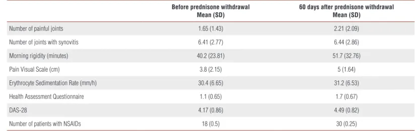
Clinical characteristics of the RA group were evalu-ated before and after medication withdrawal (Table 1). The mean (SD) RA duration was 7.5(2.6) years. Only 2/32 patients were negative for Rheumatoid Factor and 4/32 did not present erosions on the x-rays of the hands and feet. Seventeen patients (53.1%) used three or more disease-modifying anti-rheumatic drugs (DMARDs), 13 (40.6%) used 2 DMARDs and 2 (6.3%) used only one DMARD.
Disease activity was estimated using the Disease Ac-tivity Score 28 joints (DAS-28) where variables include erythrocyte sedimentation rate (ESR), number of painful joints and those with synovitis and visual scale for global evaluation of the patient. We applied a Health Assessment Questionnaire (HAQ) with visual scales of pain and the duration of morning rigidity and use of NSAIDs and anal-gesics were also reported.
The control group was composed of healthy volunte-ers, graduating students at the Santa Casa de São Paulo- Faculty of Medical Sciences, paired to the study group in relation to BMI and gender.
VERY LOW DOSE INTRAVENOUS DEXAMETHASONE SUPPRESSION TEST (IV- VLD-DST)
All patients and controls were submitted to the very low dose (20µg/m2) intravenous dexamethasone
suppres-sion test. We recently described how this new test can be used to evaluate individual sensitivity to GC in diffe-rent age groups (33). Briefly, after a fasting period of 10-12 hours, individuals from both groups received IV dexamethasone disodium phosphate (Decadron2mg/ mL – Prodome Chemical and Pharmaceutical, Brazil) at a dose of 20 µg/m2 BSAin bolus, and 30 minutes of
rest Later, a blood sample was taken for cortisol measu-rement and separation of peripheral mononuclear cells. The subsequent blood sample collections were performed every 30 minutes up to 150 minutes for cortisol measurement by RIA (BRIDGE, Adaltis, Casalecchio di Remo, Italy).
GRa DETERMINATION BY QRT-PCR
Mononuclear cells were separated from a 20mL venous blood sample collected in two 10mL tubes containing sodium heparin (Vacutainer, Becton-Dickinson), after addition of 20mL of Histopaque -1077 (Sigma, USA)
and centrifugation protocol for 30 minutes at 800g, ac-cording to manufacturer’s recommendation. Cell viabi-lity was verified in a hemocytometer (Neubauer cham-ber), using the Trypan blue dye (Trypan Blue Solution 0.4%, Sigma, USA).
Total RNA was isolated from cells using guanidinium thiocyanate-chloroform extraction (Trizol, Gibco, USA). After extraction, total RNA was diluted in 40µL of water (DNase/RNase-free water, GIBCO, USA). Complemen-tary DNA (cDNA) was synthesized from 1µg total RNA using a reverse transcription reaction (TaqMan Reverse Transcription Reagents, Applied Biosystems).
Table 1. Clinical and laboratory data before and after prednisone withdrawal in RA patients.
Before prednisone withdrawal Mean (SD)
60 days after prednisone withdrawal Mean (SD)
Number of painful joints 1.65 (1.43) 2.21 (2.09)
Number of joints with synovitis 6.41 (2.77) 6.44 (2.86)
Morning rigidity (minutes) 40.2 (23.81) 51.7 (32.76)
Pain Visual Scale (cm) 3.8 (2.15) 5 (1.64)
Erythrocyte Sedimentation Rate (mm/h) 30.4 (6.65) 31.2 (6.53)
Health Assessment Questionnaire 1.1 (0.65) 1.7 (0.67)
DAS-28 4.17 (0.86) 4.49 (0.82)
Copyright
© ABE&M todos os dir
eitos r
eser
vados.
GRα expression was determined according to the pro-tocol previously described by our group (23). Briefly, a real-time PCR was performed for GRα and BCR (Bre-akpoint Cluster Region) as a normalizing gene. Primers and probes were as follows: GRα Sense Primer GAA-GGAAACTCCAGCCAGAA; GRα Anti-sense Primer CAGCTAACATCTCGGGGAAT (Product size: 151bp);
GRα Probe
6-FAM-GCTTCCAAACATTTTTGGATA-AGACCAT-TAMRA; BCR Sense Primer CCTTCGA-CGTCAATAACAAGGAT; BCR Anti-Sense Primer CC-TGCGATGGCGTTCAC (Product size: 67bp); BCR Probe: 6-FAM-TCCATCTCGCTCATCATCACCGA-CA-TAMRA.
In each PCR run, we used a standard curve using serial dilutions of cDNA obtained from a standardized Jurkat (E6-1 clone, ATCC) cell culture. PCR conditions were equal for both genes, using TaqMan PCR Core kit (Applied Biosystems, USA). Briefly, 1X TaqMan buffer A, 500µM each dNTP, 4.5mM MgCl2, 200nM of each primer, 100nM of probe, 0.025U/µL of AmpliTaq Gold, 5µL of cDNA and water were incubated in a total volume of 25µL. Cycle con-ditions on an ABI 7500 (Applied Biosystems) were: 95oC
for 10 minutes (AmpliTaq Gold activation) followed by 45 cycles of 95oC for 15 seconds (denaturation) and 60oC for
90 seconds (annealing and extension). The ratio between GRα and BCR expression represents the number of GRα Expression Units (EU GRα of each sample.
STATISTICAL ANALYSIS
The comparison between cortisol concentrations from the same individual, before and after DEX suppression test was performed by a paired t-test. The comparison between RA patients and controls in relation to baseli-ne concentrations, lower concentrations and percent of cortisol suppression was performed using the Student-t test or the Mann-Whitney Rank Sum Test, according to data distribution evaluated by the Kolmogorov-Smir-nov test. Cortisol concentration at different time points during DEX suppression test was analyzed by Kruskal-Wallis One Way Analysis of Variance on Ranks (ANOVA on Ranks). Correlations between the percentage of cor-tisol suppression (F%) and the expression of GR were analyzed using linear regression equations (SigmaStat for Windows, v3.05). Linear regression equations of the standard curves and expression units of GRα (EU GRα) calculations were performed using MS-Excel 2000 for Windows software (Microsoft). A p-value <0.05 was considered statistically significant.
RESULTS
CLINICAL CHARACTERISTICS OF RA PATIENTS
At the time prednisone was interrupted, 18 patients were receiving 5mg per day and 11 had been using between 2.5mg and 5mg per day. Only 3 patients had not been previously receiving prednisone. There was a minimal but significant difference of GR expression (ANOVA, p=0.034, r2=0,169) but not of F% reduction (p=0.165)
according to previous prednisone dose.
Clinical worsening was observed when patients were clinically evaluated after at least 60 days of prednisone withdrawal. There was an increase in Health Assessment Questionnaire results from 1.1 to 1.7 after two months of GC-free period (p<0.001, paired t-test). The time period of morning rigidity increased from a median of 30 minu-tes to 51.6 minuminu-tes (p<0.001, Wilcoxon signed-rank minu-test; mean values are shown in Table 1) and the intensity of pain also increased, in visual pain scale (VPS), from mean (SD) values of 3.8cm (2.1) to 5cm (1.6) (p<0.001, paired t-test), the mean (SD) DAS-28 varied from 4.2 (0.9) to 4.5 (0.8) (p<0.001, paired t-test). There was no change in ESR after prednisone discontinuation (30.4 to 31.2mm/h, p=0.531, paired t-test). After two months without prednisone, all patients were only using NSAIDs and analgesics.
INTRAVENOUS VERY LOW DOSE DEXAMETHASONE SUPPRESSION TEST (IV-VLD-DST)
Mean (SD) baseline cortisol concentrations in the RA group was 12.5 (3.6) µg/dL. A significant reduction was observed when comparing baseline cortisol concen-trations with the lowest cortisol concentration obtai-ned after IV-VLD-DST (7.0 [2.2] µg/dL) (p<0.001). Mean (SD) baseline cortisol concentration in the con-trol group was 19.8 (4.4) µg/dL, also showing a sig-nificant reduction to 6.4 (1.8)µg/dL (p<0.001) after IV-DEX.
Copyright
© ABE&M todos os dir
eitos r
eser
vados.
GRa QUANTITATION BY REAL TIME PCR
Mean (SD) GRα expression was similar between RA patients (1.2 [0.19] EU GRα) and control individu-als (1.24 [1.7] EU GRα; p=0.54). GRα expression observed for three patients in the RA group that did not receive prednisone previously was similar to the remainder of the group (mean 1.12 EU and 1.22 EU, respectively).
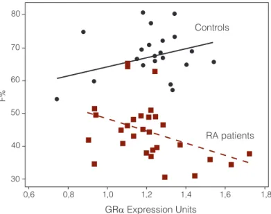
The relationship between GRα expression and the per-centage of cortisol suppression is illustrated in Figure 1. In control individuals, although no correlation was observed between GRα expression and the percentage of cortisol suppression, the angular coefficient of the regression line was positive. On the contrary, an inverse correlation was observed between GRα expression and cortisol suppres-sion in the RA group (p=0.034; r2=0.157).
GRα Expression Units Controls
80
F%
70
60
50
40
30
RA patients
0,6 0,8 1,0 1,2 1,4 1,6 1,8
Figure 1. Relationship between GRα expression and percentage of
cortisol suppression (F%) after IV-DEX. There is an inverse relationship of F% and GRα expression in RA patients (F% = 66.7 – 18.4 GRα; r2=0.157; p=0.034); however, in controls there is a non-significant relationship in the opposite direction (F% = 50.9 + 13.5 GRα; r2=0.108; p=0.158).
CORRELATION BETWEEN GRa EXPRESSION AND DISEASE ACTIVITY
A trend of positive correlation was observed between GRα expression and DAS-28 values 60 days after pred-nisone withdrawal (p=0.05; GRα (EU) = 0.827 + 0.084 DAS; r2=0.135) and an inverse correlation between
GRα expression and the absolute variation of DAS-28 (expressed as DDAS; p=0.03; GRα (EU) = 1.27 – 0.2 DAS; r2=0.163).
DISCUSSION
Overall, clinical characteristics of RA patients demons-trate that the group is formed by individuals with seve-re RA with elevated inflammatory potential: moseve-re than 93% of the patients used more than one DMARD, 53% more than 3 DMARDs, 94% presented high titers of Rheumatoid Factor and 88% presented erosions on the radiological exam. This, possibly reflects a severity bias of RA at our Institution, a tertiary care hospital in São Paulo, Brazil. After prednisone withdrawal, there was an increase in disease activity, measured by the DAS-28, HAQ and use of NSAIDs. This demonstrates that, despite the use of 2, 3 or more DMARDs, even the additional use of low dose prednisone is able to impro-ve disease control. It was possible to obtain the release of the HPA axis sixty days after prednisone withdrawal, considering as normal the baseline cortisol levels greater than 7µg/dL. Although only three patients were not previously receiving prednisone, there was no difference in GRα expression or F% reduction after there was no difference in GR > IV-VLD-DST when we compared the three patients who do not received prednisone with those prednisone was withdrawal before the study. This suggests that the HPA axis was not supressed at the time the study was performed.
Intravenous very low dose dexamethasone significantly reduced cortisol concentration and allowed the recogni-tion of individual glucocorticoid sensitivity by identifying a spectrum of cortisol reduction in both RA and control groups. This is the first time this test was used in patients with RA in order to evaluate HPA integrity and to deter-mine GC sensitivity.
Copyright
© ABE&M todos os dir
eitos r
eser
vados.
concentrations in individuals with RA during insulin tole-rance tests, despite increased IL-6 concentration. Despite high HPA stimulatory concentrations of pro-inflammatory cytokines, its activity in RA is still lower than expected.
Accepting that in RA patients there is a HPA dysfunc-tion in response to stressors, the differences between ba-seline cortisol concentrations in RA patients and control individuals could be related to this abnormal condition. The test itself may cause additional anxiety and stress.
Although cortisol reduction after IV-DEX was signifi-cant in both groups, it was less intense in the RA group, possibly representing a partial resistance to the GC negati-ve feedback at the CNS lenegati-vel.
Although an initial hypothesis for this resistance would be a decreased expression of GR, secondary to the homologous down-regulation exerted by exogenous GC upon its own re-ceptor, it was previously described that this mechanism begins 4 hours after the exposure of the GR to GC, with maximum activity between 18 and 24 hours, and with subsequent disa-ppearance 48 hours after reducing the GC to half of its initial dose (37). Therefore, since in our study the patients remained 60 days without exposure to GC, this was a sufficient period to avoid interference in GR expression.
A potential mechanism involved in GC resistance ob-served in RA patients would be an intrinsic decrease on GR expression. On the other hand, we observed that GRα ex-pression was similar in both RA and control groups. Using more accurate methods, such as qRT-PCR, our findings are comparable to the results described by Onda and cols. (24), demonstrating that GRα of RA patients are similar to controls. This corroborates the hypothesis that after 60 days without prednisone, down-regulation of GR was not the predominant mechanism influencing HPA sensitivity. Additionally, we observed in RA patients that the greater GRα expression, the lower the amplitude of cortisol sup-pression during IV-DEX (Figure 1).
Furthermore, we observed a direct relationship between disease activity (DAS-28; Disease Activity Score 28 joints) and the level of GRα expression. These two observations suggest a post-receptor resistance mechanism, in which the highly expressed GRα is not enough to reduce the inflam-matory activity of the disease, recognized by DAS-28 and despite its greater expression there is lower (and not hi-gher, as would be expected) in vivo sensibility to DEX.
The inverse correlation between GRα expression and the amplitude of variation in DAS-28, before and after prednisone withdrawal also suggests a resistance related to abnormalities at post-receptor level, in which individuals with higher GRα expression also presented a more sig-nificant increase of DAS-28 after prednisone withdrawal.
If the resistance to GC was simply inversely correlated to GRα expression, the increase in disease activity would be more evident in those individuals with lower GRα expres-sion, but this condition was not observed.
Abnormal translocation of the activated GRα to the nucleus, co-inactivation of GRα with NF-κB and AP-1 protein, heterodimerization of GRα with GRβ and altered expression and/or function of co-activators, could all be together post-receptor interferents increasing the resistan-ce to GC (16,38-42).
Future studies should address the disequilibrium in the expression of GRα (and GRβ) and NF-κB or AP-1 in RA patients as a potential source of a post-receptor resistance mechanism. Therefore, our data support the hypothesis that the resistance mechanisms to GC observed in patients with RA are not secondary to a reduced expression of GRα but rather to alterations at post-receptor level.
Acknowledgements: We thank Fundação ao Amparo à Pesquisa do Estado de São Paulo (Fapesp) (grant # 04/03208-2) and FAP – Fun-do de Amparo à Pesquisa da Faculdade de Ciências Médicas da Santa Casa de São Paulo) (grant # 03/309) for the financial support.
Disclosure: We declare there are no conflicts of interest in this article.
REFERENCES
Manolios N, Geczy C, Schrieber L. Lymphocyte migration in heal-1.
th and inflammatory rheumatic disease. Semin Arthritis Rheum. 1991;209:339-52.
Postigo AA, Garcia-Vicuña R, Diaz-Gonzalez F, Arroyo AG, de Landazuri 2.
MO, Chi-Rosso G, et al. Increased binding of synovial T lymphocytes from rheumatoid arthritis to endothelial-leukocyte adhesion molecu-le-1 (ELAM-1) and vascular cell adhesion molecumolecu-le-1 (VCAM-1). J Clin Invest. 1992;89(5):1445-52.
Yamasaki S, Kawakami A, Nakashima T, Nakamura H, Kamachi M, 3.
Honda S, et al. Importance of NF-kappaB in rheumatoid synovial tis-sues: in situ NF-kappaB expression and in vitro study using cultured synovial cells. Ann Rheum Dis. 2001;60(7):678-84.
Baeuerle PA, Henkel T. Function and activation of NF-kappa B in the 4.
immune system. Annu Rev Immunol. 1994;12:141-79.
Pan J, McEver RP. Regulation of the human P-selectin promoter by Bcl-5.
3 and specific homodimeric members of the NF-kB/Rel family. J Biol Chem. 1995;270(39):23077-83.
Wang CY, Mayo MW, Korneluk GR, Goeddel EV, Baldwin Jr AS. NF-ka-6.
ppaB antiapoptosis: induction of TRAF1 and TRAF2 and IAP1 and c-IAP2 to suppress caspase-8 activation. Science. 1998;281(5383):1680-3.
Eggert M, Kluter A, Rusch D, Schmidt KL, Dotzlaw H, Schulz M, et al. 7.
Expression analysis of the glucocorticoid receptor and the nuclear fac-tor-kB subunit p50 in lymphocytes from patients with rheumatoid arthri-tis. J Rheumatol. 2002;29(12):2500-6.
Hammaker D, Sweeney S, Firestein GS. Signal transduction networks 8.
in rheumatoid arthritis. Ann Rheum Dis. 2003;62 Suppl 2:ii86-9.
Northrop JP, Crabtree GR, Mattila PS. Negative regulation of in-9.
Copyright
© ABE&M todos os dir
eitos r
eser
vados.
Chikanza LC, Panayi GS. The effects of hydrocortisone on in vitro 10.
lymphocyte proliferation and interleukin-2 and -4 production in corticosteroid sensitive and resistant subjects. Eur J Clin Invest. 1993;23(12):845-50.
Eggert M, Schulz M, Neeck G. Molecular mechanisms of glucocorticoid 11.
action in rheumatic autoimmune diseases. J Steroid Biocherm Mol Biol. 2001;77(4-5):185-91.
Cronstein BN, Kimmel SC, Levin RI, Martiniuk F, Weissmann G. A 12.
mechanism for the antiinflammatory effects of corticosteroids: the glucocorticoid receptor regulates leukocyte adhesion to endothe-lial cells and expression of endotheendothe-lial-leukocyte adhesion molecu-le 1 and intercellular adhesion momolecu-lecumolecu-le 1. Proc Natl Acad Sci USA. 1992;89(21):9991-5.
Gil B, Pajares MA, Mato JM, Alvarez L. Glucocorticoid regulation of he-13.
patic S-adenosylmethionine synthetase gene expression. Endocrinolo-gy. 1997;138(3):1251-8.
Ray KP, Farrow S, Daly M, Talabot F, Searle N. Induction of the E-se-14.
lectin promoter by interleukin 1 and tumour necrosis factor alpha, and inhibition by glucocorticoids. Biochem J. 1997;328(Pt 2):707-15.
Chang DJ, Ji C, Kim KK, Casinghino S, McCarthy TL, Centrella M. 15.
Reduction in transforming growth factor beta receptor I expression and transcription factor CBFa1 on bone cells by glucocorticoid. J Biol Chem. 1998;273(9):4892-6.
Ray A, Prefontaine KE. Physical association and functional antagonism 16.
between the p65 subunit of transcription factor NF-kappa B and the glucocorticoid receptor. Proc Natl Acad Sci USA. 1994;91(2):752-6.
Scheinman RI, Gualberto A, Jewell CM, Cidlowski JA, Baldwin Jr AS. 17.
Characterization of mechanisms involved in transrepression of NF-kappa B by activated glucocorticoid receptors. Mol Cell Biol.1995;15(2): 943-53.
McKay LI, Cidlowski JA. Cross-talk between nuclear factor-kappa B and 18.
the steroid hormone receptors: mechanisms of mutual antagonism. Mol Endocrinol. 1998;12(1):45-56.
Lee EB, Kim JY, Lee YJ, Song YW. Glucocorticoid receptor polymor-19.
phisms in Korean patients with rheumatoid arthritis. Ann Rheum Dis. 2005;64:503-4.
Schlaghecke R, Kornely E, Wollenhaupt J, Specker C. Glucocorticoid 20.
receptors in rheumatoid arthritis. Arthritis Rheum. 1992;35(7):740-4.
Schlaghecke R, Beuscher D, Kornely E, Specker C. Effects of glucocorti-21.
coids in rheumatoid arthritis. Diminished glucocorticoid receptors do not result in glucocorticoid resistance. Arthritis Rheum. 1994;37(8):1127-31.
Van Everdingen AA, Huisman AM, Wenting MJ, Van Reesema S, 22.
Jacobs JW, Bijlsma JW. Down regulation of glucocorticoid recep-tors in early-diagnosed rheumatoid arthritis. Clin Exp Rheumatol. 2002;20(4):463-8.
Melo MR, Faria CD, Melo KC, Reboucas NA, Longui CA. Real-time PCR 23.
quantitation of glucocorticoid receptor alpha isoform. BMC Mol Biol. 2004;5(1):19.
Onda K, Rimbara E, Hirano T, Oka K, Abe H, Tahara K, et al. Role of 24.
mRNA expression of transcription factors in glucocorticoid sensitivi-ty of peripheral blood mononuclear cells and disease state in rheu-matoid arthritis. J Rheumatol. 2004;31(3):464-9.
Chikanza IC, Roux-Lombard P, Dayer JM, Panayi GS. Tumour necro-25.
sis factor soluble receptors behave as acute phase reactants following surgery in patients with rheumatoid arthritis, chronic osteomyelitis and osteoarthritis. Clin Exp Immunol. 1993;92(1):19-22.
Straub RH, Paimela L, Peltomaa R, Schlomerich J, Leirisalo-Repo 26.
M. Inadequately low serum levels of steroid hormones in relation
to interleukin-6 and tumor necrosis factor in untreated patients with early rheumatoid arthritis and reactive arthritis. Arthritis Rheum. 2002;46(3):654-62.
Straub RH, Kittner JM, Heijnen C, Schedlowski M, Schmidt RE, Jaco-27.
bs R. Infusion of epinephrine decreases serum levels of cortisol and 17-hydroxyprogesterone in patients with rheumatoid arthritis. J Rheu-matol. 2002;29(8):1659-64.
Gutierrez MA, Garcia ME, Rodriguez JA, Mardonez G, Jacobelli S, Rive-28.
ro S Hypothalamic-pituitary-adrenal axis function in patients with acti-ve rheumatoid arthritis: a controlled study using insulin hypoglycemia stress test and prolactin stimulation. J Rheumatol. 1999;26(2):277-81.
Eijsbouts AM, van den Hoogen FH, Laan RF, Hermus AR, Sweep CG, 29.
van de Putte LB. Hypothalamic-pituitary-adrenal axis activity in patients with rheumatoid arthritis. Clin Exp Rheumatol. 2005;23(5):658-64.
Nieman LK. Diagnostic tests for Cushing’s syndrome. Ann N Y Acad 30.
Sci. 2002;970:112-8.
Maguire KP, Schweitzer I, Biddle N, Bridge S, Tiller JW. The dexame-31.
thasone suppression test: importance of dexamethasone concentra-tions. Biol Psychiatry. 1987;22(8):957-67.
Longui CA, Giusti MM, Calliari LE, Katiki T, Kochi C, Monte O. Partial gluco-32.
corticoid resistance in obese children detected by very low dose dexame-thasone suppression test. J Pediatr Endocrinol Metab. 2003;16,1277-82.
Faria CDC, Cobra JF, Silva TS, Melo MR, Rocha MN, Hayashi LS, et al. 33.
A Very Low Dose Intravenous Dexamethasone Suppression Test as an Index of Glucocorticoid Sensitivity. Horm Res 2008;69:357-62.
Yehuda R, Southwick SM, Krystal JH, Bremner D, Charney DS, Mason JW. 34.
Enhanced suppression of cortisol following dexamethasone administra-tion in posttraumatic stress disorder. Am J Psychiatry. 1993;150(1):83-6.
Arnett FC, Edworthy SM, Bloch DA, McShane DJ, Fries JF, Cooper 35.
NS, et al. The American Rheumatism Association 1987 revised crite-ria for the classification of rheumatoid arthritis. Arthritis Rheum. 1988; 31(3):315-24.
Crofford LJ, Kalogeras KT, Mastorakos G, Magiakou MA, Wells J, Kanik 36.
KS, et al. Circadian relationships between interleukin (IL)-6 and hypotha-lamic-pituitary-adrenal axis hormones: failure of IL-6 to cause sustained hypercortisolism in patients with early untreated rheumatoid arthritis. J Clin Endocrinol Metab. 1997;82(4):1279-83.
Boss B, Neeck G. Correlation of IL-6 with the classical humoral disease 37.
activity parameters ESR and CRP and with serum cortisol, reflecting the activity of the HPA axis in active rheumatoid arthritis. Z Rheumatol. 2000;59 Suppl 2:62-4.
Okret S, Dong Y, Brönnegard M, Gustafsson JA. Regulation of gluco-38.
corticoid receptor expression. Biochimie. 1991;73(1):51-9.
Oakley RH, Jewell CM, Yudt MR, Bofetiado DM, Cidlowski JA. The 39.
dominant negative activity of the human glucocorticoid receptor beta isoform. Specificity and mechanisms of action. J Biolol Chem. 1999;274(39):27857-66.
Webster JC, Oakley RH, Jewell CM, Cidlowski JA. Proinflammatory 40.
cytokines regulate human glucocorticoid receptor gene expression and lead to the accumulation of the dominant negative beta isoform: a mechanism for the generation of glucocorticoid resistance. Proc Natl Acad Sci USA. 2001;98(12):6865-70.
Neeck G, Klüter A, Dotzlaw H, Eggert M. Involvement of the glucocor-41.
ticoid receptor in the pathogenesis of rheumatoid arthritis. Ann N Y Acad Sci. 2002;966:491-5.
Faria CDC, Longui CA. Molecular aspects of glucocorticoid sensitivity. 42.