Copyright
© ABE&M todos os dir
eitos r
eser
vados.
IGF-I, insulin and prostate cancer
IGF-I, insulina e câncer de próstata
Giovanna A. Balarini Lima1, Lívia L. Corrêa1, Rafael Gabrich2,
Luiz Carlos D. de Miranda2, Mônica R. Gadelha1,3
ABSTRACT
Prostate cancer is the second most frequent malignancy diagnosed in adult men. Androgens are considered the primary growth factors for prostate normal and cancer cells. However, other non-androgenic growth factors are involved in the growth regulation of prostate cancer cells. The association between IGF-I and prostate cancer risk is well established. However, there is no evidence that the measurement of IGF-I enhances the specificity of prostate cancer detection beyond that achievable by serum prostate-specific antigen (PSA) levels. Until now, there is no consensus on the possible association between IGFBP-3 and prostate cancer risk. Although not well established, it seems that high insulin levels are particularly associated with risk of aggres-sive prostatic tumours. This review describes the physiopathological basis, epidemiological evidence, and animal models that support the association of the IGFs family and insulin with prostate cancer. It also describes the potential therapies targeting these growth factors that, in the future, can be used to treat patients with prostate cancer. Arq Bras Endocrinol Metab. 2009;53(8):969-75
Keywords
Insulin; IGF-I; prostate; cancer
RESUMO
O câncer de próstata é a segunda neoplasia mais frequentemente diagnosticada em homens adultos. Os androgênios são considerados fatores de crescimento primários para células pros-táticas normais e malignas. Entretanto, outros fatores de crescimento não androgênicos estão envolvidos na regulação do crescimento das células prostáticas malignas. Associação entre IGF-I e risco de câncer de próstata é bem estabelecida. No entanto, não há evidência de que a dosagem do IGF-I melhore a especificidade na detecção do câncer de próstata, além daque-la alcançada pelos níveis de antígeno prostático específico (PSA). Até hoje, não há consenso sobre a possível associação entre IGFBP-3 e risco de câncer de próstata. Apesar de não estar estabelecido, altos níveis de insulina parecem particularmente associados ao risco de tumores prostáticos agressivos. Esta revisão descreveu base fisiopatológica, evidências epidemiológi-cas e modelos animais que apoiam a associação da família das IGFs e insulina com câncer de próstata. Também foram descritas terapias potenciais que têm como alvo esses fatores de cres-cimento, os quais, no futuro, poderão ser usados para tratar pacientes com câncer de próstata. Arq Bras Endocrinol Metab. 2009;53(8):969-75
Descritores
Insulina; IGF-I; próstata; câncer
1 Serviço de Endocrinologia, Hospital Universitário Clementino Fraga Filho (HUCFF), Universidade Federal do Rio de Janeiro (UFRJ), Rio de Janeiro, RJ, Brasil 2 Serviço de Urologia, HUCFF, UFRJ, Rio de Janeiro, RJ, Brasil 3 Serviço de Endocrinologia, Instituto Estadual de Diabetes e Endocrinologia Luiz Capriglione (IEDE), Rio de Janeiro, RJ, Brasil
Correspondence to:
Giovanna A. Balarini Lima HUCFF, UFRJ
Rua Professor Rodolpho Paulo Rocco, 255, 9E23 – Cidade Universitária – Ilha do Fundão 21941-913 – Rio de Janeiro, RJ, Brasil
Received on Oct/13/2009 Accepted on Nov/8/2009
INTRODUCTION
P
rostate cancer is the second most frequent malig-nancy diagnosed in adult men and the second cause of cancer death among Brazilian men (1). Androgens are considered the primary growth factors for prosta-te epithelial cells (2). However, other non-androgenic growth factors are involved in the growth regulation ofCopyright
© ABE&M todos os dir
eitos r
eser
vados.
epidemiological evidence and animal models that sup-port the association of the IGFs family and insulin with prostate cancer. We also described the new perspectives in anticancer therapies that, in the future, can be used to treat patients with prostate cancer.
PHYSIOPATHOLOGY OF IGFS FAMILY, INSULIN
AND PROSTATE CANCER
The physiological development and growth of the prostate is primarily dependent on testosterone and dihydrotestosterone (2). However, the evidence that low androgen levels do not exclude prostate carcinoma suggests that other growth factors, such as insulin and the IGFs family, also have a role in the prostate carci-nogenesis (12).
The effects of growth hormone (GH) are media-ted primarily through the hepatic production of IGF-I. However, GH also has direct effects in vivo that are
in-dependent of IGF-I, many of which are exerted throu-gh local production of IGFs rather than under the in-fluence of a factor in the circulation (13). Considering the prostate gland, the effects of the GH-IGF-I axis are exerted via: (a) direct action of GH; (b) hepatic IGF-I; and (c) IGF-I produced locally on the prostate tissue, through autocrine-paracrine action, possibly mediated by increase in the free IGF-I levels due to the action of prostate-specific antigen (PSA) and other proteases.
IGF-I is a potent mitogen for normal and cancerous cells (14) and exerts the mitogenic action by increasing DNA synthesis and by stimulating the cell cycle pro-gression (15). In addition, IGF-I also inhibits apoptosis (16). Both IGF-I and, to a less extent, IGF-II have di-rect mitogenic and anti-apoptotic effects on normal and transformed prostate epithelial cells and have been im-plicated in the pathogenesis of prostate cancer (17-19). The actions of IGF-I on cell proliferation and apop-tosis are mediated via a specific cell-membrane receptor, IGF-I receptor (IGF-IR). Binding of IGF-I to IGF-IR activates the receptor’s tyrosine kinase activity, which tri-ggers a cascade of reactions among a number of mo-lecules involved in the signal transduction pathway. Two distinct main signal transduction pathways have been identified. One pathway activates Ras protein, Raf protein and mitogen-activated protein kinase (MAPK). The other pathway involves activation of phosphati-dylinositol 3-kinase (PIK3), AKT (or protein kinase B), mTOR (mammalian target of rapamycin) and S6 kinase (S6K) (20,21) (Figure 1).
The interaction between IGF-I and IGF-IR is re-gulated by the IGF-binding proteins (IGFBPs), so that under circumstances in which IGFBP levels are low, IGF mitogenic activity would be expected to be high (22). Of the circulating binding proteins, IGFBP-3 is the most abundant one (23). At the tissue level, IGFBP-3 serves an IGF carrier function, prolonging the half-life of the IGFs and regulating their availability to the cell surface receptors (24). In addition to its role as an IGF-I modulator, IGFBP-3 also has IGF-inde-pendent antiproliferative actions, such as inhibition of cell growth and stimulation of apoptosis (25,26). Many proteases that are present in prostate cancer microen-vironment, including PSA, human kallicrein 2, trypsin and cathepsin D, can digest IGFBPs and release free IGF-I (27,28). The production of proteases by a cancer might therefore increase IGF-IR signaling.
Structurally, IGF-IR resembles the insulin receptor, and there is more than 50% homology between them (29). Because of this homology, insulin and IGF-I are able to cross-bind to each other’s receptor or to the hybrid receptors (formed by an insulin half receptor and an IGF-I half receptor), albeit with much weaker binding affinity than that for the preferred ligand (30). Therefore, besides the classical metabolic actions of insulin, it also exerts proliferative and anti-apoptotic effects. The insulin signaling pathways are the same as the IGF-I’s (Figure 1). The demonstration that both IR and IGF-IR are commonly expressed in human prostate cancer specimens (19) implicates insulin and IGF-I signaling in the initiation/progression of prosta-te cancer. In addition, the demonstration that the GHR
IGF-I, IGF-II, insulin
IGF-IR, IR, hybrid receptor
IRS
PI3K-PDK/AKT-TOR-S6K pathway Ras-raf-MAPK pathway
Cell proliferation Anti-apoptosis
Metabolic actions Cell survival Anti-apoptosis
Figure 1. IGF-I and insulin receptor activation and downstream signaling.
Copyright
© ABE&M todos os dir
eitos r
eser
vados.
is also expressed in prostate carcinoma tissues and that its mRNA levels are higher in carcinoma tissues than in benign prostate hyperplasia suggests that GH may play an important role in human prostate cancer (31).
The IGFs family also seems to be important in an-drogen-independent progression of prostate cancer. Activation of alternative autocrine, paracrine, or endo-crine stimulatory pathways after androgen withdrawal initiate intracellular signal transduction pathways that can replace androgens for growth and survival of pros-tate tumor cells, either by bypassing the androgen receptor (AR) all together or by activating the AR. IGF-I, MAPK and AKT promotes non-ligand activation of AR, con-tributing to proliferation in an androgen depleted en-vironment (32).
IGFS FAMILY AND PROSTATE CANCER:
EPIDEMIOLOGICAL EVIDENCE
Retrospective and prospective epidemiological studies conducted in distinct populations have demonstrated that elevated serum IGF-I level is associated with an increased risk of prostate cancer. In 1997, a small Greek case-control study (n = 52) reported a multivariate re-lative risk (RR) of prostate cancer of 1.91 per 60 ng/ mL increase in IGF-I level (95% confidence interval, CI: 1.00-3.73) (33). A larger case-control study (n = 210) from Sweden reported a similar positive associa-tion between IGF-I level and prostate cancer risk (34). A RR of 1.51 per 100 ng/mL increase in IGF-I level (95%CI: 1.00-2.26) was observed. In this study, the authors also examined IGFBP-3 and prostate cancer risk, but no significant association was observed.
In the Physicians’ Health Study, a prospective epi-demiological study, the associations between IGF-I and
IGFBP-3 levels and subsequent prostate cancer risk among 152 patients and 152 age-matched controls were investigated (17). A RR of 4.32 (95%CI: 1.76-10.6), if comparing men in the highest and lowest quartiles of IGF-I, when controlling for IGFBP3, was found. The corresponding RR for IGFBP3 was 0.41 (95%CI: 0.17-1.03). There was a significant linear trend between IGF-I and prostate cancer risk (RR: 2.1 per 100 ng/mL increase in IGF-I level; 95%CI: 1.3-3.2). No substantial change in this association was ob-served when adjusting for quartiles of weight, height, body mass index (BMI), plasma lycopene, and plasma hormone levels (estradiol, testosterone, dihydrotestos-terone, sex hormone binding globulin (SHBG) and an-drostenediol glucuronide).
To date, five meta-analyses evaluating the associa-tion between IGF-I level, IGFBP-3 level and prosta-te cancer were published (Table 1). The first one (35) analyzed 14 case-control studies, and the combined data showed that circulating levels of IGF-I were sig-nificantly higher in prostate cancer patients. The odds ratio (OR) for prostate cancer was 1.47 (95%CI: 1.23-1.77) among men with high IGF-I compared to tho-se with low IGF-I. A meta-analysis of nine prospective studies, that included 1,512 men with prostate cancer, found a similar risk (OR: 1.31; 95%CI: 1.03-1.71) (36). These findings were less significant than that reported by another meta-analysis that estimated a summary OR of 1.83 (95%CI: 1.03-3.26) for the association of the uppermost categories of serum IGF-I compared to the lowermost (6).
Recently, a collaborative reanalyzes of individual data from 12 prospective studies (n = 3,700 prostate cancer cases and 5,200 control participants) on the re-lationships between circulating levels of IGFs and
sub-Table 1.Meta-analyses evaluating IGF-I and IGFBP-3 levels (comparison of highest and lowest quantiles) and prostate cancer risk
Reference IGF-I IGFBP-3
Studies (n) Cases (n) OR (95%CI) Studies (n) Cases (n) OR (95%CI)
Shi and cols. (35) 14 1,460 1.47
(1.27-1.71)
14 1,460 1.26
(1.03-1.54)
Renehan and cols. (6) 6 904 1.49
(1.14-1.95)
6 904 0.88
(0.61-1.28)
Morris and cols. (36) 9 1,512 1.31
(1.03-1.71)
9 1,512 1.05
(0.82-1.35)
Roddam and cols. (37) 12 3,671 1.38
(1.19-1.60)
12 3,600 1.23
(1.06-1.43)
Rowlands and cols. (4) 42 7,481 1.21
(1.07-1.36)
29 6,541 0.88
(0.79-0.98)
Copyright
© ABE&M todos os dir
eitos r
eser
vados.
sequent prostate cancer risk was conducted. A 38% in-creased odds of prostate cancer risk, comparing highest
versus lowest quintiles of IGF-I (OR: 1.38; 95%CI:
1.19-1.60), was observed (37).
The largest systematic review of studies reporting on the association of IGF-I with the risk of prostate can-cer was recently published (4). Unlike previous meta-analyzes, this one included both retrospective and pros-pective studies (n = 42 studies) and demonstrated that the published literature is consistent with an average 21% increase risk of prostate cancer per standard devia-tion increase in IGF-I (OR: 1.21; 95%CI: 1.07-1.36). They also showed a stronger association of IGF-I with more aggressive (OR: 1.21; 95%CI: 0.97-1.51) and ad-vanced (OR: 1.41; 95%CI: 1.07-1.85) cancers, com-pared to nonaggressive (OR: 1.05; 95%CI: 0.99-1.12) and localized (OR: 1.10; 95%CI: 0.98-1.11) ones.
Differently from IGF-I, the association between IGFBP-3 and prostate cancer risk is not a consensus. Considering the five meta-analyses previously men-tioned, two of them unexpectedly suggested that IGFBP-3 is positively associated with prostate cancer risk (35,37). The probable inverse association between IGFBP-3 and prostate cancer risk was only seen in re-trospective, but not prospective studies, so that, when analyzed together, the overall data suggest no eviden-ce for an association of prostate caneviden-cer with IGFBP-3 (4). Also, there is little evidence for a role of IGF-II, IGFBP-1 or IGFBP-2 in prostate cancer risk (4).
Although there is association between higher levels of IGF-I and prostate cancer risk (4,6,35-37), the addi-tion of IGF-I measurement to the PSA level or free/to-tal PSA index does not seem to enhance the specificity of prostate cancer detection in clinical practice (4,36). Also, IGF-I levels are not a useful marker of prostate cancer in men with elevated PSA levels (38,39).
The association of acromegaly – a disease characterized by high GH and IGF-I levels – and prostate cancer is des-cribed elsewhere in this issue (refer to Corrêa and cols.).
INSULIN AND PROSTATE CANCER:
EPIDEMIOLOGICAL EVIDENCE
Prospective cohort studies showed either no associa-tion or prostate cancer risk reducassocia-tion with elevated C-peptide or insulin levels (40,41). However, one study observed a higher risk of prostate cancer mortality for men with C-peptide levels in the highest versus the
lo-west quartile (RR: 2.38; 95%CI: 1.31-4.30) (42).
Type 2 diabetes mellitus (T2DM), characterized by
both hyperinsulinemia and hyperglycemia, has been related to cancer development in many epidemiologi-cal studies. Diabetic patients experience a higher risk of developing colorectal (7), endometrial (43), pan-creatic (44) and postmenopausal breast (45) cancer compared with non-diabetic individuals. On the con-trary, a history of T2DM has been related to a reduc-tion of prostate cancer risk (46). However, in a study that included more than 2,000 prostate cancer cases, the inverse association was confined to non-aggressive prostate tumours (47). This inverse association may be explained by the frequent occurrence of hypogonadism in patients with T2DM (48).
INSULIN, IGFS FAMILY AND PROSTATE CANCER:
ANIMAL MODELS
The possibility of studying human prostatic cancer in in vitro and in vivo environment has allowed the
develo-pment of important tools to study many aspects of the biology of this cancer.
Transgenic mice expressing human IGF-I in basal epithelial cells of prostate have been characterized (49). Transgene expression led to activation of the IGF-IR and spontaneous tumorigenesis in prostate epithelium. Typical and atypical hyperplasias, prostatic intraepithe-lial neoplasia and well differentiated adenocarcinomas appeared in these mice. They offer an animal model for prostate cancer that will allow study of the stepwise de-velopment of this disease and the mechanisms whereby IGF-I mediates this process.
The Transgenic Adenocarcinoma of Mouse Prostate (TRAMP) model has been used to investigate the role of the GH-IGF-I axis on in vivo prostate carcinogenesis
and neoplastic progression. This model was developed using a prostate-specific fragment of the rat probasin regulatory sequence to specifically target expression of the simian virus 40 (SV40) large tumor antigen-coding region in the prostate epithelium (50). The changes that occur during prostate carcinogenesis in these mice highly resemble human prostate transformation (50) and take place in the setting of normal serum androgen levels (51). Using the TRAMP model, a mice homo-zygous for lit, a mutation that inactivates the GHRH
receptor (GHRH-R) and reduces circulating levels of GH and IGF-I, was generated (52). The lit mutation
Copyright
© ABE&M todos os dir
eitos r
eser
vados.
and was also associated with improved survival. The re-sults suggest that prostate carcinogenesis and progres-sion may be influenced by germ line variation of genes encoding signaling molecules in the GH-IGF-I axis.
Also in the TRAMP model, it was found that the expression of IGF-I mRNA in the prostate is elevated in early cancer progression. Nonetheless, the expres-sion of IGF mRNA is not increased in advanced and metastatic disease, suggesting that only organ-confined disease seems to be IGF-I dependent (53).
Using a murine model of prostate cancer genera-ted with LNCaP human prostate cancer cells, it was demonstrated that tumors from mice on the high car-bohydrate/high fat diet had higher levels of activated AKT and modestly higher insulin receptor levels than tumors from mice on the low carbohydrate/high fat diet. Serum from mice on the high carbohydrate/high fat diet was more mitogenic for LNCaP cells in vitro
than serum from mice fed the low carbohydrate/high fat diet. A high diet in refined carbohydrates is associa-ted with increased tumor growth and with activation of signaling pathways distal to the insulin receptor (54).
NEW PERSPECTIVES IN PROSTATE CANCER
THERAPY TARGETING THE IGF FAMILY AND
INSULIN
Converging data from laboratory, clinical and popula-tion studies suggest that the IGFs family is implicated in different cancers. These findings led to the develop-ment of novel anticancer therapies targeting this family.
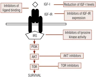
In the last ten years, several drug development re-searches to design novel agents to target IGF signaling were proposed. Many drug candidates demonstrated antineoplastic activity in laboratory models (55-57). Since 2008, dozens of drug candidates are being eva-luated in clinical trials. Strategies include a reduction of IGF-I levels or bioactivity, inhibition of IGF-IR func-tion and inhibifunc-tion of signaling pathways downstream to IGF-IR (Figure 2).
Reduction of IGF-I levels can be achieved with the use of GH releasing hormone antagonists, somatostatin analogs (e.g. octreotide, pasireotide) and GHR antago-nist (pegvisomant). Octreotide, used together with com-plete androgen blockade, in patients with prostate carci-noma has beneficial results (58). In vitro, pegvisomant
inhibits the proliferation of prostatic cancer cells (59). Inhibition of IGF-IR function can be accom-plished with the use of inhibitors of IGF-I binding
Reduction of IGF-I levels IGF-I
IGF-IR
IRS
PI3K
AKT
TOR
SURVIVAL
Inhibitors of IGF-IR expression
Inhibitors of tyrosine kinase activity Inhibitors of
ligand binding
AKT inhibitors
TOR inhibitors
Figure 2. Therapeutic strategies targeting the IGF-I - IGF-IR signaling.
IRS: insulin-receptor substrate; PI3K: phosphatidylinositol 3-kinase; TOR: target of rapamycin.
(e.g. antireceptor antibodies), inhibitors of IGF-IR ex-pression (e.g. antisense oligonucleotides, small inter-fering RNA) and inhibitors of tyrosine-kinase activity (60). Moreover, inhibition of IGF-IR downstream sig-naling pathways can be achieved with the use of AKT inhibitors and mTOR inhibitors (61).
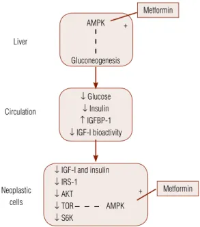
Meanwhile, due to reports documenting insulin re-ceptor on neoplasms and to evidence that higher serum insulin levels are associated with adverse cancer outco-mes (42,62), the use of metformin in cancer patients is being investigated. Actually, population studies provi-ded preliminary evidence that it might have antineoplas-tic or chemo preventing activity (63). Although often referred to as insulin-sensitizer, recent evidence suggests that the key mechanism of action of metformin is as an activator of the AMP-activated protein kinase (AMPK) pathway (64). By activating AMPK in the liver, it su-ppresses gluconeogenesis, leading to decreased hepatic glucose output and therefore to reduced blood glucose and insulin levels. In addition, metformin can increase AMPK activity in neoplastic cells, leading to downstre-am effects that include inhibition of mTOR signaling, protein synthesis and cell proliferation (Figure 3).
CONCLUSIONS
Copyright
© ABE&M todos os dir
eitos r
eser
vados.
8. Yu H, Spitz MR, Mistry J, Gu J, Hong WK, Wu X. Plasma levels of insulin-like growth factor-I and lung cancer risk: a case-control analysis. J Natl Cancer Inst. 1999;91(2):151-6.
9. Wakai K, Ito Y, Suzuki K, Tamakoshi A, Seki N, Ando M, Ozasa K, Watanabe Y, Kondo T, Nishino Y, Ohno Y; JACC Study Group. Se-rum insulin-like growth factors, insulin-like growth factor-binding protein-3, and risk of lung cancer death: a case-control study nes-ted in the Japan Collaborative Cohort (JACC) Study. Jpn J Cancer Res. 2002;93:1279-86.
10. Hankinson SE, Willett WC, Colditz GA, Hunter DJ, Michaud DS, Deroo B, et al. Circulating concentrations of insulin-like growth factor-I and risk of breast cancer. Lancet. 1998;351(9113):1393-6. 11. Muti P, Quattrin T, Grant BJ, Krogh V, Micheli A, Schunemann HJ,
et al. Fasting glucose is a risk factor for breast cancer: a prospec-tive study. Cancer Epidemiol Biomarkers Prev. 2002;11(11):1361-8. 12. Morgentaler A, Bruning CO, DeWolf WC. Occult prostate
cancer in men with low serum testosterone levels. JAMA. 1996;276(23):1904-6.
13. Kaplan SA, Cohen P. The somatomedin hypothesis 2007: 50 years later. J Clin Endocrinol Metab. 2007;92(12):4529-35.
14. Macaulay VM. Insulin-like growth factors and cancer. Br J Cancer. 1992; 65(3):311-20.
15. Qu BH, Karas M, KovaI A, Le Roith D. Insulin receptor substrate-4 enhances insulin-like growth factor-I-induced cell proliferation. J Biol Chem. 1999; 274(44):31179-84.
16. Párrizas M, Saltiel AR, Le Roith D. Insulin-like growth factor I inhibits apoptosis using the phosphatidylinositol 3’kinase and mitogen-activated protein kinase pathways. J Biol Chem. 1997;272(1):154-61.
17. Chan JM, Stampfer MJ, Giovannucci E, Gann PH, Ma J, Wilkinson P, et al. Plasma insulin-like growth factor–1 and prostate cancer risk: a prospective study. Science. 1998;279(5350):563-6. 18. Pollak M. Insulin-like growth factors (IGFs) and prostate cancer.
Epidemiol Rev. 2001;23(1):59-66.
19. Cox ME, Gleave ME, Zakikhani M, Bell RH, Piura E, Vickers E, et al. Insulin receptor expression by human prostate cancers. Prostate. 2009;69(1):33-40.
20. Le Roith D, Werner H, Beitner-Johnston D, Roberts CT Jr. Molecu-lar and celluMolecu-lar aspects of the insulin-like growth factor-I receptor. Endoc Rev. 1995;16(2):143-63.
21. Le Roith D. Regulation of proliferation and apoptosis by the in-sulin-like growth factor I receptor. Growth hormone & IGF-I Res. 2000;10 Suppl:S12-3.
22. Pollak M. Insulin and insulin-like growth factor signaling in neo-plasia. Nat Rev Cancer. 2008;8(12):915-28.
23. Jones JI, Clemmons DR. Insulin-like growth factors and their bin-ding proteins: biological actions. Endocr Rev. 1995;16(1):3-34. 24. Baxter RC, Butt AJ, Schendlich LJ, Martin JL. Antiproliferative and
apoptotic activities of insulin-like growth factor binding pro-tein-3. Growth hormone & IGF-I Res. 2000;10 Suppl A:S10-1. 25. Butt AJ, Firth SM, Baxter RC. The IGF axis and programmed cell
death. Immunol Cell Biol. 1999;77(3):256-62.
26. Hong J, Zhang G, Dong F, Rechler MM. Insulin-like growth factor (IGF)-binding protein-3 mutants that do not bind IGF-I or IGF-II stimulate apoptosis in human prostate cancer cells. J Biol Chem. 2002;277(12):10489-97.
27. Cohen P, Graves HCB, Peehl DM, Kamarei M, Giudice LC, Rosen-feld RG. Prostate-specific antigen (PSA) is an insulin-like growth factor binding protein-3 protease found in seminal plasma. J Clin Endocrinol Metab. 1992;75(4):1046-53.
28. Koistinen H, Paju A, Koistinen R, Finne P, Lövgren J, Wu P, et al. Prostate-specific antigen and other prostate-derived proteases cleave IGFBP-3, but prostate cancer is not associated with prote-olytically cleaved circulating IGFBP-3. Prostate. 2002;50(2):112-8. 29. Mynarcik DC, Williams PF, Schaffer L, Yu GO, Whittaker J.
Identifi-cation of common ligand binding determinants of the insulin and
AMPK
Liver
Circulation
Neoplastic cells
Metformin Metformin
↓ Glucose
↓ Insulin ↑ IGFBP-1
↓ IGF-I bioactivity
↓ IGF-I and insulin ↓ IRS-1 ↓ AKT
↓ TOR AMPK ↓ S6K
Gluconeogenesis +
+
Figure 3. Metformin actions that may be important for prostate cancer treatment
AMPK: AMP-activated protein kinase; IRS: insulin-receptor substrate; TOR: target of rapamycin; S6K: S6 kinase. ---: inhibition; +: stimulation.
currently used serum PSA levels. Until now, there is no consensus on the possible association between IGFBP-3 and prostate cancer risk. Although not well established, it seems that high insulin levels are particularly associated with risk of aggressive prostatic tumours. The develop-ment of drugs targeting IGF family and insulin repre-sents a new perspective in the prostate anticancer therapy.
Disclosure: no potential conflict of interest relevant to this article was reported.
REFERENCES
1. Instituto Nacional do Câncer (INCA). Ministério da Saúde, Brasil. Available from: http://www.inca.gov.br.
2. Cuhna GR, Donjacour AA, Cooke PS, Mee S, Bigsby RM, Higgins SJ, et al. The endocrinology and developmental biology of the prostate. Endoc Rev. 1987;8(3):338-62.
3. Wilson JD. The pathogenesis of benign prostatic hyperplasia. Am J Med. 1980;68(5):745-56.
4. Rowlands M, Gunnell D, Harris R, Vatten LJ, Holly JMP, Matin RM. Circulating insulin-like growth factor peptides and prostate cancer risk: a systematic review and meta-analysis. Int J Cancer. 2009;124(10):2416-29.
5. Ma J, Pollak MN, Giovannucci E, Chan JM, Tao Y, Hennekens CH, et al. Prospective study of colorectal cancer risk in men and plas-ma levels of insulin-like growth factor (IGF)-I and IGF binding pro-tein-3. J Natl Cancer Inst. 1999;91(7):620-5.
6. Renehan AG, Zwahlen M, Minder C, O’Dwyer ST, Shalet SM, Eg-ger M. Insulin-like growth factor (IGF)-I, IGF binding protein-3, and cancer risk: systematic review and meta-regression analysis. Lancet. 2004;363(9418):1346-53.
Copyright
© ABE&M todos os dir
eitos r
eser
vados.
insulin-like growth factor 1 receptors. Insights into mechanisms of ligand binding. J Biol Chem. 1997;272(30):18650-5.
30. Werner H, Weinstein D, Bentov I. Similarities and differences be-tween insulin and IGF-I: structures, receptors, and signaling pa-thways. Arch Physiol Biochem. 2008;114(1):17-22.
31. Weiss-Messer E, Merom O, Adi A, Karry R, Bidosee M, Ber R, et al. Growth hormone (GH) receptors in prostate cancer: gene ex-pression in human tissues and cell lines and characterization, GH signaling and androgen regulation in LNCaP cells. Mol Cell Endo-crinol. 2004;220(1-2):109-23.
32. So A, Gleave M, Hurtado-Col A, Nelson C. Mechanisms of the de-velopment of androgen independence in prostate cancer. World J Urol. 2005;23(Part 1):1-9.
33. Mantzoros CS, Tzonou A, Signorello LB, Stampfer M, Tricho-poulos D, Adami HO. Insulin-like growth factor 1 in relation to prostate cancer and benign prostatic hyperplasia. Br J Cancer. 1997;76(9):1115-8.
34. Wolk A, Mantzoros CS, Andersson SO, Bergström H, Signorello LB, Lagiou P, et al. Insulin-like growth factor 1 and prostate cancer risk: a population-based, case-control study. J Natl Cancer Inst. 1998;90(12):911-5.
35. Shi R, Berkel HJ, Yu H. Insulin-like growth factor-I and prostate cancer: a meta-analysis. Br J Cancer. 2001;85(7):991-6.
36. Morris JK, George LM, Wu T, Wald NJ. Insulin-like growth factors and cancer: no role in screening. Evidence from the BUPA study and meta-analysis of prospective epidemiological studies. Br J Cancer. 2006;95(1):112-7.
37. Roddam AW, Allen NE, Appleby P, Key TJ, Ferrucci L, Carter HB, et al. Insulin-like growth factors, their binding proteins and prostate cancer risk: analysis of individual patient data from 12 prospecti-ve studies. Ann Intern Med. 2008;149(7):461-71, W83-8.
38. Finne P, Auvinen A, Koistinen H, Zhang W-M, Määttänen L, Ran-nikko S, et al. Insulin-like growth factor I is not a useful marker of prostate cancer in men with elevated levels of prostate-specific antigen. J Clin Endocrinol Metab. 2000;85(8):2744-7.
39. Ismail AH, Pollak M, Behlouli H, Tanguay S, Bégin LR, Aprikian AG. Insulin-like growth factor-1 and insulin-like growth factor binding protein-3 for prostate cancer detection in patients under-going prostate biopsy. J Urol. 2002;168(6):2426-30.
40. Chen C, Lewis SK, Voigt L, Fitzpatrick A, Plymate SR, Weiss NS. Prostate carcinoma incidence in relation to prediagnostic circu-lating levels of insulin-like growth factor I, insulin-like growth factor binding protein 3, and insulin. Cancer. 2005;103(1):76-84. 41. Hubbard JS, Rohrmann S, Landis PK, Metter EJ, Muller DC,
An-dres R, et al. Association of prostate cancer risk with insulin, glu-cose, and anthropometry in the Baltimore longitudinal study of aging. Urology. 2004;63(2):253-8.
42. Ma J, Li H, Giovannucci E, Mucci L, Qiu W, Nguyen PL, et al. Pre-diagnostic body-mass index, plasma C-peptide concentration, and prostate cancer-specific mortality in men with prostate cancer: a long-term survival analysis. Lancet Oncol. 2008;9(11):1039-47. 43. Friberg E, Orsini N, Mantzoros CS, Wolk A. Diabetes mellitus
and risk of endometrial cancer: a meta-analysis. Diabetologia. 2007;50(7):1365-74.
44. Huxley R, Ansary-Moghaddam A, Berrington de GA, Barzi F, Woo-dward M. Type-II diabetes and pancreatic cancer: a meta-analysis of 36 studies. Br J Cancer. 2005;92(11):2076-83.
45. Larsson SC, Mantzoros CS, Wolk A. Diabetes mellitus and risk of breast cancer: a meta-analysis. Int J Cancer. 2007;121(4):856-62. 46. Kasper JS, Giovannucci E. A meta-analysis of diabetes mellitus
and the risk of prostate cancer. Cancer Epidemiol Biomarkers Prev. 2006;15(11):2056-62.
47. Leitzmann MF, Ahn J, Albanes D, Hsing AW, Schatzkin A, Chang SC, et al. Diabetes mellitus and prostate cancer risk in the Prosta-te, Lung, Colorectal, and Ovarian Cancer Screening Trial. Cancer Causes Control. 2008;19(10):1267-76.
48. Dhinsa S, Prabhakar S, Sethi M, Bandyopadhyay A, Chaudhu-ri A, Dandona P. Frequent occurrence of hypogonadotropic hypogonadism in type 2 diabetes. J Clin Endocrinol Metab. 2004;89(11):5462-8.
49. DiGiovanni J, Kiguchi K, Frijhoff A, Wilker E, Bol DK, Beltran L, et al. Deregulated expression of insulin-like growth factor 1 in pros-tate epithelium leads to neoplasia in transgenic mice. Proc Natl Acad Sci USA. 2000;97(7):3455-60.
50. Greenberg NM, DeMayo F, Finegold MJ, Medina D, Tilley WD, As-pinale JO, et al. Prostate cancer in a transgenic mouse. Proc Natl Acad Sci USA. 1995;92(8):3439-43.
51. Wang J, Eltoum IE, Lamartiniere CA. Genistein alters growth fac-tor signaling in transgenic prostate model (TRAMP). Mol Cell En-docrinol 2004;219(1-2):171-80.
52. Majeed N, Blouin MJ, Kaplan-Lefko PJ, Barry-Shaw J, Greenberg MN, Gaudreau P, et al. A germ line mutation that delays prostate cancer progression and prolongs survival in a murine prostate cancer model. Oncogene. 2005;24(29):4736-40.
53. Foster BA, Kaplan PJ, Greenberg NM. Peptide growth factors and prostate cancer: new models, new opportunities. Cancer Metas-tasis Rev. 1998-1999;17(4):317-24.
54. Venkateswaran V, Haddad AQ, Fleshner NE, Fan R, Sugar LM, Nam R, et al. Association of diet-induced hyperinsulinemia with accelerated growth of prostate cancer (LNCaP) xenografts. J Natl Cancer Inst. 2007;99(23):1793-800.
55. Goya M, Miyamoto S, Nagai K, Ohki Y, Nakamura K, Shitara K, et al. Growth inhibition of human prostate cancer cells in human adult bone implanted into non obese diabetic/severe combined immunodeficient mice by a ligand-specific antibody to human insulin-like growth factors. Cancer Res. 2004;64(17):6252-8. 56. Haluska P, Carboni JM, Loegering DA, Lee FY, Wittman M,
Saul-nier MG, et al. In vitro and in vivo antitumor effects of the dual insulin-like growth factor-I/insulin receptor inhibitor, BMS-554417. Cancer Res. 2006;66(1):362-71.
57. Ji QS, Mulvihill MJ, Rosenfeld-Franklin M, Cooke A, Feng L, Mak G, et al. A novel, potent, and selective insulin-like growth factor-I receptor kinase inhibitor blocks insulin like growth factor-I re-ceptor signaling in vitro and inhibits insulin-like growth factor-I receptor dependent tumor growth in vivo. Mol Cancer Ther. 2007;6(8):2158-67.
58. Sciarra A, Panebianco V, Ciccariello M, Salciccia S, Gentilucci A, Lisi D, et al. Complete response to the combination therapy with androgen blockade and somatostatin analogue in a patient with advanced prostate cancer: magnetic resonance imaging with 1H-spectroscopy. Eur Urol. 2008;53(3):652-5.
59. Schally AV, Varga JL. Antagonistic analogs of growth hormone-releasing hormone: new potential antitumor agents. Trends En-docrinol Metab. 1999;10(10):383-91.
60. Hewish M, Chau I, Cunningham D. Insulin-like growth factor 1 re-ceptor targeted therapeutics: novel compounds and novel treat-ment strategies for cancer medicine. Recent Pat Anticancer Drug Discov. 2009;4(1):54-72.
61. Engelman JA. Targeting PI3K signaling in cancer: opportunities, challenges and limitations. Nat Rev Cancer. 2009;9(8):550-62. 62. Wolpin BM, Meyerhardt JA, Chan AT, Ng K, Chan JA, Wu K, et
al. Insulin, the insulin-like growth factor axis, and mortality in patients with nonmetastatic colorectal cancer. J Clin Oncol. 2009;27(2):176-85.
63. Evans JM, Donnelly LA, Emslie-Smith AM, Alessi DR, Morris AD. Metformin and reduced risk of cancer in diabetic patients. BMJ. 2005;330(7503):1304-5.