108
www.scielo.br/rsbmt Address to: Dr. Fabio Ribeiro Braga. Depto. de Veterinária/UFV. Av. Ph Rolfes s/n,
36570-000 Viçosa, MG, Brazil.
Phone: 55 31 3899-1458
e-mail: [email protected]
Received in 05/02/2011
Accepted in 30/09/2011
In vitro
predatory activity of conidia of fungal isolates
of the
Duddingtonia lagrans
on
Angiostrongylus vasorum
irst-stage larvae
Fabio Ribeiro Braga
[1], Juliana Milani Araujo
[1], Jackson Victor de Araújo
[1],
Filippe Elias de Freitas Soares
[2], Alexandre de Oliveira Tavela
[1], Luiza Neme Frassy
[1],
Walter dos Santos Lima
[3]and Lanuze Rose Mozzer
[3][1]. Departamento de Veterinária, Universidade Federal de Viçosa, Viçosa, MG. [2]. Departamento de Bioquímica e Biologia Molecular, Universidade Federal de Viçosa, Viçosa, MG. [3]. Departamento de Parasitologia Animal, Universidade Federal de Minas Gerais, Belo Horizonte, MG.
ABSTRACT
Introduction: Angiostrongylus vasorum is a nematode that parasitizes molluscs, dogs, and even man. Methods: The objective was to evaluate the predatory activity of the conidia of two fungal isolates of Duddingtonia lagrans (AC001 and CG722) on irst-stage larvae (L1) of A. vasorum in laboratory conditions. Results: At the end of the experiment, there were signiicant reductions (p<0.01) of 74.5% and 63.2%, on average, in the A. vasorum L1 recovered in the AC001 and CG722 treatment conditions, respectively. Conclusions: The two isolates of fungi were eficient in the capture and destruction of A. vasorum L1.
Keywords: Nematophagous fungi. Duddingtonia lagrans. Angiostrongylus vasorum. Parasites of the genus Angiostrongylus (A. vasorum,
A. cantonensis, and A. costaricensis) infect aquatic and terrestrial molluscs, which are the intermediate hosts. The life cycle of Angiostrongylus, although not understood completely, demonstrates a complexity of situations in which man may appear as a potential host1,2. According to Saeed
et al.1, A. vasorum is a protostrongylid nematode with
a biological cycle of the heteroxenic type, occurring in several regions where they were previously considered harmless3. Molluscs (intermediate hosts) become infected
by ingesting irst-stage larvae (L1) of the parasite left in the
feces of infected definitive hosts (dogs and wild canids). The larvae go through two changes in the mollusc until the third evolutionary stage (L3), when it becomes infectious to
the deinitive host4. Dogs infected with A. vasorum may have
a variety of symptoms, including neurological ones5, that can
have severe consequences. In addition, due to close contact of humans with pets, especially dogs, there is a possibility of human contamination with A. vasorum. Because of the medical importance of angiostrongyliasis in humans and animals, studies have been carried out to determine the eficacy of treatment with albendazole6. However, there are no oficially approved drugs
for the treatment of dogs with angiostrongyliasis7.
The use of nematophagous fungi and ovicidal predators can help in the environmental decontamination of infective forms (or eggs and larvae) of potentially zoonotic parasites and therefore reduce the recurrence of helminth infections8. Duddingtonia
lagrans is considered the most promising nematode-trapping
species in the control of nematodiasis in domestic animals due to its large chlamydospore production. In laboratory conditions, different fungal isolates of D. flagrans have previously been successfully utilized to control gastrointestinal nematodes in domestic animals, especially the isolates AC001 and CG7229,10. However, this is the irst report comparing
the in vitro predatory activity of the conidia of different isolates of the fungus D. lagrans on irst-stage (L1) larvae of A. vasorum.
The objective of the present study was to evaluate the predatory activity of the conidia of two fungal isolates of D. lagrans (AC001 and CG722) on irst-stage larvae (L1) of A.
vasorum in laboratory conditions.
Two isolates of the nematophagous fungus D. lagrans (AC001 and CG722) were used. These isolates were obtained from Brazilian agricultural soil. After growth of the isolates in 2% cornmeal agar, new culture disks 4mm in diameter were transferred to 9cm-diameter Petri dishes containing 20ml of 2% water agar (2% WA). Then, for a period of 21 days, 1ml of distilled water containing 1,000 larvae of Panagrellus sp. was added daily to induce fungal conidia formation. When complete fungal development was observed, 5ml of distilled water were added to each Petri dish, and the conidial and mycelial fragments were removed using the technique described by Araújo et al.11.
The suspension present in the plates was screened through a sieve attached to a plastic container to remove the mycelium fragments.
The strain used in the assays was originally isolated from feces of two naturally infected dogs from the Brazilian City of Caratinga in the State of Minas Gerais12. This strain had been
maintained by successive passages in dogs. Feces of infected dogs were collected and placed in a modified Baermann funnel for 12h for L1 recovery13. After this period, the tube was
removed and centrifuged at 200 × g for 2min. The supernatant
Revista da Sociedade Brasileira de Medicina Tropical 46(1):108-110, Jan-Feb, 2013
109
www.scielo.br/rsbmt
Braga FR et al - In vitro predatory activity of conidia of fungal isolates
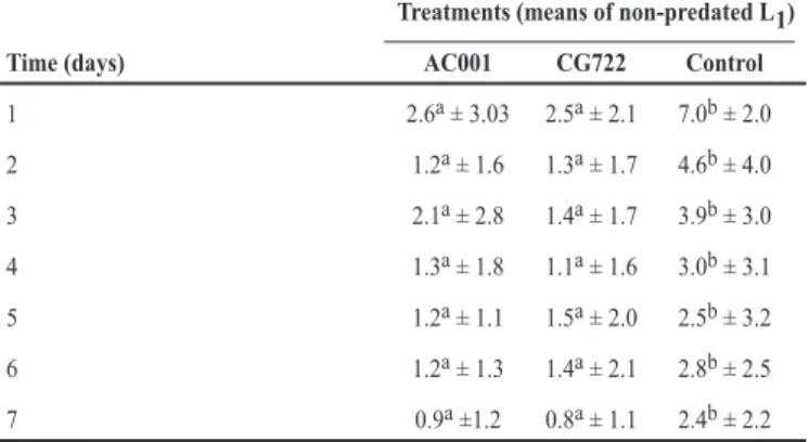
TABLE 1 - Daily means and standard deviations of non-predated irst-stage larvae (L1) of Angiostrongylus vasorum per 4mm-diameter ield in 2% water agar for the group treated for seven days with the fungus Duddingtonia lagrans (AC001 and CG722) and for the control group (without fungi).
Treatments (means of non-predated L1)
Time (days) AC001 CG722 Control
1 2.6a ± 3.03 2.5a ± 2.1 7.0b ± 2.0 2 1.2a ± 1.6 1.3a ± 1.7 4.6b ± 4.0 3 2.1a ± 2.8 1.4a ± 1.7 3.9b ± 3.0 4 1.3a ± 1.8 1.1a ± 1.6 3.0b ± 3.1 5 1.2a ± 1.1 1.5a ± 2.0 2.5b ± 3.2 6 1.2a ± 1.3 1.4a ± 2.1 2.8b ± 2.5 7 0.9a ±1.2 0.8a ± 1.1 2.4b ± 2.2
AC001: Duddingtonia lagrans;CG722: Duddingtonia lagrans.
Means with a superscript letter indicate that the lines are not statistically different (p>0.01), Tukey’s test.
was discarded, and the pellet containing A. vasorum L1 was resuspended in 5ml of 0.85% saline solution. The content was homogenized, and three 10µl samples were removed and distributed in 7.5 × 2.5cm glass slides. A count of the larvae was carried out under a stereomicroscope (25×). The total larval number was estimated by a simple rule of three.
The predation test was conducted on the surface of the Petri dishes according to a modiied technique previously described by Braga et al.8. Three groups were formed on
9cm-diameter Petri dishes containing 20ml of 2% WA: two treatment groups (AC001 and CG722) and one control group (without fungi). Six repetitions were made for each group. The Petri dishes were previously marked into 4mm-diameter ields. In the treated groups, each Petri dish contained A. vasorum L1 and 500 conidia of the fungal isolate AC001 or CG722 in
2% WA. Each Petri dish in the control group contained only 500 L1 in 2% WA. Every 24 h for 7 days, ten 4mm-diameter random
ields on each plate in the treated and control groups were observed under an optical microscope at 10× magniication; the number of L1 that had not been preyed on was counted on each plate. At the
end of the 7-day period, the non-predated L1 were recovered from
the Petri dishes using the Baermann apparatus with water at 42ºC. The data obtained were examined by analysis of variance at 1 and 5% probability levels using the BioEstat 3.0 software (Manuel Ayres, Brazil). The eficiency of the predation activity was evaluated using Tukey’s test at the 1% probability level. The percentage reduction in the mean larval recovery was calculated by the following equation:
Reduction (%) = (mean L1 recovery from control – mean L1 recovery from treatment) × 100.
Mean L1 recovery from control
In the present study, even with the administration of conidia, the fungus produced optimum results as early as in the irst 24 hours of reading (Table 1). The mean number of A. vasorum L1 not preyed on per 4mm-diameter ield in the control condition was signiicantly different (p<0.01) from that on the plates treated with the fungus D. flagrans (AC001 and CG722) throughout the experiment. In addition, there were typical fungal structures (conidia and traps) on the boards of the treated groups during the test. At the end of the experiment, the following A. vasorum L1 percentage reductions were observed: 74.5% for
AC001 and 63.2% for CG722 (Figure 1).
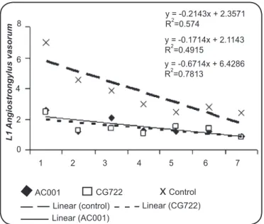
The linear regression coeficients calculated by the analysis of the mean number of A. vasorum L1 per 4mm-diameter ield
in the treated and control groups were: 0.21 (AC001), 0.17 (CG722), and 0.67 (control) (Figure 2).
It is estimated that close to 1 billion people are currently infected by geohelminths, mainly due to contact with common soil, indicating that this is an important route of human infection, which itself is associated with varied grave health consequences if left untreated. In addition, studies on parasites infesting domestic animals have provoked increasing interest due to the intimate relationship that exists between man and these animals, which may be a public health concern14.
The genera Ancylostoma and Toxocara, which are parasites of dogs and cats, and possibly the species A. vasorum stand out among helminths with zoonotic potential. Besides showing medical–veterinary importance, A. vasorum as a cardiopulmonary parasite of domestic and wild dogs requires special attention and investigation also because it can infect humans as well6,14. Few
studies have mentioned the in vitro predatory activity of different nematophagous fungi on larvae of nematode parasites of dogs.
In the present study, the isolate CG722 demonstrated a mean value of 0.8 in relation to viable L1 in the treated Petri dishes
at the end of the 7-day period. This result is concordant with Campos et al.9, who, using the same isolate (CG722) preying on
L3 of Strongiloydes papillosus, demonstrated a mean of 10.41
in vitro at the end of 7 days. However, the changes observed here may have occurred as a result of various factors. According to Mendoza De Gives et al.15, the composition of the cuticle
of nematodes can be a determining factor in the ability of fungi to prey on the nematodes. Also, antigenic variations in different nematodes or in the fungal isolate used may
FIGURE 1 - Means and standard deviations (bars) of infective non-predated Angiostrongylus vasorum larvae recovered from 2% water-agar plates by the Baermann method on the seventh day of treatment with the following fungal isolates: Duddingtonia lagrans (AC001 and CG722) and control group (without fungi).
The asterisk denotes a signiicant difference (p<0.05) between the fungus-treated group and the control; Tukey's test at 1% probability level.
100
80
60
40
20
0
AC001 CG722 Control
L1
110
www.scielo.br/rsbmt
Rev Soc Bras Med Trop 46(1):108-110, Jan-Feb, 2013
REFERENCES
inluence the rate of predation. Braga et al.10 demonstrated
that AC001 grown in Petri dishes containing a solid culture (2% WA) did prey on and consequently destroyed 80.3% of A. vasorum L1 at the end of 7 days. However,
comparing this predatory activity with the observation in the present study, it can be noted that the eficiency (74.5%) in this same strain was similar since the predation of larvae occurred after 24h. In this study, there was no statistically signiicant difference in the predatory ability of isolates of D. lagrans, as evidenced by the number of A. vasorum larvae recovered from the plates with the strains CG722 and AC001. However, differences in the inter- and intraspeciic activity of predatory nematophagous fungi are common and have been observed in experiments with other fungal isolates11.
The negative coeficients of correlation indicate a downward behavior of the regression curves for the treatments with the fungal isolates AC001 (0.21) and CG722 (0.17). This was caused by the reduction in the mean numbers of non-predated A. vasorum L1 per 4mm-diameter ield during the experimental assay, mainly due to the capture of L1 in fungal traps. The reduction in the number of L1 per 4mm-diameter ield in the
control group during the study, however, was caused by the migration of larvae to the periphery of the Petri dishes, where the moisture level was higher. This inding was also reported by Araújo et al.15, who carried out in vitro tests in Petri dishes.
The results of this study confirm previous works on the eficiency of D. lagrans in the control of larvae of nematode parasites of dogs. As the larvae are free in the environment, there exists the possibility of human infection since other parasites of the genus Angiostrongylus have been proven to be zoonotic2. In
this context, we suggest the application of nematophagous fungi, especially any of the isolates of the species D. lagrans tested (AC001, CG722, and CG768), that are capable of destroying L3 of potentially zoonotic gastrointestinal nematode parasites.
1. Saeed I, Maddox-Hyttel C, Monrad J, Kapel CMO. Helminths of red fox (Vulpes vulpes) in Denmark. Vet Parasitol 2006; 139:168-179.
2. Caldeira RL, Carvalho OS, Mendonça CLFG, Graeff-Teixeira C, Silva MCF, Ben R, et al. Molecular differentiation of Angiostrongyluscostaricensis, A. cantonensis, and
A. vasorum by polymerase chain reaction-restriction fragment length polymorphism. Mem Inst Oswald Cruz 2003; 98:1039-1043.
3. Morgan ER, Shaw SE, Brennan SF, De Waal TD, Jones BR, Mulcahy G.
Angiostrongylus vasorum:a real heartbreaker. Trends Parasitol 2005; 21:49-51. 4. Guilhon J. Role des limacidés dans le cycle évolutif d´Angiostrongylus vasorum
(Baillet, 1866). CR Acad Sci 1960; 251:2252-2253.
5. Cury MC, Guimarães MP, Lima WS, Caldeira MCM, Couto TR, Murta K, et al. Biochemical serum proiles in dogs experimentally infected with Angiostrongylus vasorum (Baillet, 1866). Vet Parasitol 2002; 128:121-127.
6. Eckert J, Lämmler G. Angiostrongylose bei Mensch und Tier. Parasitol Res 1972; 39:303-322.
7. Conboy GA. Angiostrongylus vasorum in dogs in Atlantic Canada and their treatment with milbemycin oxime. Vet Rec 2004; 155:16-18.
8. Braga FR, Silva AR, Araujo JM,Carvalho RO, Araujo JV,Frassy LN. Atividade predatória dos fungos nematófagos Duddingtonia lagrans, Monacrosporium thaumasium e Artrobotrys robusta sobre larvas infectantes de Strongyloides stercoralis. Rev Soc Bras Med Trop 2010; 43:588-590.
9. Campos AK, Araújo JV, Guimarães MP. Interaction between the nematophagous fungus Duddingtonia lagrans and infective larvae of Haemonchus contortus
(Nematoda: Trichostrongyloidea). J Helminthol 2008; 82:337-341.
10. Braga FR, Carvalho RO, Araujo JM, Silva AR, Araújo JV, Lima WS,
et al.Predatory activity of the fungi Duddingtonia lagrans, Monacrosporium thaumasium, Monacrosporium sinense and Arthrobotrys robusta on
Angiostrongylus vasorum irst-stage larvae. J Helminthol 2009; 83:303-308. 11. Araújo JV, Santos MA, Ferraz S, Maia AS. Antagonistic effect of predacious
Arthrobotrys fungi on infective Haemonchus placei larvae. J Helminthol 1993; 67:136-138.
12. Lima WS, Costa HMA, Guimarães MP, Leite ACR. Angiostrongylus vasorum
(Baillet, 1866) Nematoda: Prothostrongylidae em cães de Minas Gerais, Brasil. Mem Inst Oswaldo Cruz 1985; 80:233-235.
13. Barçante JMP, Barçante TA, Dias SRC, Vieira LQ, Lima WS, Negrão-Corrêa D. A method to obtain axenic Angiostrongylus vasorum irst-stage larvae from dog feces. Parasitol Res 2003; 89:89-93.
14. Patteson MW, Gibbs C, Wotton PR, Day MJ. Angiostrongylus vasorum infection in seven dogs. Vet Rec1993;4:565-570.
15. Mendoza De Gives P, Davies KG, Clark SJ, Behnke JM. Predatory behavior of trapping fungi against srf mutants of Caenorhabditis elegans and different plant and animal parasitic nematodes. Parasitology 1999; 119:95-104.
FINANCIAL SUPPORT
CNPq (Conselho Nacional de Desenvolvimento Cientíico e Tecnológico; National Council of Technological and Scientiic Development), FAPEMIG (Fundação de Amparo à Pesquisa do Estado de Minas Gerais; Research Support Foundation of Minas Gerais), and CAPES (Coordenação de Aperfeiçoamento de Pessoal de Nível Superior; Coordination for Improvement of Higher Education Personnel).
The authors declare that there is no conlict of interest.
CONFLICT OF INTEREST
FIGURE 2 - Linear regression curves calculated using the mean Angiostrongylus vasorum larvae (L1)per 4mm-diameter ield for the group treated with the fungus Duddingtonia lagrans (AC001 and CG722) and the control group (without fungi) as a function of time (1 to 7 days).
8
6
4
2
0
1 2 3 4 5 6 7 y = -0.2143x + 2.3571
R2=0.574
y = -0.1714x + 2.1143
R2=0.4915
y = -0.6714x + 6.4286
R2=0.7813
L1
Angiostrongylus vasorum
In conclusion, the fungal conidia of D. lagrans (AC001 and CG722) have predatory activity on irst-stage larvae of A. vasorum and could be used as a possible alternative method of biological control of A. vasorum larvae.
x
AC001 CG722 Control
Linear (control) Linear (CG722)