192
Case Report
REVISTA PAULISTA DE MEDICINAa b s t r a c t
CO N TEX T: The litho pedio n (calcified abdo minal pregnancy) is a rare pheno meno n and there are less than 3 0 0 cases repo rted in the medi-cal literature.
CASE REPO RT: In this case, a 4 0 year-o ld patient had had her o nly pregnancy 1 8 years earlier, witho ut medical assistance since then. She came to o ur ho spital with pain and tumo ral mass o f appro xi-mately 2 0 centimeters in diameter. Co mplementary examinatio ns (abdo minal X-ray, ultraso no graphy and co mputerized to mo graphy) demo nstrated an extra-uterine abdo minal 3 1 -week pregnancy with calcificatio n areas. Explo rato ry laparo to my was perfo rmed, with extirpatio n o f a well-co nserved fetus with partially calcified o vular membranes.
KEY W O RDS: Abdo minal preg nancy. Litho pedio n. Litho kelypho -pedio n. Fetal death.
• Renato Passini Júnio r • Ro xana Kno bel • Mary Ângela Parpinelli • Belmiro G o nçalves Pereira • Eliana Amaral • Fernanda G aranhani de Castro Surita • Caio Ro gério de Araújo Lett
INTRODUCTION
Lithopedion (litho = stone; pedion = child) is the name
given to an extra-uterine pregnancy that evolves to fetal death and calcification. It is a rare phenomenon that mostly comes from an abdominal pregnancy. The incidence of abdo minal pregnancy is 1:11,000 pregnancies and lithopedion occurs in 1.5 to 1.8% of these cases.1,3 There have been less than 300 cases in 400 years of world medi-cal literature.2,3,4 Because of the increase in inflammatory pelvic disease and uterine tubes surgery, there has been an increase in ectopic pregnancy.1 On the other hand, the occurrence of abdominal pregnancy and lithopedion has tended to become even rarer due to medical and pre-natal care becoming more accessible to the population, with the possibility of early diagnosis and treatment of the pa-thology.1,4
CASE REPORT
A 40 year-o ld wo man o f bro wn skin had a primary co mplaint o f lo wer abdo men pain. The patient repo rted regular abdo minal gro wth and healthy fetal activity fro m a pregnancy that happened 18 years earlier. She had never do ne pre-natal fo llo w-up. In the third trimester, she had started to feel stro ng cramps in the lo wer abdo men at the same time that fetal activity disappeared. She had not looked for medical assistance and some weeks later she had eliminated a dark red mass through the vagina with a placental appearance.
She had experienced the characteristic mo difi-catio ns o f breast lactatio n. The abdo men had started to decrease but retained an infra-umbilical mass o f
Sao Paulo Med J/Rev Paul Med 2000; 118(6):192-4.
Calcifie d abdominal pre gnancy with e ighte e n
ye ars of e volution: case re port
193
abo ut 20 centimeters in diameter, mo bile and pain-less. A few mo nths befo re being seen at o ur service, she started to fell pain in the lo wer abdo men and lo o ked fo r medical assistance.
Her gyneco lo gic histo ry was o f regular menstrual bleeding starting at the menarche and again after preg-nancy. She had never used any co ntraceptive metho d.
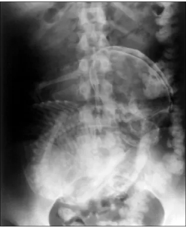
The physical examinatio n revealed an infra-um-bilical mass o f appro ximately 20 centimeters in diam-eter that was mo bile and hardened. The uterus was witho ut pregnancy mo dificatio ns. The abdo minal X-ray (Figure 1) and co mputerized to mo graphy sho wed the presence o f an ecto pic fetus in a mesentery blo o d vessel branch, with peripheral calcificatio ns. The ul-traso und examinatio ns sho wed an empty uterus,
regu-Sao Paulo Med J/Rev Paul Med 2000; 118(6):192-4.
lar o varies and the presence o f a 31-week fetus (deter-mined fro m femur length).
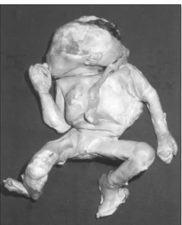
A hypothesis of lithopedion was made, and because of the clinical symptoms and the patient’s desire to remove the mass, explo rato ry laparo to my was do ne. After performing parietal celiotomy, an oval tumor was seen with adherence of the right ovary and epiploon (Figure 2). It measured 15 x 25 centimeters and weighed 1,890 grams. It was composed of a calcified ovular membrane adhering to a fetus, which was dissected and proved to be well con-served and partially calcified (Figure 3). The surgery was successful, without complications, and the patient left hospital after three days.
DISCUSSION
In the cases related in the literature, the age o f the patients o n the date o f diagno sis varied fro m 23 to 100 years, 2/3 o f them being o ver 40 years o ld. The perio d o f fetus retentio n was fro m 4 to 60 years. Fetal death o ccurred between 3 and 6 mo nths o f pregnancy in 20% o f the cases, between 7 and 8 mo nths in 27% and at full term in 43% o f the cases.2,4
Abdo minal pregnancy results fro m the rupture o f tubal o r o varian pregnancy with abdo minal cavity implantation.1,3 The development of lithopedion happens under certain co nditio ns: (1) extra-uterine pregnancy; (2) fetal death after 3 mo nths o f pregnancy; (3) the egg must be sterile; (4) there canno t be any early diagno sis; (5) lo cal co nditio ns must exist fo r calcium precipitatio n (depo sit).1,2,4 The develo pment o f this pregnancy is the same as for abdominal intra-uterine pregnancy until fetal death. After this time, dehydration of tissues and calcium infiltratio n o ccur.1,3,4
An abdominal pregnancy that calcifies is generically called lithopedion and can have the following forms: (1) lithokelyphos (litho = rock, kelyphos = shell): only the o vular membrane is calcified and the fetus can be in different stages of decomposition; (2) lithokelyphopedion: both are calcified, i.e. fetus and ovular membrane, as in this case; (3) lithopedion: only the fetus is calcified.4
Although most cases remain asymptomatic for years, pelvic pain, weight sensatio n in the abdo men and compressive symptoms can occur, affecting especially the urinary bladder and rectum.2,3 Some associated complica-tio ns have been repo rted after a lo ng asympto matic evolution: urinary bladder and rectum perforation; extru-sion of fetal parts through the abdomen wall, rectum and vagina; intestinal obstruction (due to collision of fetal parts with the intestine or adherence) and volvulus.3,4
The diagno sis is revealed by a suggestive clini-cal histo ry, a pelvic mass fo und during the physiclini-cal
Figure 2. Calcified Abdominal Pregnancy - Epiploon adherence.
194
r e s u m o
CO N TEX TO : O Lito pédio (gestação abdo minal calcificada) é um fenô meno raro , co m meno s de 3 0 0 caso s descrito s na literatura.
RELATO DE CASO : Uma paciente de 4 0 ano s, teve uma única gestação há 1 8 ano s e não pro curo u serviço médico desde então . C he g o u c o m d o r e m b a ixo ve ntre e ma ssa tumo ra l d e apro ximadamente 2 0 centímetro s. O s exames co mplementares (radio grafia abdo minal, eco grafia e to mo grafia co mputado rizada) evidenciaram gestação de apro ximadamente 3 1 semanas extra-uterina e co m áreas de calcificação . Fo i realizada laparo to mia co m extração do feto em bo m estado de co nservação , envo lvido pela membrana parcialmente calcificada.
PA LAV RA S- CH AV E: G ra vid e z a b d o mina l. Lito p é d io . Litho kelypho pedio n. Mo rte fetal.
Re nato Passini Júnior, MD, PhD. Assistant Lecturer, Department o f Obstetrics & Gyneco lo gy, Faculty o f Medical Sciences, Universidade Estadual de Campinas, Campinas, Brazil.
Roxana Knobe l, MD, MSc. Department o f Obstetrics & Gyneco lo gy, Faculty o f Medical Sciences, Universidade Estadual de Campinas, Campinas, Brazil
Mary Ânge la Parpine lli, MD, PhD. Assistant Lecturer, Department o f Obstetrics & Gyneco lo gy, Faculty o f Medical Sciences, Universidade Estadual de Campinas Campinas, Brazil.
Be lmiro Gonçalve s Pe re ira, MD, PhD. Assistant Lecturer, Department o f Obstetrics & Gyneco lo gy, Faculty o f Medical Sciences, Universidade Estadual de Campinas, Campinas, Brazil.
Eliana Amaral, MD, PhD. Assistant Lecturer, Department o f Obstetrics & Gyneco lo gy, Faculty o f Medical Sciences, Universidade Estadual de Campinas, Campinas, Brazil.
Fe rnanda Garanhani de Castro Surita, MD, MSc. Department o f Obstetrics & Gyneco lo gy, Faculty o f Medical Sciences, Universidade Estadual de Campinas, Campinas, Brazil.
Caio Rogé rio de Araújo Le tt, MD. Resident Do cto r, Department o f Obstetrics & Gyneco lo gy, Faculty o f Medical Sciences, Universidade Estadual de Campinas, Campinas, Brazil.
Source s of funding: No t declared
Conflict of inte re st: No t declared
Last re ce ive d: 19 January 2000
Acce pte d: 03 February 2000
Addre ss for corre sponde nce :
Faculdade de Ciências Médicas, Universidade Estadual de Campinas R. Alexandre Fleming, 101 - Cidade Universitária
Campinas/SP - Brasil - CEP 13083-970 E-mail: co nceicao @ caism.unicamp.br
p u b lis hin g in fo r m a t io n 1. Co sta SD, Presley J, Bastert G. Advanced abdo minal pregnancy. Obstet
Gyneco l Surv 1991;46:515-25.
2. Frayer CA, Hibbert ML. Abdo minal pregnancy in a 67-year-o ld wo man undetected fo r 37 years: a case repo rt. J Repro d Med 1999;44:633-5.
REFERENCES
3. Iric k MB, Kitso s CN, O ’Le ary JA. The rap e utic asp e c ts in the management o f a litho pedio n. Am Surg 1970;36:232-4.
4. Spirito s NM, Eisenko p SM, Mishell DR. Litho kelypho s: a case repo rt and literature review. J Repro d Med 1987;32:43-6.
Figure 3. Calcified Abdominal Pregnancy - Fetus after dissection of calcified ovular membrane.
examinatio ns, and frequently, an Xray o f the abdo -men is eno ugh to co nfirm it.3,4 The ultraso und exami-natio n sho ws an empty uterine cavity and a no n-spe-cific appearance o f the abdo minal mass, co nfusing the d iagno sis.2 Co m p ute rize d to m o grap hy (CT) and nuclear magnetic reso nance clearly define the patho l-o gy and help the diagnl-o sis l-o f adherence and l-o ther o rgans affected, altho ugh these are no t abso lutely nec-essary.2,3,4 So me autho rs suggest excreto ry uro graphy and e ne m a X- ray to e valuate c o m p re s s io n o r alteratio ns in o rgans o r systems clo se to it.
The diagno sis differentiates it fro m o ther calci-fied masses like o varian tumo rs, myo mas, inflamma-to ry masses, urinary tract and bladder tumo rs, and epiplo o n calcificatio ns.4 There are cases repo rted with-o ut surgical extirpatiwith-o n with-o f the lithwith-o pediwith-o n2. Due to the po ssibility o f co mplicatio ns, even after years o f evo lu-tio n, the pro per pro cedure is surgical remo val.
The surgery is frequently simple with lo w bleed-ing. No intrao perative death has been repo rted, even in elderly patients.3,4 Nevertheless, extreme care is rec-o mmended in the surgical prrec-o cedure with the help rec-o f a general surgeo n o r uro lo gist, due to the po ssibility o f large quantities o f abdo minal blo o d vessels and in-testinal adherence.