Sao Paulo Med J. 2009; 127(6):379-81
379
Case report
Finger burns caused by concentrated hydroluoric acid, treated
with intra-arterial calcium gluconate infusion: case report
Queimadura digital por ácido luorídrico concentrado tratada com infusão intra-arterial de
gluconato de cálcio: relato de caso
Eduardo Mello De Capitani
I, Elcio Shiyoti Hirano
II, Isabela de Souza Cortez Zuim
III, Laura Bertanha
III, Ronan José Vieira
IV,
Paulo Roberto Madureira
V, Fábio Bucaretchi
VIPoison Control Center, University Hospital, Universidade Estadual de Campinas (Unicamp), Campinas, São Paulo, Brazil
IMD, PhD. Associate professor, Department of Internal Medicine, Poison Control Center, School of Medicine, University Hospital, Universidade Estadual de Campinas (Unicamp),
Campinas, São Paulo, Brazil.
IIMD. Medical doctor, Emergency Department, University Hospital, Universidade Estadual de Campinas (Unicamp), Campinas, São Paulo, Brazil.
IIIUndergraduate medical student, Poison Control Center, School of Medicine, University Hospital, Universidade Estadual de Campinas (Unicamp), Campinas, São Paulo, Brazil. IVMD, PhD. Assistant professor, Department of Internal Medicine, Poison Control Center, School of Medicine, University Hospital, Universidade Estadual de Campinas (Unicamp),
Campinas, São Paulo, Brazil.
VMD, PhD. Assistant professor, Department of Preventive and Social Medicine, Poison Control Center, School of Medicine, University Hospital, Universidade Estadual de Campinas
(Unicamp), Campinas, São Paulo, Brazil.
VIMD, PhD. Assistant professor, Department of Pediatrics, Poison Control Center, School of Medicine, University Hospital, Universidade Estadual de Campinas (Unicamp), Campinas,
São Paulo, Brazil.
ABSTRACT
CONTEXT: Hydroluoric acid (HF) is widely used in industry and at home. Severe lesions can occur after contact with highly concentrated solutions, leading to tissue necrosis and bone destruction. Speciic treatment is based on neutralization of luoride ions with calcium or magnesium solutions.
CASE REPORT: A 41-year-old male was seen at the emergency department 35 minutes after skin contact with 70% HF, showing whitened swollen lesions on the middle and fourth ingers of his right hand with severe pain starting immediately after contact. 2.5% calcium gluconate ointment was applied. Twenty-four hours later, the patient was still in severe pain and the lesions had worsened. Considering the high concentration of the solution, early start of severe pain, lesion characteristics and impossibility of administering calcium gluconate subcutaneously because of the lesion location, the radial artery was catheterized and 2% calcium gluconate was administered via infusion pump for 36 hours, until the pain subsided. No adverse effects were seen during the procedure. Ten days later, the lesions were stable, without bone abnormalities on X-rays. Six months later, a complete recovery was seen.
CONCLUSIONS: Intra-arterial calcium gluconate might be considered for inger burns caused by concentrated HF. Complete recovery of wounded ingers can be achieved with this technique even if started 24 hours after the exposure. However, controlled clinical trials are needed to conirm the effectiveness and safety of this intervention.
RESUMO
CONTEXTO: Ácido luorídrico é largamente usado na indústria e no ambiente doméstico. Lesões graves podem ocorrer depois de contato com soluções altamente concentradas levando a necrose tecidual e destruição óssea. O tratamento especíico é baseado na neutralização dos íons de lúor com soluções de cálcio ou magnésio.
RELATO DE CASO: Homem de 41 anos foi atendido na sala de urgência 35 minutos depois de contato da pele com ácido luorídrico a 70%, apresentando lesões esbranquiçadas e edemaciadas nos dedos médio e quarto da mão direita com dor intensa que iniciou logo após o contato. Pomada de gluconato de cálcio a 2,5% foi aplicada. Depois de 24 horas, o paciente continuava com dor mais intensa e as lesões haviam piorado. Considerando a concentração da solução, o início precoce da dor intensa, as características das lesões e a impossibilidade de administrar gluconato de cálcio no subcutâneo devido ao local da lesão, foi inserido cateter na artéria radial para infusão de gluconato de cálcio a 2% com bomba de infusão por 36 horas até melhora da dor. Nenhum efeito adverso foi observado durante o procedimento. Dez dias depois as lesões encontravam-se estáveis, sem alterações dos ossos vistas nos raios-X. Seis meses depois houve recuperação completa.
CONCLUSÃO: Gluconato de cálcio intra-arterial pode ser considerado em queimaduras digitais por ácido luorídrico. Recuperação completa dos dedos acometidos pode ser obtida com essa técnica mesmo que iniciada 24 horas após a exposição. Porém, ensaios clínicos controlados são necessários para conirmar a efetividade e a segurança desta intervenção.
KEY WORDS: Hydroluoric acid. Fingers. Calcium gluconate. Infusions, intra-arterial. Caustics.
PALAVRAS-CHAVE: Ácido luorídrico. Dedos.
Sao Paulo Med J. 2009; 127(6):379-81
De Capitani EM, Hirano ES, Zuim ISC, Bertanha L, Vieira RJ, Madureira PR, Bucaretchi F
380
INTRODUCTION
Hydroluoric acid (HF) is widely used in many industrial ields, in-cluding chemicals, fertilizers, pesticides, plastics, dyes, leather tanning, electrical sets and semiconductor manufacture. It is also used for do-mestic purposes like marble, brick and stone cleaning, rust removal and glass etching.1,2
Pain and erythematous lesions can take more than 24 hours to ap-pear when the HF concentration is less than 20%. he symptoms may be delayed by eight hours at intermediate HF concentrations (20 to 50%). Acute intense pain always occurs at concentrations greater than 50%, even without immediate skin lesions.1,2 Accidents commonly
af-fect ingers during applications of highly concentrated HF to stone, bricks, marble and glass surfaces. Severe local pain with possible de-struction of the distal phalanx when inger burns are not adequately treated is frequently observed.1 he skin may appear normal initially,
with only mild erythematous lesions, thus giving the false impression that the outcome would be favorable and erroneously downplaying the need for care.1 We describe a case of inger burns caused by highly
con-centrated HF that was treated with intra-arterial calcium gluconate 24 hours after the accident, with complete recovery.
CASE REPORT
A 41-year-old Caucasian male was seen at the emergency depart-ment 35 minutes after skin contact with 70% HF solution in an occu-pational accident. He had been cleaning a slate loor around a pool us-ing special gloves to do the job, but afterwards he touched the contami-nated cork of the bottle without protection.
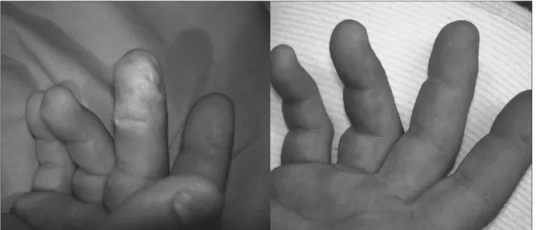
He presented lesions on the middle inger and part of the fourth inger of his right hand (Figure 1). He started feeling severe local pain immediately after contact (numerical rating scale, NRS = 8). He washed the lesions profusely with water for at least 15 minutes and
then went to the emergency department. On arrival, the skin of the middle inger was already whitened and swollen, and he complained of severe local pain (NRS = 10). 2.5% calcium gluconate ointment was applied, and he was asked to continue to use it every two hours for the next 24 hours.
Twenty-four hours later, the patient was still in severe pain (NRS = 10) and the lesions had worsened. Serum calcium was 9.5 mg/dl. A bolus of 10% calcium gluconate (10 ml) was administered intravenously without any improvement of the pain. Considering the high concentration of the HF solution, the early start and mainte-nance of severe pain 24 hours after the accident, the whitened and swollen lesion suggesting progression to necrosis and the impossibility of subcutaneous administration of calcium gluconate because of the lesion location, it was decided to administer the drug intra-arterially through catheterization of the radial artery at wrist level. A solution of 2% calcium gluconate in 5% dextrose was given by means of an in-fusion pump every four hours for 36 hours, until the pain subsided (NRS = 3).
No adverse efects were seen, and the catheter was withdrawn with-out local problems. Ten days later, the lesions were stable with no sign of necrosis. Radiography on the right hand showed no bone abnormali-ties. Six months later, the patient presented complete recovery from the lesions (restitutio ad integrum) (Figure 1).
DISCUSSION
HF is a fairly weak acid compared with sulfuric (H2SO4), hydro-chloric (HCl) and nitric (HNO3) acids. Due to its very low dissociation constant (1000 times lower than HCl), HF presents rather weak release of hydrogen ions. he toxic mechanism relates mostly to its non-disso-ciated and uncharged chemical form, which can penetrate through the skin and subcutaneous tissues by means of non-ionic difusion, target-ing calcium-rich tissues like bones.2
Finger burns caused by concentrated hydroluoric acid, treated with intra-arterial calcium gluconate infusion: case report
Sao Paulo Med J. 2009; 127(6):379-81
381
After dissociation, the strong electronegativity of the luoride ionallows it to bind tightly to any endogenous cation, particularly calcium (Ca) and magnesium (Mg), to produce insoluble salts.2 Soluble salts can
also be formed with other cations that dissociate rapidly, thereby releas-ing luoride ions again and leadreleas-ing to further tissue destruction. his toxic action can produce hypocalcemia, hypomagnesemia, cell necrosis, bone decalciication and destruction and cell dehydration with release of potassium ions (hyperkalemia).2 Diferently from strong acids, it can
take several days for HF to complete the process of tissue destruction and neutralization.
As acid penetrates, cell necrosis occurs and the skin can become whitened, eventually developing vesicles and progressing to bone de-struction. Disproportionate pain in relation to the apparently benign skin lesion is quite typical of HF burns, and serves to alert to the need for aggressive treatment and careful follow-up.1
he typical presentation of more than 50% of HF lesions is whit-ened tissue surrounded by erythema, accompanied by severe pain.3 Such
accidents should always be investigated for systemic efects, indepen-dently of the extent of the lesions.3
Topical therapy with application of 2.5% calcium gluconate gel can be attempted in mild or moderate cases.4 For inger burns, the ointment
must be applied in a latex glove.
Intradermal or subcutaneous application of 10% calcium gluconate solution (never calcium chloride), around and into the afected area, can be of help in cases of lesions in which the subcutaneous tissue is loose enough to support gluconate deposits without interfering with normal blood circulation.3,5 Obviously, this cannot be used for inger lesions.
Intravenous calcium gluconate or magnesium sulfate must be given when serum calcium and magnesium imbalance is detected. Regional intravenous infusion of calcium can be used for inger lesions using a Bier block technique.6
Intra-arterial perfusion of calcium gluconate is a good alternative in cases of swollen and painful injured ingers. he advantages that can be listed for its use include rapid pain relief and delivery of a greater amount of calcium, with better distribution to tissues because of cal-cium induction of vasodilatation. Adverse efects and complications are rare and, for moderate to severe burns of ingers and hands (general-ly with HF concentrations > 10%), intra-arterial calcium infusion is thought to be more efective than local therapy.3,5
CONCLUSION
Intra-arterial calcium gluconate might be considered for inger burns caused by highly concentrated HF, when topical treatment is con-sidered useless, or when intradermal and subcutaneous calcium injec-tions cannot be performed. Complete recovery of wounded ingers can be achieved with this technique, even if it is started 24 hours after the exposure, as seen in this report. Controlled clinical trials must be carried out to assess the efectiveness and safety of this intervention.
REFERENCES
1. Anderson WJ, Anderson JR. Hydroluoric acid burns of the hand: mechanism of injury and treatment. J Hand Surg Am. 1988;13(1):52-7.
2. Sheridan RL, Ryan CM, Quinby WC Jr, Blair J, Tompkins RG, Burke JF. Emergency management of major hydroluoric acid exposures. Burns. 1995;21(1):62-4.
3. Lin TM, Tsai CC, Lin SD, Lai CS. Continuous intra-arterial infusion therapy in hydroluoric acid burns. J Occup Environ Med. 2000;42(9):892-7.
4. Roblin I, Urban M, Flicoteau D, Martin C, Pradeau D. Topical treatment of experimental hydroluoric acid skin burns by 2.5% calcium gluconate. J Burn Care Res. 2006;27(6): 889-94.
5. Vance MV, Curry SC, Kunkel DB, Ryan PJ, Ruggeri SB. Digital hydroluoric acid burns: treat-ment with intraarterial calcium infusion. Ann Emerg Med. 1986;15(8):890-6.
6. Graudins A, Burns MJ, Aaron CK. Regional intravenous infusion of calcium glucona-te for hydroluoric acid burns of the upper extremity. Ann Emerg Med. 1997;30(5): 604-7.
Meeting, date and place where the paper was presented: Clinical case presented at the 28th International Congress of the European Association of Poison Centres and Clinical Toxicologists, May 6-9, 2008, in Seville, Spain
Sources of funding: None
Conlict of interest: None
Date of irst submission: August 7, 2008
Last received: November 26, 2009
Accepted: November 30, 2009
Address for correspondence:
Eduardo Mello De Capitani
Centro de Controle de Intoxicações, Hospital das Clínicas, Faculdade de Ciências Médicas da Universidade Estadual de Campinas (FCM/Unicamp)