ABSTRACT
Mariana da Cunha Lopes de Lima Allan de Oliveira Santos Celso Darío Ramos Luiz Ricardo Gonzalez José Inácio Oliveira Edwaldo Eduardo Camargo
ORIGINAL AR
repetitive strain injury
Division of Nuclear Medicine, Department of Radiology, Faculdade
de Ciências Médicas, Universidade Estadual de Campinas (Unicamp),
Campinas, São Paulo, Brazil
INTRODUCTION
Repetitive strain injury (RSI) is a group of diseases that occur in workers who perform repetitive movements.1 Many types of RSI have been described, such as clerk’s palsy, bricklayer’s shoulder, carpenter’s elbow, jan-itor’s elbow, telegraphist’s cramp and writer’s cramp.2-4 Imprecise associations between RSI and age, gender, fi tness and weight have been made.4 Some injuries can be related to RSI, such as tendinitis, bursitis, epicondylitis, teno-synovitis and carpal tunnel syndrome.
It is very important to correctly diagnose RSI because such injuries may cause the pa-tient to have temporary or even permanent incapacity to perform the same function or similar functions. In addition, RSI may cause legal consequences, such as dismissal from work and changing of jobs.
RSI is diffi cult to diagnose, and clinical signs, physical examination and complementa-ry tests are the basis for such diagnoses. There is no “gold standard” method for diagnosing this disorder.
OBJECTIVE
The purpose of this study was to eva-luate the usefulness of three-phase bone scintigraphy (TPBS) in diagnosing RSI in the upper limbs.
METHODS
Population Population
Seventy-three patients with clinical suspi-cion of RSI in the upper limbs were selected from January 1997 to December 1998, and were prospectively studied in the Occupa-tional Medicine Division of our university hospital (Universidade Estadual de Campi-nas, Unicamp). Forty-seven were males and 24 were females, and their ages ranged from 19 to 50 years (mean age: 31.2 years). The
inclusion criteria consisted of clinical signs of RSI in the patient’s history and physical examination, but without abnormalities seen on radiographs or computed tomography. Some patients had clinical suspicion of RSI in more than one joint; thus, 127 joints with suspected RSI were studied. The joints under investigation were the shoulders (77/127), elbows (16/127) and wrists (34/127). These patients had been working in their present jobs for between seven months and 10 years (mean: 7.9 years) and the time elapsed be-tween the beginning of joint symptoms and the RSI diagnosis ranged from three months to 10 years (mean: 24 months). The majority of the patients were machine operators, and other professions represented were keyboard operators, cooks, cashiers and tailors.
Forty healthy volunteers (22 females and 18 males) with a mean age of 35.5 years were used as a control group for the shoulders. An additional group of 40 normal individuals (25 females and 15 males) with a mean age of 35.2 years was used as a control group for the elbows and wrists. Individuals were only included in these control groups if they pre-sented absence of pain or other symptoms and did not perform repetitive movements using their upper limbs.
Three-phase bone Three-phase bone scintigraphy (TPBS) scintigraphy (TPBS)
Acquisition protocol Acquisition protocol
TPBS was performed using a scintillation camera-computer system after bolus injection of 1,110 MBq (25 mCi) of 99m Tc-methylene-diphosphonate (99mTc-MDP).
During the fl ow phase, sequential images were acquired at the rate of two seconds per frame for 80 seconds with the painful joint in the fi eld of view. Blood pooling images for 500,000 counts in the same position were ac-quired fi ve minutes after radiotracer injection.
CONTEXT AND OBJECTIVE: The diagnosis of repetitive strain injury (RSI) is subjective and solely based on clinical signs and physical ex-amination. The aim of this paper was to assess the usefulness of three-phase bone scintigraphy (TPBS) in diagnosing RSI.
DESIGN AND SETTING: Prospective study at the Division of Nuclear Medicine, Department of Radiology, School of Medical Sciences, Univer-sidade Estadual de Campinas (Unicamp).
METHODS: Seventy-three patients (mean age 31.2 years; 47 males) with clinical suspicion of RSI in the upper limbs were studied. A total of 127 joints with suspicion of RSI were studied. The shoulders, elbows and wrists were ana-lyzed semi-quantitatively, using the shafts of the humeri and ulnae as references. The results were compared with a control group of 40 normal individuals. The patients’ signs and symptoms were used as the “gold standard” for calculating the probabilities.
RESULTS: From visual analysis, abnormalities were observed in the fl ow phase for four joints, in the blood pool phase for 11 joints and in the delayed images for 26 joints. Visual analysis of the joints of the control group did not show any abnormalities. Semi-quantitative analysis showed that most of the patients’ joint ratios were normal. The exceptions were the wrists of patients with left-sided RSI (p = 0.0216). However, the sensitiv-ity (9%) and accuracy (41%) were very low.
CONCLUSION: TPBS with semi-quantitative analysis has very low sensitivity and accuracy in the detection of RSI abnormalities in the up-per limbs.
Delayed planar images with 300,000 counts each were obtained two hours after injection in the same position. Additional whole-body images in the anterior and posterior positions were also acquired.
Visual analysis Visual analysis
Two nuclear physicians blindly inter-preted the studies. In the event of disagree-ment, both observers re-analyzed the study. The blood fl ow, blood pooling and delayed images from TPBS were considered to be either positive or negative. The images were considered positive when increased activity in the suspicious joint in comparison with the contralateral asymptomatic joint, or increased activity in bilateral joints in comparison with adjacent structures, was noted.
Semi-quantitative analysis Semi-quantitative analysis
Semi-quantitative analysis was performed on both shoulders, elbows and wrists joints of all patients and also in the control group, whether or not RSI was suspected.
For the evaluation of the shoulders, re-gions of interest (ROIs) were outlined on each of the following structures: acromioclavicular joints, coracoid processes and humeral heads. The humeral shaft was used as the reference. Counts per pixel obtained from the ROIs on the shoulders were divided by the counts per pixel from the ROIs on the humeral shafts. Ratios were obtained for all three structures (acromioclavicular joints, coracoid processes and humeral heads) in both shoulders of all patients and control individuals (Figure 1).
For the evaluation of the elbows and wrists, ROIs were outlined around the joints, bilaterally. The ulnar shaft was used as the reference. Counts per pixel obtained from the ROIs on the elbows and wrists were divided by counts per pixel from the ROIs on the ulnar shafts (Figure 2).
Statistical analyses Statistical analyses
For the patients with unilateral complaints of RSI, the semi-quantitative values obtained from the symptomatic joint were compared with those obtained from the patients’ asymptomatic contralateral side. The Wilcoxon test was used for comparisons between the two sides, for each joint. The level of signifi cance set was 5%. The joints of patients with bilateral symptoms were not compared with the patients’ contralateral joints for obvious reasons.
The semi-quantitative values obtained from the symptomatic joints were also com-pared with those obtained from the joints of the control group. The Mann-Whitney test was performed in order to compare differences between the patient and the control groups. The level of signifi cance set was 5%.
The sensitivities, specifi cities, positive and negative predictive values, accuracies and like-lihood ratios for positive and negative results from TPBS, in relation to RSI diagnoses, were calculated using visual and semi-quantitative analysis. The patients’ signs and symptoms were used as the “gold standard”.
RESULTS
Visual analysis Visual analysis
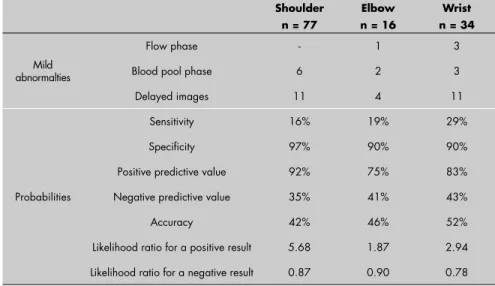
Table 1 displays the results from the visual analysis. Mild abnormalities in the blood fl ow phase were observed in four joints and mild abnormalities in the blood pool phase were observed in 11 joints. There was mildly in-creased blood fl ow and blood pooling without abnormalities on the delayed images from one wrist and one shoulder.
Mildly increased uptake was observed on the delayed images for 26 joints (11 shoulders, 11 wrists and four elbows).
Visual analysis of the joints from the con-trol group showed a wide range of symmetric uptake. However, none of these joints were considered pathological, since this group did not present with any signs or symptoms.
Table 1 also displays the probabilities re-lating to the usefulness of TPBS for diagnosing RSI for each joint. The probabilities that the visual analysis of TPBS would be diagnostic for RSI were as follows: sensitivities: 16% to 29%; specifi cities: 90-97%; positive predictive values: 75-92%; negative predictive values:
Table 1.Mild abnormalities and probabilities obtained by visual analysis in three-phase bone scintigraphy of repetitive strain injury
Shoulder n = 77
Elbow n = 16
Wrist n = 34
Mild abnormalties
Flow phase - 1 3
Blood pool phase 6 2 3
Delayed images 11 4 11
Probabilities
Sensitivity 16% 19% 29% Specifi city 97% 90% 90% Positive predictive value 92% 75% 83% Negative predictive value 35% 41% 43%
Accuracy 42% 46% 52%
Likelihood ratio for a positive result 5.68 1.87 2.94 Likelihood ratio for a negative result 0.87 0.90 0.78 Figure 1. Regions of interest (ROIs) for the
shoulders were outlined for the acromio-clavicular joints, coracoid processes and humeral heads, and the humeral shaft was used as the reference in the analysis with three-phase bone scintigraphy.
Table 2.Mean and standard deviation (SD) of ratios from semi-quantitative analysis of the shoulders, elbows and wrists in the patient (with symptoms of repetitive strain injury) and control groups by three-phase bone scintigraphy. The p-values represent the comparison between the suspected repetitive strain injury joints with both joints of the control group
Patient group
(side of complaint) Control group p-values* Right Left
Joint Mean SD Mean SD Mean SD Right Left
AC 2.83 0.63 2.72 0.63 2.82 0.61 0.7304 0.3455 CP 3.28 0.59 3.16 0.70 3.28 0.61 0.8852 0.4418 HH 2.82 0.66 2.76 0.64 3.10 0.71 0.0176 0.0176
E 3.13 0.51 3.00 0.77 2.86 0.80 0.2426 0.5090 W 2.21 0.46 2.26 0.47 1.95 0.53 0.0529 0.0216
AC = acromioclavicular joint; CP = coracoid process; HH = humeral head; E = elbow; W = wrist; *Mann-Whitney test (values in boldface were statistically signifi cant).
35-43%; accuracies: 42-52%; likelihood ratio for a positive result: 1.87-5.68; and likelihood ratio for a negative result: 0.78-0.90. Semi-quantitative analysis Semi-quantitative analysis
Table 2 displays the ratios obtained for each joint in the patient and control groups. In the control group, there was a wide variation in the intensity of uptake in normal joints of the upper limbs.
The probabilities relating to the useful-ness of TPBS for diagnosing RSI for each joint are displayed in Table 3. The prob-abilities that the semi-quantitative analysis of TPBS would be diagnostic for RSI were as follows: sensitivities: 0% to 9%; specifi cities: 95-100%; positive predictive values: 0-100%; negative predictive values: 28-38%; accura-cies: 28-41%; likelihood ratio for a positive result: 0-1.76; and likelihood ratio for a negative result 0.96-1.03.
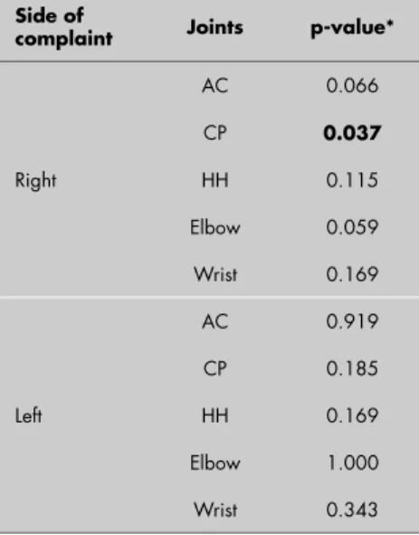
Among the patients with unilateral RSI complaints, the semi-quantitative uptake values obtained for the suspected joints were compared with the asymptomatic contralat-eral joint (Table 4). There was a signifi cant difference between the ratio in the coracoid process of patients with a right-side complaint and the ratio in the asymptomatic left side. The other affected joints did not show any difference in uptake when compared with the contralateral joints.
The semi-quantitative uptake values obtained from the joints with suspected RSI were also compared with the ratios from both joints of the control group (Table 2). Both the patients with symptoms in the left and right shoulders showed signifi cant differences in the humeral heads (p = 0.0176 for both groups) when compared with the control group. How-ever, the uptake values for the affected joint (mean values for patients: 2.82 for the right side and 2.76 for the left side) were lower than those for the asymptomatic joints (mean value for the control group: 3.10).
Among the patients with RSI complaints in their elbows, no differences were observed.
Patients with left wrist symptoms had a signifi cantly higher uptake ratio than did the control group (p = 0.0216). However, the sensitivity (9%) and accuracy (41%) were very low.
DISCUSSION
RSI is a disease of great clinical impor-tance, because it may cause incapacity of the affected limb (especially when not well treated) and the diagnosis may lead to legal consequences. The complaints include muscle fatigue, stiffness and aching, weakness,
pares-Table 3. Probabilities of the semi-quantitative analysis of three-phase bone scintigraphy in the diagnosis repetitive strain injury
AC CP HH Elbow Wrist
Sensitivity 0% 4% 1% 0% 9%
Specifi city 97% 100% 100% 100% 95% Positive predictive value 0% 100% 100% * 75% Negative predictive value 31% 32% 31% 28% 38%
Accuracy 30% 34% 32% 28% 41%
Likelihood ratio for a positive result 0 * * * 1.76 Likelihood ratio for a negative result 1.03 0.96 0.99 1.00 0.96
AC = acromioclavicular joint; CP = coracoid process; HH = humeral head; *In these cases it was not possible to calculate the values because there were no cases with false positive results.
Table 4. p-values for the comparison of ratios between the side with the uni-lateral repetitive strain injury (RSI) and the patient’s contralateral asymptomatic side, in analysis with three-phase bone scintigraphy
Side of
complaint Joints p-value*
Right
AC 0.066 CP 0.037
HH 0.115 Elbow 0.059 Wrist 0.169
Left
AC 0.919 CP 0.185 HH 0.169 Elbow 1.000 Wrist 0.343
*Wilcoxon’s test; AC = acromioclavicular joint; CP = cora-coid process; HH = humeral head.
thesia and lack of coordination. The treatment for RSI usually depends on careful analysis and modifi cation of work techniques and may include rest, anti-infl ammatory drugs and local injections. The clinical diagnosing of RSI is diffi cult because it is mainly based on the patient’s complaint and on non-specifi c clinical signs from physical examination.3 The diagnosis is usually based on patient complaints. Imaging tests are not suffi ciently accurate to help signifi cantly in the fi nal di-agnosing of RSI.1,5
1. Bywaters EGL. Lesions of bursae, tendons and tendon sheaths. Clin Rheum Dis. 1979;5(3):883-926.
2. Buckle PW. Work factors and upper limb disorders. BMJ. 1997;315(7119):1360-3.
3. Bird HA, Hill J. Repetitive strain disorder: towards diagnostic criteria. Ann Rheum Dis. 1992;51(8):974-7.
4. Panush RS. Occupational and Recreational Musculoskeletal Disorders. In: Ruddy S, Harris ED Jr, Sledge CB, Budd RC, Sergent JS, editors. Kelley’s Textbook of Rheumatology. 6th ed. Philadelphia: WB Saunders; 2001. p. 429-38.
5. Evans DM, Lawton DS. Assessment of hand function. Clin Rheum Dis. 1984;10(3):697-725.
6. Leclerc A, Chastang JF, Niedhammer I, Landre MF, Roquelaure Y, Study Group on Repetitive Work. Incidence of shoulder pain in repetitive work. Occup Environ Med. 2004;61(1):39-44.
7. Hagberg M. Clinical assessment, prognosis and return to work with reference to work related neck and upper limb disorders. G Ital Med Lav Ergon. 2005;27(1):51-7.
8. Tsauo JY, Lee HY, Hsu JH, Chen CY, Chen CJ. Physical exercise and health education for neck and shoulder complaints among sedentary workers. J Rehabil Med. 2004;36(6):253-7. 9. Pozderac RV. Longitudinal tibial fatigue fracture: an
uncom-mon stress fracture with characteristic features. Clin Nucl Med. 2002;27(7):475-8.
10. Drubach LA, Connolly LP, D’Hemecourt PA, Treves ST. Assessment of the clinical signifi cance of asymptomatic lower extremity uptake abnormality in young athletes. J Nucl Med. 2001;42(2):209-12.
11. al-Nahhas AM, Jawad AS, McCready VR, Kedar R. Detection of increased blood fl ow to the affected arm in repetitive strain
injury with radionuclide and Doppler ultrasound studies. A case report. Clin Nucl Med. 1995;20(7):615-8.
12. Mühlen CA, Santos CS, Paiva CC, et al. Lesões por esforços repetitivos em digitadores: inefi cácia do método cintilográfi co na detecção de periartrites dos membros superiores. [Lesions due to repetitive efforts in keyboard operators: ineffi cacy of scintigraphic method in detection of periarthritis of the upper limbs]. Rev Bras Reumatol. 1991;31(6):195-8.
Sources of funding: None Confl icts of interest: None
Date of fi rst submission: April 4, 2005 Last received: May 26, 2006 Accepted: May 29, 2006
REFERENCES RSI continues to be an important disease
and, over the last few years, some papers have been published about its incidence,6 clinical evaluation7 and treatment.7,8 There are some studies using TPBS to investigate RSI in athletes, mainly related to the lower limbs.9,10 Some authors have reported on the successful use of bone scans for diagnosing RSI or similar diseases.9,11
There have not been many descriptions of TPBS studies for investigating RSI among workers. al-Nahhas et al.11 investigated a keyboard operator with a clinical diagnosis of right-hand and forearm RSI. The radiographs were normal and the patient underwent TPBS and Doppler ultrasonography. TPBS showed increased blood fl ow to the right forearm and no uptake in the delayed im-ages. Doppler ultrasonography confi rmed the increased fl ow to the right forearm. The authors suggested that detection of increased blood fl ow by these techniques could play a role in diagnosing such patients.
The present study did not confi rm the fi ndings of al-Nahhas et al.11 and found only a few cases of blood fl ow and/or blood pooling abnormalities. Their study was a case report
and probably the patient had unusually marked infl ammatory process. Our fi ndings are in agreement with the study by Mühlen et al.,12 who also reported the low sensitivity of TPBS for diagnosing RSI. They studied 21 keyboard operators with and without a clinical suspicion of RSI in the forearms. Visual analysis demonstrated a correlation with the clinical data in only one patient, who had abnormalities in all three phases of TPBS. Six patients with a clinical suspicion of tenosynovitis or epicondylitis had normal scans and four patients without complaints had abnormalities in all three phases. These authors concluded that TPBS was not helpful for diagnosing RSI in keyboard operators.
In the present study, the finding of two patients with increased blood fl ow and blood pooling but without abnormalities in the delayed images was interpreted as soft-tissue infl ammation. The majority of the abnormalities were observed in the delayed images and were considered mild by visual analysis in all cases.
The semi-quantitative analysis performed on the delayed images did not demonstrate abnormalities in patients with elbow RSI.
Signifi cantly increased uptake was observed in the coracoid processes of patients with right-sided complaints, in comparison with the contralateral asymptomatic side.
Among the patients with bilateral humeral head RSI, an unusual fi nding was observed. There was signifi cantly decreased uptake in the humeral heads, in comparison with the control group. This may have been secondary to unde-ruse of this region due to bilateral RSI.
Regardless of the high specificity of TPBS in detecting RSI, the sensitivity and accuracy were very low both by visual and semi-quantitative analyses, for all joints, including the wrists.
CONCLUSIONS
AUTHOR INFORMATION
Bárbara Juarez Amorim, MD. Attending physician in the Division of Nuclear Medicine, Department of Radiology, Faculdade de Ciências Médicas, Universidade Estadual de Campinas (Unicamp), Campinas, São Paulo, Brazil. Elba Cristina Sá de Camargo Etchebehere, PhD.
Attending physician in the Division of Nuclear Medicine, Department of Radiology, Faculdade de Ciências Médicas, Universidade Estadual de Campinas (Unicamp), Campinas, São Paulo, Brazil.
Graciella Dalla Torre, BSc. Division of Statistics, Math Institute, Universidade Estadual de Campinas (Unicamp), Campinas, São Paulo, Brazil.
Mariana Cunha Lopes Lima, MD. Attending physician in the Division of Nuclear Medicine, Department of Radiology, Faculdade de Ciências Médicas, Universidade Estadual de Campinas (Unicamp), Campinas, São Paulo, Brazil. Allan Oliveira Santos, MD. Attending physician in the
Division of Nuclear Medicine, Department of Radiology, Faculdade de Ciências Médicas, Universidade Estadual de Campinas (Unicamp), Campinas, São Paulo, Brazil. Celso Darío Ramos, PhD. Professor in the Division of
Nuclear Medicine, Department of Radiology, Faculdade de Ciências Médicas, Universidade Estadual de Campinas (Unicamp), Campinas, São Paulo, Brazil.
Luiz Ricardo Gonzalez, MSc. Attending physician in the Division of Occupational Medicine, Department of Preventive and Social Medicine, Faculdade de Ciências Médicas, Universidade Estadual de Campinas (Unicamp), Campinas, São Paulo, Brazil.
José Inácio Oliveira, PhD. Professor in the Division of Occupational Medicine, Department of Preventive and Social Medicine, Faculdade de Ciências Médicas, Uni-versidade Estadual de Campinas (Unicamp), Campinas, São Paulo, Brazil.
Edwaldo Eduardo Camargo, PhD. Director of the Division of Nuclear Medicine, Department of Radiology, Faculdade de Ciências Médicas, Universidade Estadual de Campinas (Unicamp), Campinas, São Paulo, Brazil.
Address for correspondence: Bárbara Juarez Amorim
Serviço de Medicina Nuclear
Hospital das Clínicas da Universidade Estadual de Campinas (Unicamp)
Caixa Postal 6.142
Campinas (SP) — Brasil – CEP 13083-888 Tel. (+55 19) 3788-7801
Fax (+55 19) 3788-7821 E-mail: [email protected]
Copyright © 2006, Associação Paulista de Medicina
Resumo
Baixa sensibilidade da cintigrafi a óssea trifásica no diagnóstico de lesão por esforço repetitivo
CONTEXTO E OBJETIVO: O diagnóstico das lesões por esforço repetitivo (LER) é difícil e baseado em sinais clínicos, exame físico e testes complementares. O objetivo deste trabalho foi avaliar a utilidade da cintilografi a óssea trifásica (COT) no diagnóstico das lesões por esforço repetitivo.
TIPO DE ESTUDO E LOCAL: Estudo prospectivo realizado no Serviço de Medicina Nuclear, Departamento de Radiologia, Faculdade de Ciências Médicas, Universidade Estadual de Campinas (Unicamp), Campi-nas, Brasil.
MÉTODOS:Foram estudados 73 pacientes (47 homens; idade média 31,2 anos) com diagnóstico clínico de LER nos membros superiores. Um total de 127 articulações com diagnóstico clínico de LER foram estudadas. As imagens foram submetidas à análise semi-quantitativa dos ombros, cotovelos e punhos, usando as diáfi ses dos úmeros e ulnas como referência. Os resultados foram comparados com um grupo controle de 40 indivíduos normais. O quadro clínico dos pacientes foi usado como “padrão ouro” para calcular as probabilidades.
RESULTADOS: Na análise visual dos pacientes foram observadas alterações em quatro articulações na fase do fl uxo sangüíneo, em 11 articulações na fase de equilíbrio e em 26 articulações nas imagens tardias. A análise visual das articulações do grupo controle não mostrou alterações. Na análise semi-quantitativa, a maioria dos valores dos pacientes foram normais. As exceções foram os punhos dos pacientes com lesões por esforço repetitivo no lado esquerdo (p = 0,0216). Entretanto, a sensibilidade (9%) e a acurácia (41%) foram muito baixas.
CONCLUSÃO: A COT com análise semi-quantitativa apresenta sensibilidade e acurácia muito baixas para detectar LER nos membros superiores.