Activity in the
Drosophila
Circadian Oscillator
A´ ron Szabo´1,2, Christian Papin1,2, Daniela Zorn3, Prishila Ponien4,5, Frank Weber3, Thomas Raabe6, Franc¸ois Rouyer1,2*
1Institut de Neurobiologie Alfred Fessard, Centre National de la Recherche Scientifique Unite´ Propre de Recherche 3294, Gif-sur-Yvette, France,2De´partement de Biologie, Universite´ Paris Sud, Orsay, France,3Heidelberg University, Biochemistry Center (BZH), Im Neuenheimer Feld 328, Heidelberg, Germany,4Institut de Chimie des Substances Naturelles, CNRS UPR2301, Gif-sur-Yvette, France,5IMAGIF, Centre de Recherche de Gif, Gif-sur-Yvette, France,6University of Wuerzburg, Institute of Medical Radiation and Cell Research, Wuerzburg, Germany
Abstract
Phosphorylation is a pivotal regulatory mechanism for protein stability and activity in circadian clocks regardless of their evolutionary origin. It determines the speed and strength of molecular oscillations by acting on transcriptional activators and their repressors, which form negative feedback loops. InDrosophila, the CK2 kinase phosphorylates and destabilizes the PERIOD (PER) and TIMELESS (TIM) proteins, which inhibit CLOCK (CLK) transcriptional activity. Here we show that CK2 also targets the CLK activator directly. Downregulating the activity of the catalyticasubunit of CK2 induces CLK degradation, even in the absence of PER and TIM. Unexpectedly, the regulatorybsubunit of the CK2 holoenzyme is not required for the regulation of CLK stability. In addition, downregulation of CK2aactivity decreases CLK phosphorylation and increasesper andtimtranscription. These results indicate that CK2 inhibits CLK degradation while reducing its activity. Since the CK1 kinase promotes CLK degradation, we suggest that CLK stability and transcriptional activity result from counteracting effects of CK1 and CK2.
Citation:Szabo´ A´, Papin C, Zorn D, Ponien P, Weber F, et al. (2013) The CK2 Kinase Stabilizes CLOCK and Represses Its Activity in theDrosophilaCircadian Oscillator. PLoS Biol 11(8): e1001645. doi:10.1371/journal.pbio.1001645
Academic Editor:Ueli Schibler, University of Geneva, Switzerland
ReceivedJune 11, 2013;AcceptedJuly 19, 2013;PublishedAugust 27, 2013
Copyright:ß2013 Szabo´ et al. This is an open-access article distributed under the terms of the Creative Commons Attribution License, which permits unrestricted use, distribution, and reproduction in any medium, provided the original author and source are credited.
Funding:This work was funded by Agence Nationale de la Recherche ‘‘DrosoClock’’, ‘‘ClockGene’’ and ‘‘FunGenDroso’’, ‘‘Equipe FRM’’ program of Fondation pour la Recherche Me´dicale, and European Union 6th Framework Programme ‘‘EUCLOCK’’. AS was partly supported by Association pour la Recherche sur le Cancer and FR is supported by Institut National de la Sante´ et des Etudes et Recherches Me´dicales. TR was funded by the DFG SFB1047 grant from Deutsche Forschungsgemeinschaft. The funders had no role in study design, data collection and analysis, decision to publish, or preparation of the manuscript.
Competing Interests:The authors have declared that no competing interests exist.
Abbreviations:CT, circadian time; DD, constant darkness; LD, light:dark.
* E-mail: [email protected]
Introduction
Circadian oscillations of gene expression, physiology, and behavior are found in a wide range of organisms. They are governed by temporally regulated feedback loops in which transcription factors activate the expression of their own inhibitors. In the Drosophila circadian oscillator, the CLOCK (CLK) and CYCLE (CYC) bHLH-PAS domain transcription factors activate expression of theperiod(per) andtimeless(tim) genes at the end of the day. The delayed accumulation of PER and TIM and their transfer to the nucleus leads to transcriptional repression of CLK/ CYC during the late night. The repression phase is also shaped by other repressors/activators such as CLOCKWORK ORANGE (CWO) and KAYAK-a[1]. Subsequent degradation of PER and TIM repressors in the morning allows transcription to resume towards the evening [2,3]. Controlled phosphorylation, ubiquity-lation, and proteasome-dependent degradation of PER and TIM set the timing of their delayed accumulation and clearance. The PER protein is phosphorylated by the DOUBLETIME (DBT, CK1d/e), CK2, and NEMO kinases and polyubiquitylated by the SCFSlimb ubiquitin ligase complex [4–12]. TIM associates with PER, preventing its degradation, but TIM itself is subjected to phosphorylation and subsequent breakdown. TIM phosphoryla-tion involves the CK2 and SHAGGY (SGG, GSK-3) kinases and
TIM degradation also depends on SCFSlimb and a CULLIN-3-based ubiquitin ligase complex [7,8,13–15]. Phosphatase activity counterbalances the effects of the aforementioned and probably also of other kinases: PP2A regulates PER abundance, while PP1 targets both PER and TIM [16,17].
CLK phosphorylation cycles with a peak in the morning and a minimum in the early night [18–21]. Similarly, CLK immunore-activity in head extracts or brain tissue seems to oscillate in phase with its phosphorylation [21–23], although harsh extraction liberates chromatin-bound CLK, which results in relatively constant CLK levels [20,24,25]. Whether oscillations of CLK immunoreactivity in neurons reflect rhythmic changes of total CLK protein amount is still unclear [23,26]. Due to the cyclic regulation of CLK as opposed to constitutive expression of CYC, the CLK protein appears to represent the key rhythmic component of the circadian activator in Drosophila [27]. CLK DNA-binding and transcriptional activity show a robust oscillation with an evening peak that is associated with the rapid increase of
NEMO destabilizes CLKin vivoand might thus be a CLK kinase [29]. CLK transcriptional activity in cultured cells is affected by calcium/calmodulin-dependent kinase II and mitogen-activated protein kinase [30]. Ubiquitylation is also involved in the regulation of CLK and BMAL1, the CYC ortholog in mammals [31,32]. In Drosophila, USP8 was recently reported to decrease CLK activity by deubiquitylation [25].
The CK2 kinase has a key function in the clockwork of various organisms [33]. InNeurospora, CK1 and CK2 phosphorylate both the White Collar Complex (WCC) transcriptional activator as well as its inhibitor FREQUENCY (FRQ) to control their activity, subcellular localization, and stability [34–36]. In mammals, CK2 and CK1 destabilize PER2, although phosphorylation at specific CK2 target sites stabilizes the protein [37,38]. The CK2 holoenzyme is formed by a tetrameric complex consisting of two catalytic (a) and two interacting regulatory (b) subunits [39]. Theb subunits stabilize the a subunits that possess constitutive kinase activity. Phosphorylation of most substrates is enhanced by CK2b, while some substrates are more efficiently phosphorylated by free CK2a in the absence of CK2b [40]. In Drosophila, CK2a and CK2baffect PER and TIM abundance and subcellular localiza-tion, which correlates with a direct phosphorylation of both proteins by the CK2 holoenzyme in vitro [8,9,41–43]. The dominant-negative CK2aTik
mutation strongly increases TIM stability even in the absence of PER, supporting TIM as the main target of CK2 [15]. The CK2aTikprotein overexpression induces hyperphosphorylation of TIM that could be explained by enhanced phosphorylation or reduced dephosphorylation of TIM by other kinases and phosphatases [15].
Since the identity of the kinases involved in the control of CLK phosphorylation remains unclear, we asked whether CK2 plays a role in the phosphorylation and regulation of CLK. Our results indicate that inhibition of CK2a activity strongly increases CLK degradation, whereas CK2b does not affect CLK stability. The CK2 holoenzyme is recruited onto PER, TIM, and CLK mainly during late night, inducing CLK hyperphosphorylationin vivoand
CK2 phosphorylates CLK in vitro. Specific CLK activity is increased in dominant-negative CK2aTik
-expressing flies indicat-ing repression of CLK by CK2a. Our findindicat-ings define, to our knowledge, the firstbona fidekinase ofDrosophilaCLK that plays a role in its degradation and hyperphosphorylation. The unstable but strongly active CLK acquired by CK2a inhibition joins the club of other circadian transcription factors with similar properties such as the WCC complex inNeurospora.
Results
CK2aActivity Promotes CLK Protein Phosphorylation and Stability
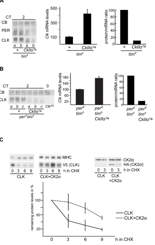
A putative role of CK2 in CLK regulation was first addressed by analyzing head extracts of flies expressing a dominant-negative version of the CK2a catalytic subunit. As previously reported [15,42],w;tim-gal4;UAS-CkIIaTik flies (hereafter tim.Tik flies) were behaviorally arrhythmic (Table 1) and displayed weak and strongly delayed PER and TIM oscillations, with high levels of mildly phosphorylated PER and highly phosphorylated TIM (Figure 1A). As CLK efficiently binds to DNA in the evening, the estimation of CLK levels through the circadian cycle is affected by extraction conditions. In sonicated head extracts, CLK protein has been shown to stay at constant levels, in contrast to a robust cycle of its phosphorylation [19,20]. However, the existence of oscillations in CLK levels remains discussed [22–25]. In our hands, sonicated extracts of control flies showed weak cycling of CLK levels, although peak time was rather variable between experiments (Figures 1A and S1A). Nonsonicated extracts always showed CLK levels cycling with a trough in the evening (Figure S1B). Importantly, both sonicated and nonsonicated extracts oftim.Tikflies showed very low CLK levels with reduced phosphorylation on the first day of constant darkness (DD) (Figures 1A and S1A and S1B). In order to better estimate CLK levels intim.Tikflies, sonicated extracts were treated withlprotein phosphatase (Figure S1C). Again, a strong decrease of unpho-sphorylated CLK abundance was observed in tim.Tik animals. Moreover,ClkmRNA levels were about 1.5-fold higher intim.Tik
flies than in controls (Figure 1B), indicating that low CLK protein levels are not a consequence of reducedClkexpression. Consequent-ly, the protein/mRNA ratio for CLK decreased to approximately 10% in tim.Tik (Figure S1D). Immunolabeling of whole-mount brains oftim.Tik flies also supported a strong reduction of CLK levels in the small ventral lateral neurons (s-LNvs) (Figure 1C), with no change in its nuclear-only localization (not shown).
To independently analyze the effect of decreasing CK2aactivity on CLK, a UAS-CkIIaRNA interference (RNAi) construct was expressed undertim-gal4control. Adult flies were kept at 29uC to increase Gal4-dependent expression. CLK in sonicated head extracts of w;tim-gal4/+;UAS-CkIIa-RNAi (tim.CkIIa-RNAi) flies showed a similar phenotype to that oftim.Tikflies, with reduced phosphorylation and levels throughout the cycle (Figure 1D). Dampened and delayed PER and TIM oscillations were observed with increased protein levels during the day. In support of its specificity, the induction of RNAi reduced CK2a protein levels (Figure S1E). Although high mortality of tim.CkIIa-RNAi flies after long incubation at high temperature prevented the assess-ment of their locomotor activity rhythms at 29uC, they displayed long period rhythms at 25uC (Table 1). Similar long period rhythms are observed in heterozygousw;tim-gal4/+;UAS-CkIIaTik
/ +flies (Table 1), as previously reported [42].
CK2aStabilizes CLK in the Absence of PER and TIM
TIM was described as the likely primary target of CK2ain the circadian clock, effects elicited on PER being only secondary [15].
Author Summary
The CK2 kinase is an ancient enzyme known to be at the heart of self-sustaining circadian clocks in animals, plants, and fungi. Circadian clocks are responsible for daily circadian rhythms in molecular, physiological, and behav-ioral processes. Their mechanism relies on transcriptional activators and repressors that constitute a feedback loop. The CLOCK (CLK) activator is required to initiate the transcription of theperiod(per) andtimeless(tim) genes in the late day. The PER and TIM repressors accumulate in a delayed manner and translocate to the nucleus to repress their own gene. Degradation of these repressors allows the activator to start a new cycle. In the fruit fly Drosophila melanogaster, CK2-mediated phosphorylation of the PER and TIM repressors targets them for degradation. Here we find that the CLK activator is also regulated by CK2. In contrast to PER and TIM, CLK is stabilized by CK2a
We thus asked whether TIM was required for CK2a effects on CLK, by comparing the profile of CLK protein in sonicated head extracts oftim01andtim01tim.Tikflies. In the absence of TIM, the CK2aTik protein induced a prominent reduction in CLK phosphorylation and a significant decrease of protein levels (Figure 2A). Furthermore,ClkmRNA levels were about four times higher in tim0 tim.Tik compared to tim0, supporting a strong degradation of the CLK protein intim0tim.Tikflies (Figure 2A). Since a PER/DBT complex controls CLK phosphorylation [20,28], we asked whether PER was required for CLK modifica-tions by CK2a, even in the absence of TIM. Effects of the CK2aTik protein were thus analyzed inper0tim0double mutants, where CLK appeared minimally phosphorylated in a CkIIa+
background (Figures 2B and S2A). CLK levels and phosphorylation were further diminished in the presence of the CK2aTik protein (Figures 2B and S2A). Since the CK2aTikprotein overproduction increased ClkmRNA levels by about twofold inper0 tim0double mutants, the CLK protein/mRNA ratio was reduced just as intim0
mutants (Figure 2B). A similar decrease of CLK protein levels was observed in per0 tim.Tik flies (Figure S2B) despite increasedClk
mRNA levels, suggesting that CLK protein was again strongly destabilized in the absence of PER. These observations reveal that CK2astabilizes CLK in the absence of PER and/or TIM. Since
CLK phosphorylation is further decreased by CK2aTik
expression, CK2a is important for the PER/TIM-independent minimal phosphorylation program of CLK.
The up-regulation ofClkmRNA levels intim.Tikflies suggested that CK2a could repress Clktranscription. To test whether Clk
transcription was affected in thetim.Tikgenotype,Clkpre-mRNA levels were estimated. They were not increased in tim.Tik flies compared to controls, although a reduced antiphasic cycling was observed at DD1 (Figure S2C). The antiphasic oscillation was reminiscent of PER and TIM oscillations persisting in these flies (see Figure 1A). The increase of mature Clk mRNA levels in
tim.Tikflies thus seems not to be the consequence of higherClk
gene transcription and rather supports a posttranscriptional control of ClkmRNA by CK2a. In agreement with a posttran-scriptional control, the VRI and PDP1 regulators of Clk
transcription were not affected inper0tim.Tikflies (Figure S2D). Finally, since the transcriptional regulation of thecryptochrome(cry) gene is similar to the one of theClkgene [44], we testedcrymRNA levels inper0tim.Tik flies. No increase of cry mRNA levels was observed in the presence of the CK2aTik
protein (Figure S2E), supporting a specific control ofClkmRNA levels by CK2a.
The data from tim.Tik flies strongly suggested that CK2a controls CLK stability independently from PER and TIM. To
Table 1.Locomotor activity rhythms in constant darkness of flies.
Genotype Number of flies Rhythmic flies (%) Period length (h) Power
w; tim-GAL4/+ 12 100 23.660.1 198613
yw;; UAS-CkIIaTik 16 81 24.0
60.1 72613
w; tim-GAL4/+;UAS-CkIIaTik/+ 21 100 30.960.1 17868
w; tim-GAL4;UAS-CkIIaTik 16 18* 27.8
62.9 3163
w; UAS-CkIIa/+ 9 88 24.160.1 144622
w; tim-GAL4/UAS-CkIIa 16 100 25.660.1 21169
w;; UAS-CkIIaRNAi 17520 R-2 14 92 24.860.1 133613
w; tim-GAL4/CyO; UAS-CkIIaRNAi 17520 R-2 14 93 32.160.3 143610
w; UAS-FLAG-CkIIa/+ 16 100 23.660.1 185617
w; tim-GAL4/UAS-FLAG-CkIIa 8 100 24.660.1 142619
w; UAS-CkIIbRNAi#106845/+; UAS-CkIIbRNAi#32377/+ 14 100 23.460.1 207616
w; tim-GAL4/UAS-CkIIbRNAi#106845; UAS-CkIIbRNAi#32377/+ 26 42* 31.261.6 3465
w;; gal1118/+ 15 100 24.460.1 18768
w;; gal1118/UAS-CkIIbRNAi#32377 29 93 33.160.3 8968
w; UAS:CkIIb-VIIb(4)/+ 15 86 24.560.1 145614
w; UAS:CkIIb-VIIb(5)/+ 14 92 24.260.1 129615
w; UAS:CkIIb-VIIc(6)/+ 16 93 23.860.2 141614
w; UAS:CkIIb-VIIa(5)/+;gal1118/UAS-CkIIbRNAi#32377 17 100 24.860.1 220610
w; UAS:CkIIb-VIIb(4)/+;gal1118/UAS-CkIIbRNAi#32377 24 100 24.560.1 21566
w; UAS:CkIIb-VIIb(5)/+;gal1118/UAS-CkIIbRNAi#32377 16 100 25.060.1 21767
w; UAS:CkIIb-VIIc(2)/+;gal1118/UAS-CkIIbRNAi#32377 15 100 25.460.1 226610
w; UAS:CkIIb-VIIc(6)/+;gal1118/UAS-CkIIbRNAi#32377 16 100 25.360.1 22066
w; UAS-gfp/+;gal1118/+ 16 100 23.660.1 180615
w; UAS-gfp/+;gal1118/UAS-CkIIbRNAi#32377 30 90 32.560.1 8568
w; UAS-FLAG-CkIIb/+ 13 92 24.060.3 138617
w; tim-GAL4/UAS-FLAG-CkIIb 15 100 24.760.1 166614
w; UAS-FLAG-CkIIb/+;gal1118/UAS-CkIIbRNAi#32377 14 93 24.660.1 219622
The mean values of period (in hours) and associated power (see Materials and Methods) are given6s.e.m. *Genotypes considered arrhythmic (see Materials and Methods).
obtain direct evidence for this, CLK degradation kinetics were analyzed in a cycloheximide (CHX) chase-based assay in Drosophila Schneider 2 (S2) cells. Since transfected V5-tagged CLK induced bothperandtimexpression in S2 cells in our hands, we used RNAi againstperandtimto eliminate any effect of PER and TIM proteins. After blocking protein synthesis with CHX, CLK showed robust degradation during the following 9 h (Figure 2C). When FLAG-HA-tagged CK2a was co-expressed, CLK degradation proceeded very slowly. The increase of CK2a levels by exogenous expression was rather limited in these conditions, indicating that a small increase in total CK2aprotein can have substantial effects on CLK degradation. These results confirm the in vivoobservations and strongly support a role for CK2ain the inhibition of CLK breakdown.
CK2bDoes Not Influence CLK Stability
Since inhibition of CK2aaffected CLK stability and phosphor-ylation, we asked whether CK2bknockdown would have similar effects. Pdf-gal4 UAS-CkIIb-RNAi/+ flies have been reported to display long period rhythms [45]. Driving two CkIIb-RNAi
transgenes under the control of tim-gal4 (hereafter tim.CkIIb -RNAi flies, see Materials and Methods) induced behavioral arrhythmicity (Table 1). The specificity of the CkIIb RNAi was first behaviorally assessed by rescue experiments involvingCkIIb
RNAi under the control of the strong PDF+
cell driver gal1118
[46] and the co-expression of different CK2b isoforms. The strongly altered behavior ofw;; gal1118/UAS-CkIIb-RNAicould be rescued by overexpression of the VIIa, VIIb, and VIIc CK2b isoforms (see [47]) (Table 1). Western blots against CK2brevealed a reduction in two isoforms intim.CkIIb-RNAi animals, while a third isoform remained unaffected (Figure S3A). TIM and PER cycling was profoundly altered in head extracts oftim.CkIIb-RNAi
flies at DD1 (Figures 3A and S3B). In contrast, CLK oscillations were only slightly affected. In particular,tim.CkIIb-RNAiflies did not show the pronounced decrease in CLK levels that was observed intim.Tikflies. Furthermore, CK2bdepletion in aper0 background did not result in a marked reduction of CLK phosphorylation or quantity (Figure 3B). Since equivalent levels of ClkmRNA were observed in per0 flies with or withoutCkIIb
RNAi expression, their protein/mRNA ratios were identical (Figure 3C and D), in contrast totim.Tik flies. Similarly, CLK was not affected when CkIIb RNAi was expressed in a tim0
background (not shown). In conclusion, although CK2a and CK2bproteins similarly affect TIM and PER accumulation and phosphorylation, the CK2bsubunit does not seem to be required for CK2ato control CLK degradation and phosphorylation.
CK2aPreferentially Forms Complexes with Highly Phosphorylated CLK in the Morning
The strong effects of CK2ainhibition on CLK suggested that the two proteins might physically interact. Flies expressing a FLAG-tagged CK2a protein under tim-gal4 control displayed strong behavioral rhythms with a 1 h period lengthening (Table 1). Anti-FLAG immunoprecipitation experiments were performed from FLAG-CK2a-expressing fly head extracts at different circadian times and showed co-immunoprecipitation of TIM, PER, and CLK mostly at the end of the subjective night and in the subjective morning when these proteins are mainly
hyperpho-Figure 1. CK2a inhibition triggers CLK degradation. (A, D) Western blot of sonicated head extracts from flies collected at DD1. Time (h) is indicated as CT. Gray and black bars represent subjective day and subjective night, respectively. A Coomassie Blue (CB) stained band in the size range of CLK is used as a loading control for blots run on 4% gels. Brackets indicate hypo- and hyperphosphorylated forms of CLK. At least two independent experiments were performed for each blot. (A) Two copies oftim-gal4and two copies ofUAS-CkIIaTiktransgene were used for the experimental genotype. (B) Quantitative RT-PCR measure-ments ofClkmRNA levels in heads of flies collected at DD1. Results were averaged from at least four independent experiments. Error bars indicate the s.e.m. Averaged values were normalized to the CT2 control averaged value set to 100. (C) Quantification of CLK immunofluores-cence in the PDF-expressing s-LNvs. Fluoresimmunofluores-cence index is given in
arbitrary units. Error bars indicate s.e.m. (D) Flies were entrained and collected at 29uC. One copy oftim-gal4and two copies of theCkIIaRNAi construct were used for the experimental genotype.
sphorylated (Figure 4A). Although relatively abundant medium-phosphorylated clock proteins were observed in the extracts at CT16, they were poorly co-immunoprecipitated with CK2a. The CK2asubunit thus appears to preferentially make complexes with highly phosphorylated forms of TIM, PER, and CLK. Flies expressing FLAG-tagged CK2b were also behaviorally rhythmic with a slightly lengthened period (Table 1), and FLAG-CK2b expression could rescue the severe period lengthening induced by
CkIIb RNAi (Table 1), indicating that the tagged protein was functional. Similarly to CK2a, CK2bwas found to be associated with hyperphosphorylated TIM, PER, and CLK in the late subjective night and in the subjective morning, whereas little amounts of proteins were co-immunoprecipitated at other circadian times (Figure 4B). The results thus suggest that CK2 holoenzyme is involved in the hyperphosphorylation of CLK, PER, and TIM in the late night/morning part of the cycle.
Since CK2astrongly influences CLK stability in the absence of PER, anti-FLAG immunoprecipitations were also done in per0 tim.FLAG-CkIIaflies (Figure 4C). Minute amounts of hypopho-sphorylated CLK were co-immunoprecipitated in per0 extracts, nevertheless indicating that CLK-CK2acomplexes may exist in the absence of PER. Conversely, CK2b did not co-immunopre-cipitate with CLK in a per0 background (Figure 4C). The poor detection of CK2a–CLK complexes in the absence of PER suggested a very labile interaction between the two proteins or indirect PER-independent effects of CK2aon CLK.
CK2 subunits preferentially associate with clock proteins at times when those are present in the nucleus. CK2awas, however, described to localize to the cytoplasm of LNv-s [8]. We therefore set out to investigate whether CK2a could be present in the nucleus of LNv-s as well. Whole-mount adult brains were stained with an anti-CK2a antibody together with PDF and anti-CLK. PDF is known to be exclusively cytoplasmic [48], while CLK is almost completely nuclear in our hands (see also [26]). Although CK2a predominantly localized to the cytoplasm of s-LNv-s, a fine cloud of CK2astaining co-localized with CLK to the nucleus (Figure 4D).
CK2aIncreases CLK Phosphorylation in a PER-Dependent Manner
To further decipher the function of CK2a in CLK phosphor-ylation, CLK protein was analyzed in flies overexpressing wild-type CK2a. As previously reported [41], CK2a overexpression induced a modest lengthening of the behavioral period (Table 1).
w;tim-gal4/UAS-CkIIa (tim.CkIIa) flies showed subtle changes of PER and TIM oscillations with a slightly delayed degradation of the TIM (CT 4–8) and PER (CT 8) proteins during daytime at DD1 (Figures 5A and S4A–B). CLK levels in CK2a overexpress-ing head extracts were higher at CT0 and lower at CT12 compared to controls, but overall protein levels were not significantly affected (Figure 5A). In contrast, CLK phosphoryla-tion was strongly altered, with forms always more phosphorylated than the wild-type minimal phosphorylation that is observed at CT12 (Figure 5A). CLK phosphorylation was not increased by CK2aoverexpression in aper0background (Figure 5B), indicating that CLK hyperphosphorylation by CK2a required PER. The results thus support a PER-dependent hyperphosphorylation of CLK by CK2a, whereas CLK hypophosphorylation and stability appears to be mostly controlled by a PER-independent CK2a function.
The CLK phosphorylation defects in flies with altered CK2a functions and the presence of CLK-CK2a/bcomplexes suggested that CK2 might directly phosphorylate CLK. We thus asked whether the CK2 holoenzyme could phosphorylate CLKin vitro.
Indeed, CLK was phosphorylated by CK2, and the presence of PER increased CLK phosphorylation by about twofold (Figure S4C). Addition of TIM protein did not affect the CK2-dependent phosphorylation of CLK. When only the CK2acatalytic subunit was used for thein vitro assay, CLK was phosphorylated with a similar efficiency and showed the same PER-mediated facilitation of its phosphorylation (Figure 5C). This confirms thein vivoresults indicating that at least some of the CK2a effects on CLK phosphorylation do not require CK2b, and supports a direct phosphorylation of CLK by CK2a.
CK2aDecreases CLK Transcriptional Activity
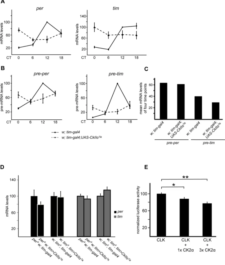
The strong influence of CK2a on CLK phosphorylation and stability suggests that CLK-dependent transcription could be affected in flies with altered CK2aactivity. As previously reported [42], intermediate levels ofperandtimmRNAs were observed in
tim.Tikflies (Figure 6A).
perand timpre-mRNA levels were measured to more directly estimate CLK transcriptional activity. Average nonoscillating levels of pre–per and pre–tim were observed in tim.Tik flies (Figure 6B,C), despite the very reduced amounts of CLK protein (see Figure 1A). It thus suggested that CLK transcriptional activity was strongly increased when CK2aactivity was diminished. Flies expressing the CK2aTik
protein in aper0,tim0, orper0tim0double mutant background revealed no statistically significant changes in
perand timmRNA levels compared to wild-type CK2acontrols (Figures 6D and S5A). However, CLK protein levels were reduced to 25–50% in CK2aTik expressing flies (see Figures 2 and S2), suggesting that specific CLK activity was still increased. Effects of CK2aon the transcriptional activity of CLK thus appears to be at least partly independent of PER and TIM.per and tim mRNA levels were also measured in tim.CkIIb-RNAi flies and showed intermediate levels compared to controls (Figure S5B). Since CLK quantity is unaffected bytim.CkIIb-RNAi(Figure 3A), CK2ß does not strongly modify CLK transcriptional activity.
Finally, dependent transcription was tested by CLK-induced reporter gene expression in S2 cells. On a CLK-binding synthetic minimal enhancer composed of three per-derived E-boxes, CLK-dependent transcription was decreased in a dose-dependent manner by CK2a co-expression (Figure 6E). Since CLK quantity was increased in the presence of CK2a overex-pression (see Figure 2C), the transcriptional decrease could hardly be a consequence of lower CLK levels.
Discussion
Temporally controlled phosphorylation of clock proteins is a key feature of the transcriptional-translational negative feedback loop underlying the Drosophila circadian clock. Although the CLK activator shows robust oscillations of its phosphorylation levels, the phosphorylation mechanisms and how they affect CLK function remain largely unknown. Our study aimed at determining whether CK2 was involved in the control of CLK phosphorylation and how it would affect CLK circadian function. Overexpression of the CK2aTik
dominant-negative enzyme or RNA interference against
Figure 2. PER and TIM-independent degradation of CLK in CK2aTikoverexpressing flies.(A, B) Western blot of sonicated head extracts from flies collected at DD1. A CB stained band in the size range of CLK is used as a loading control. At least two independent experiments were performed for each blot. (A) Comparison of CLK protein intim.Tikand control flies intim0background. (Left) Comparison betweentim.Tikand
controls intim0background for PER and CLK at CT2.w; tim0tim-gal4(tim0+) andw; tim0tim-gal4; UAS-CkIIaTik(tim0CkIIaTik) were used.aandbare different protein extracts from the same genotype at the same time point. We loaded 100mg of extracts. (Middle) Quantitative RT-PCR measurements ofClkmRNA levels in heads of flies collected at DD1. Results are means of pooled values from two time points (CT2 and 14) with at least two independent samples for each time point. Error bars indicate s.e.m. Average values were normalized to the mean of the control (w; tim0tim-gal4) set to 100. Previous analysis of separate values at CT2 and CT14 indicated that they were similar (Table S1) justifying their common treatment (see above). (Right) CLK protein/ClkmRNA ratio calculated from mean CT2–CT14 values of Western blot quantification and quantitative RT-PCR data. Ratios were normalized to the control (w; tim0; tim-gal4) set to 100. Abbreviations as in (A). (Right) CLK protein/ClkmRNA ratio calculated from mean
CK2aassociated with hyperphosphorylated forms of CLK in the morning and it was able to directly phosphorylate the CLK proteinin vitro. Effects of CK2aon CLK stability and activity did not require PER or TIM, but CLK phosphorylation by CK2a involves both PER-independent and PER-dependent functions. The results suggest that direct phosphorylation by CK2astabilizes CLK and diminishes its transcriptional activity.
CLK protein levels but notClkmRNA levels are low intim.Tik
andtim.CkIIa-RNAiflies, indicating that CK2 specifically affects CLK protein levels. Although a role of CK2a in CLK protein synthesis cannot be completely excluded, three sets of experimen-tal data support a posttranslational action of CK2aon CLK. First, CK2aassociates with CLK, PER, and TIM in protein complexes. Second, CK2a affects CLK phosphorylation state in vivo and is able to phosphorylate CLK directlyin vitro. Third, CK2astabilizes CLK even after protein synthesis blockage with CHX. Impor-tantly, tim.Tik flies show reduced CLK phosphorylation, as predicted from kinase inhibition. This is in contrast with the effects of CkIIaTik on PER and TIM, for which highly phosphorylated forms of the proteins accumulate, although PER phosphorylation remains lower than the highest state in the wild-type [8,15,41,42]. Nevertheless, CK2 is able to phosphorylate PER and TIMin vitro
[8,41,49]. The CkIIaTik
mutation affects TIM phosphorylation in the absence of PER, whereas TIM is required to observe effects of
CkIIaTikon PER [15]. TIM was thus proposed to be a direct target of CK2 that drives CK2-dependent modification of PER [15]. One might expect that TIM or PER relays the effects of CK2 on CLK. This is not supported by the strong effect ofCkIIaTik
on the CLK protein in per01, tim01, or per01 tim01 double mutants. However, PER strongly influences CLK phosphorylation by CK2. First, overexpression of wild-type CK2a induces CLK hyperphosphorylation in per+ but not in per0 flies. Second, PER enhancesin vitrophosphorylation of CLK by CK2a. Finally, the abundance of CLK/CK2 complexes observed in head extracts is much lower inper0mutants. In comparison toper0
, wild-type flies accumulate much more CLK/CK2a complexes, particularly in the morning when PER and CLK are abundant and hyperpho-sphorylated. The PER–CLK interaction is the strongest in the morning and the weakest in the early evening when PER is highly degraded. It seems unlikely that this temporal pattern of CLK interactions with PER is strongly altered in FLAG-CK2a overexpressing animals used for the immunoprecipitation since they show behavioral and molecular rhythms similar to wild-type flies. CLK/CK2a complexes strongly decrease after CT4 when high levels of CLK but not PER remain, suggesting that CLK/ CK2a interactions follow phosphorylated PER abundance. PER hence could drive a large fraction of CLK/CK2ainteractions with PER-free CLK being a weaker CK2 substrate. Since PER interacts in the late night with CLK species that no longer bind chromatin [22], it suggests that CLK/PER/CK2acomplexes are mostly unbound to DNA.
PER/DBT-dependent phosphorylation marks CLK for degra-dation [19,20]. Although the NEMO kinase destabilizes CLK [29], whether it acts as a PER/DBT-dependent CLK kinase is not known. Our results indicate that inhibiting CK2a activity increases CLK breakdown, whereas overexpressing CK2ainduces accumulation of highly phosphorylated CLK. CK2athus appears to have opposite effects on CLK stability, compared to DBT and NEMO. Since both CK1 (DBT) and CK2 show a preferential association with CLK in the morning, they might counteract each other to control CLK degradation and recycling for a next transcription cycle. Interestingly, a kinase complex that includes CK1 promotes the SUPERNUMERARY LIMBS (SLMB)-dependent proteolysis of the CUBITUS INTERRUPTUS (CI) transcription factor, whereas CK2 stabilizes CI by preventing its ubiquitylation [50,51].
As opposed totim.Tikflies, flies expressingCkIIbRNAi did not show significantly decreased CLK levels. In addition, CK2aand the CK2 holoenzyme are both able to phosphorylate Drosophila
CLKin vitro. Our data suggest that CLK, in contrast to PER and TIM, might be a substrate of CK2aalone rather than a substrate of the CK2 holoenzymein vivo. Several studies suggest that CK2a andbdo not act synergistically on a handful of substrates [52] or even play antagonist roles with CK2binhibiting CK2a-dependent phosphorylation of some target proteins (see [40]). In mammals, CK2ais more efficient than the CK2 holoenzyme to phosphor-ylate BMAL1, and can also phosphorphosphor-ylate CLK [53].
As previously reported [15,42],tim.Tik flies showed interme-diate levels of per and tim transcripts. We corroborated the involvement of transcription in this phenomenon by determining
perandtimpre-mRNA profiles. It has been proposed that the high levels of hyperphosphorylated TIM in tim.Tik would prevent normal PER-dependent transcriptional repression [15]. However, the fact that flies expressing the CK2aTik
protein in aper0,tim0, or
per0tim0double mutant background have only half dose (or less) of CLK but as high levels ofper and tim transcripts as theCkIIa+ controls supports an additional PER/TIM-independent transcrip-tional function of CK2. Importantly, the small amount of remaining CLK protein in the late night in per+
tim.Tik flies drives similarly high pre–perexpression as much more CLK in the wild-type (see Figure 6B). That also undermines CK2’s involve-ment only in PER/TIM repressor function during CLK-mediated transcription. A likely explanation is that the low-level hypopho-sphorylated CLK is extremely active in flies with reduced CK2a activity. In line with thein vivoresults, the luciferase activity assay in cultured S2 cells uncovered a dose-dependent repression of CLK activity by the CK2a subunit on a minimal enhancer-promoter element.
CK2athus appears to control CLK-dependent transcription by increasing PER/TIM repressing capacity and jointly decreasing CLK activity by some other mechanism. CK2b supports TIM-dependent repression (Figure S5B), but may not contribute to the PER/TIM-independent control of CLK activity by CK2a. Since
Comparison of CLK protein intim.Tikand control flies inper0tim0background. (Left) Genotypes:per0w;tim0(per0tim0) andper0w;tim0tim-gal4;
UAS-CkIIaTik(per0tim0CkIIaTik) as well asw;;ClkJrk.a,b, andcare different protein extracts from the same genotype at the same time point. (Middle)
Quantitative RT-PCR measurements ofClkmRNA levels in head extracts of flies collected at CT2. Mean values +/2 s.e.m. from at least three independent experiments are shown with theper0tim0control set to 100. (Right) CLK protein/ClkmRNA ratio calculated with mean values of Western
blot quantification and quantitative RT-PCR data at CT2. Ratios were normalized to the control (per0tim0) set to 100. (C) Cycloheximide chase of CLK degradation in the presence of CK2aoverexpression.perandtimdsRNA was applied to S2 cells prior to transfection. We transfected 1mg pAc-Clk-V5/ His6 with or without 3mg of the FMO02931 CK2aexpression vector. Transfections were split in four equal volumes for the degradation assay. ‘‘h in CHX’’ indicates the hours for which respective cells were incubated in cycloheximide to stop protein synthesis. Immunoreactivity against MHC (myosin heavy chain) was used as loading control. Anti-V5, anti-CK2a, and anti-HA were used to reveal CLK and CK2a, respectively. (Left) Western blots for CLK alone (CLK) and CLK cotransfected with CK2a(CLK+CK2a). (Right) CK2aexpression from the FMO02931 plasmid after 1 d of induction. (Bottom) Average degradation profiles from three independent experiments (as shown on the left). Error bars represent s.e.m.
deubiquitylation of CLK by USP8 decreases its activity [25], it will be interesting to investigate whether CK2aphosphorylation affects CLK ubiquitylation.
In the Neurospora circadian transcriptional feedback loop, the FRQ repressor recruits CK1 and CK2 to promote phosphoryla-tion of the WCC activator complex resulting in the inhibiphosphoryla-tion of its transcriptional activity [35,54–57]. Reactivation of WCC occurs through its dephosphorylation by phosphatases such as PP2A [54,56]. WCC is destabilized when turning active and gets stabilized as soon as it resumes a transcriptionally inactive state [56,57]. This is reminiscent of our finding about the role of CK2 in CLK activity regulation. Recently, BMAL1 and CLK were also shown to be ‘‘Kamikaze’’ activators in mammals in that their activity was dependent on proteasome function—highly unstable CLK and BMAL1 were the most active, while proteasome
inhibition resulted in long-lived but less potent activators [31,32,58]. Our findings indicate that CK2 might be a key player in such a mechanism, by promoting CLK stability and decreasing its activity. It remains to be seen how CK2 and DBT-dependent kinase activities interact on CLK to set CLK transcriptional activity to a proper phase in the circadian cycle.
Materials and Methods
Fly Stocks and Constructs
Drosophila melanogasterstocks were maintained on a 12 h:12 h LD cycle on standard corn meal-yeast-agar medium at 25uC.ClkJrkis a dominant allele of Clk, which results in a truncated and highly unstable CLK protein [59]. per01 [60], tim01 [61], w;tim-gal4-62
[62], w;;gal1118 [46], per01w;;13.2(per(D)-HA10His) F21 [63],
Figure 3. CK2bdoes not contribute to the inhibition of CLK degradation.(A, B) Western blot of nonsonicated head extracts from flies collected at DD1. Gray and black bars represent subjective day and subjective night, respectively. A CB stained band in the size range of CLK is used as a loading control. Brackets indicate hypo- and hyperphosphorylated forms of CLK. At least two independent experiments were performed for each blot. (A) Comparison betweentim.CkIIbRNAi(w; tim-gal4/106845; 32377/+) andtim-gal4/+controls in aper+background, for TIM, PER, and CLK proteins. (B) Comparison betweentim.CkIIbRNAiandtim-gal4/+controls in aper0background, for TIM and CLK.aandbare different protein
extracts from the same genotype at the same time point. (C) Quantitative RT-PCR measurements ofClkmRNA levels in heads of flies collected at DD1. Results shown are means of pooled values from two time points (CT2 and 14, which gave similar values) with two independent samples for each time point. Error bars indicate s.e.m. Average values were normalized to the mean control (per0w;tim-gal4/
+) values set to 100. (D) CLK protein/ClkmRNA ratio calculated from mean CT2–CT14 values of Western blot (B) and quantitative RT-PCR (C) data. Ratios were normalized to the control (per0w; tim-gal4/+) ratios set to 100.
yw;;P{UAS-CkIIa.Tik} T1 [15], yw;P{UAS-CkIIa.L} 35 [41],
w;UAS-FLAG-CkIIa [51], and lines carrying UAS transgenes encoding each of the five CK2b isoforms [64] have been previously described. The gal1118 driver line in the adult brain is expressed in the small and large LNv-s in addition to some few nonclock cells [46]. UAS-RNAi flies against CkIIb (stocks 32377 and 106845) andCkIIa(stock 17520 R-2) are described in http:// stockcenter.vdrc.at/control/main and http://www.shigen.nig.ac. jp/fly/nigfly/index.jsp, respectively. Both CkIIb RNAi lines (32377 and 106845) were induced in all the experiments using
CkIIb RNAi except specifically indicated. TheUAS-FLAG-CkIIb
construct was made by inserting a FLAG-CK2ß coding segment (kindly provided by A. Bidwai, West Virginia University) into the pUAST vector, and w;UAS-FLAG-CkIIb transgenic flies were generated by standard procedures. For in vitro phosphorylation assays,Clkconstructs with a 6-histidine fusion tag as well asperand
timwere expressed from a SP6 promoter incorporated in a pAc-5.1 vector, as described previously [30]. The FMO02931 expression plasmid was obtained from the Drosophila Genomics Resource Center (DGRC). It contains the full CkIIa ORF tagged C-terminally with FLAG and HA and driven by the metallothionein promoter. We verified theCkIIaORF and the promoter region by sequencing.
Behavioral Analysis
Behavioral assays for locomotor activity rhythms were carried out with 1- to 5-d-old males at 25uC inDrosophilaactivity monitors (TriKinetics). Illumination was provided by standard white fluorescent low-energy bulbs. Light intensity at fly level was in the range of 300–1000mW/cm2. Flies were first entrained to 12 h:12 h light-dark (LD) cycles for 4 d and then transferred to constant darkness (DD). Activity data were analyzed from the second to the ninth day in DD. Data analysis was done with the FaasX 1.16 software that is derived from the Brandeis Rhythm Package (see [65]) and is freely available upon request (Apple Mac OSX only). Rhythmic flies were defined by x2 periodogram analysis of an 8-d dataset with the following criteria (filter ON): power $20, width $1.5 h, with no selection on period value. Power and width represent the height and width of the period-ogram peak, respectively, and give the significance of the calculated period. Genotypes with a reduced number of rhythmic flies (,50%), low power (,50), and high s.e.m. of the period (.1) are considered arrhythmic. Experiments were reproduced two or three times with very similar results.
Protein Sample Preparation, Phosphatase Treatment, Sonication, and Western Blotting
We entrained 1 to 5-d-old flies to 12 h:12 h LD cycles for 4 d and transferred to DD (CT0 is 12 h after the last lights-OFF). Flies were collected on dry ice during the first day of DD (CT0–24). We homogenized 30–60 heads on ice in a modified RBS buffer [20]: 10 mM HEPES pH 7.5, 5 mM Tris-HCl pH 7.5, 50 mM KCl, 10% glycerol, 2 mM EDTA, 1% Triton X-100, 0.4% NP-40, 1 mM DTT, Complete Mini Protease Inhibitor Cocktail Tablet (Roche), Phosphatase Inhibitor Cocktail 2 and 3 (Sigma-Aldrich), and 20 mM b-glycerophosphate (3–4ml buffer/head). A Brink-mann Heidolph Mechanical Overhead Stirrer RZR1 was used for the homogenization. After 1 min of extraction, tubes were incubated in ice for 30 min, then homogenized again for another minute. If sonication was included after this step, samples were sonicated on ice with a Vibracell ultrasonic processor (Bioblock Scientific) at 4W output for 5610 s with 1 s breaks. Following
Bradford protein concentration measurement (BioRad), superna-tants were used for polyacrylamide gel electrophoresis. When
supernatants were treated withlprotein phosphatase, 1,600 units of l protein phosphatase (New England Biolabs) and 1 mM MnCl2were added to sonicated extracts prepared in phosphatase inhibitor-free buffer [10 mM HEPES pH 7.5, 100 mM KCl, 0.1 mM EDTA, 5% glycerol, 0.1% Triton X-100, 5 mM DTT, and EDTA-free Complete Mini Protease Inhibitor Cocktail Tablet (Roche)] and subsequently incubated for 30 min at 30uC. Reaction was stopped by adding 16 NuPAGE LDS sample
buffer (Life Technologies), 500 mM DTT, and incubation for 10 min at 70uC. We loaded 50mg total protein on Novex 4% Tris-Glycine precast gels (Life Technologies) for PER, TIM, and CLK immunoblotting, except specifically indicated. When indi-cated, NuPAGE Novex 3–8% Tris-Acetate gels were used for TIM and CLK immunoblotting for a better resolution of hyperphosphorylated forms. Samples (50mg) for FLAG, CK2a, and CK2b immunoblots were run on NuPAGE Novex 4–12% Bis-Tris precast gels (Life Technologies). Electrophoresis and blotting were done according to the manufacturer’s instructions except for a 3 h running time for Tris-Acetate and a 2 h running time for Tris-Glycine gels. Equal loading was verified by Ponceau S staining on blotting membranes, which were blocked in 5% nonfat dry milk in TBST (Tris-Buffered Saline with 0.1% Tween-20) for 1 h at 25uC and then incubated with the primary antibody overnight at 4uC. The following primary antibodies were used diluted in 5% milk in TBST: rabbit anti-V5 (Sigma-Aldrich V8137, Lot 019K4827) at 1:4,000, rabbit anti-myosin heavy chain (MHC, kind gift of Roger E. Karess, Institut Jacques Monod, Paris) at 1:400,000, rabbit anti-CK2a (Abcam ab81435) at 1:1,000, mouse anti-CK2b (Calbiochem 6D5 218712) at 1:1,000, rat anti-TIM [7] at 1:2,000, goat anti-CLK (Santa Cruz Biotechnology sc27070) at 1:1000, rabbit anti-PER [66] at 1:10,000, guinea pig anti-VRILLE [44] at 1:5,000, and guinea pig anti-PDP1e[67] at 1:5,000. For immunoblotting of anti-FLAG immunoprecipitations, guinea pig GP90 anti-CLK [18] at 1:1,000 was used since an aspecific IgG-derived band revealed with the SC27070 anti-CLK on the immunoprecipitates. Membranes were washed three times for 10 min, then the HRP-conjugated secondary antibodies (Santa Cruz Biotechnology) were added diluted in 5% milk in TBST: goat rabbit (1:10,000), goat anti-rat (1:20,000), goat anti-mouse (1:20,000), donkey anti-goat (1:10,000), and goat anti-guinea pig (1:10,000). In the case of anti-CK2b, TrueBlot ULTRA Anti-Mouse IgG-HRP (eBioscience, 1:2,000) was used as a secondary antibody to circumvent problems resulting from primary antibody light chain detection after immunoprecipitation.
Blots were revealed with the Amersham ECL Plus reagent (GE Healthcare). SimplyBlue SafeStain (Life Technologies) was used to stain membranes after blotting. Images were quantified with the NIH ImageJ (1.43 k) software after background subtraction. Calculations were done and histograms were generated with Microsoft Excel.
Immunoprecipitation
Technologies) without DTT for 10 min at 70uC. Supernatants were complemented with DTT (500 mM) and reduced for 10 min at 70uC.
Cell Culture Experiments
Drosophila Schneider 2 (S2) cells [kind gift of Anne Plessis (Institut Jaques Monod, Paris)] were maintained in SFX-Insect Medium (HyClone) supplemented with 10% fetal bovine serum Aldrich) and 1% penicillin-streptomycin solution (Sigma-Aldrich) as previously described [68]. Complementary single-stranded RNA-s were in vitro transcribed from purified PCR templates containing the T7 RNA polymerase promoter site on both ends, using the MEGAscript T7 Kit (Life Technologies). Reactions were purified with the MEGAclear Kit (Life Technol-ogies) and precipitated in ethanol/sodium acetate for concentra-tion followed by resuspension in 40ml H2O and annealing of the two strands (30 min at 65uC and slow cooling to room temperature). RNA quality and quantity was assessed by spectrophotometry and agarose gel electrophoresis. Primers for
per target sequence amplification by PCR were: TTAATAC- GACTCACTATAGGGAGAAAGGAGGACAGCTTCTGCT-GC and TTAATACGACTCACTATAGGGAGAGATATGAT-CCCGGTGGCCGTG and for tim were: TTAATA; CGACT-CACTATAGGGAGACTGGTTACTAGCAACTCCGCA and TTAATACGACTCA; and CTATAGGGAGAGCAGGATAT-TTCTCAGCAGCA.
pAc-Clk-V5/His6 [10], pAc-Renilla luciferase (kind gift of M. Rosbash), and p3x69-luc (containing three copies ofperE-box as enhancer element [10,69]) were already described. Transient transfection was performed with Effectene (Qiagen) using plasmids purified with the Plasmid Midi Kit (Qiagen). DNA quantities were equalized for transfection by addition of empty pAc vector. The induction ofCkIIaunder the control of metallothionein promoter was achieved by adding 500mM CuSO4 to the cells 1 d after transfection.
For luciferase activity assays, 106cells were seeded in six-well plates, left to proliferate in serum-free medium for 48 h, were transfected in serum-free medium, supplemented with serum and antibiotics 4 h later, and harvested 48 h posttransfection. Cells were washed in PBS and lysed on plate with Passive Lysis Buffer according to the Dual-Luciferase Reporter Assay System manual (Promega). Lysates were cleared by centrifugation at 4uC and 10ml of supernatant was measured for firefly and Renilla luciferase activities with the Dual-Luciferase Reporter Assay System (Promega) on a Mithras LB 940 luminometer (Berthold Technologies). Firefly luciferase activities were nor-malized to corresponding Renilla luciferase activities to control
for transfection efficiency and protein concentration. Experi-ments were made in duplicates or quadruplicates and repeated at least twice.
For degradation assays, cells were seeded in 60 mm dishes (2.56106cells/dish) and treated withperandtimdsRNA (37.5mg) in serum-free medium for 48 h followed by transfections in medium containing serum and antibiotics. One day posttransfec-tion, cells were split in four equal volumes and seeded in 12-well plates followed by induction with CuSO4. One day after induction, cycloheximide (CHX, Sigma-Aldrich) was added to each well at a final concentration of 0.58 mM, and cells were harvested 0, 3, 6, and 9 h after the beginning of CHX treatment. After harvest, cells were centrifuged for 5 min at 2,000gat 20uC, washed once with PBS, and pellets were frozen at280uC until extraction. Protein extraction was achieved by lysing cells in 40ml of HE buffer (described above) supplemented with 0.5% Triton X-100 by means of pipetting and vortexing. After centrifugation for 10 min at 14,000 rpm at 4uC, supernatants were subjected to Bradford assay. We used 20mg protein for polyacrylamide gel electrophoresis. Blots were revealed with anti-V5 for CLK and with anti-MHC as a loading control. Both blots were quantified by ImageJ. V5 reactivity was normalized to MHC reactivity for each sample, which was used for the calculations that are plotted in Figure 2E.
In VitroPhosphorylation Assays
In vitrotranscription/translation and phosphorylation reactions were carried out as described previously [30], with the following differences: CLK protein with a N-terminal 6-histidin fusion tag as well as PER and TIM were expressed in TNT SP6-Quick Coupled High Yield Wheat Germ expression system (Promega) for 2 h at 25uC with the addition of 0.2 mM staurosporine to block phosphorylation. Subsequently CLK protein was precipitated with 20ml nickel-nitrilotriacetate (Ni-NTA) agarose for 90 min at 4uC either with or without prior addition of PER or TIM expressing lysates. Affinity purified CLK protein with or without co-precipitated PER or TIM was subjected to on bead phosphory-lation reactions by human casein kinase II holoenzyme (New England Biolabs) or recombinant human CK2asubunit (Kinase-Detect, DK-5792 Aarslev, Denmark) in 50ml phosphorylation buffer (20 mM Tris-HCl, pH 7.5, 50 mM KCl, 10 mM MgCl2) with 0.5mCi/ml c–32
P-ATP at 30uC for the holoenzyme and at 37uC for CK2a. The amount of CLK-incorporated32P-phosphate was quantified by autoradiography and densitometry after SDS-page electrophoresis and blotting to nitrocellulose membrane. The intensity of the32P-signal was normalized by total CLK protein level, as quantified by Western blot analysis.
Figure 4. PER, TIM, and CLK are found in protein complexes containing CK2.Anti-FLAG immunoprecipitation (IP) from nonsonicated head extracts of flies collected at DD1. FLAG-CK2aor FLAG-CK2bwas immunoprecipitated from 1 mg of total protein extracts from heads and 50% of the precipitate was subjected to Western blot analysis. We loaded 50mg of head extracts as input controls. IgG LC indicates the immunoglobulin G light chain used for the precipitation that is well detected by the anti-mouse secondary antibody. Gray and black bars represent subjective day and subjective night, respectively. Experiments were performed at least twice. (A, Left) FLAG-CK2awas immunoprecipitated fromtim.FLAG-CkIIaflies andtim-gal4/+negative controls (control) in a per+
background. IP of CK2a(FLAG) and co-IP of CK2b, TIM, PER, and CLK were visualized by immunoblotting. «*» shows an aspecific band recognized by the anti-PER antibody. (Right) TIM, PER, and CLK immunoblots of the corresponding inputs onper+
background. The time ‘‘CT10’’ indicates a mixed population of flies harvested at CT8 and CT12. A CB stained band in the size range of CLK is used as a loading control. TIM was run on a 3–8% Tris-Acetate gel. (B, Left) FLAG-CK2bwas immunoprecipitated fromtim.FLAG-CkIIßflies andtim-gal4/+negative controls (control) in aper+
background. IP of CK2ß (with anti-CK2ß) and co-IP of CK2a, TIM, PER, and CLK were visualized by immunoblotting. (Right) TIM, PER, and CLK immunoblots of the corresponding inputs. A CB stained band in the size range of CLK is used as a loading control. (C) FLAG-CK2aor FLAG-CK2b was immunoprecipitated fromtim .FLAG-CkIIa (a) flies and tim-gal4/+negative controls (C) in aper0 background. co-IP of CLK was visualized by immunoblotting. Input samples and immunoprecipitates were run on the same gel forCanda.(D) Image showing PDF (green), CK2a(magenta), and CLK (blue) fluorescent immunolabeling in small PDF+LNv-s of an adult fly at ZT3. The fourth square is a composite picture of the three stainings. Single optical planes are shown taken by confocal microscopy.
Quantitative RT-PCR
Total RNA was prepared from adult heads (about 35) using the Promega SV Total RNA Isolation System. It was quantified using the Nanodrop ND-1000 spectrophotometer, and the integrity of the RNA was verified using the Agilent 2100 bioanalyser with the eukaryote total RNA Nano assay. RNA was treated with rDNase (NucleoSpin RNA Kit, Macherey-Nagel) in solution after RNA isolation to ensure optimal conditions for pre-mRNA detection. One mg of total RNA was reverse-transcribed in a 50ml final reaction in presence of 0.4mM oligodT(15) or random hexamer primers (for detection of pre-mRNA-s), 8 mM dNTP, 40 units of RNasine, and 400 units of M-MLV RTase H-minus (Promega), during 3 h at 37uC. Quantitative PCR was performed with a Roche LightCycler (mRNA-s) or an Applied Biosystems 7900HT
Fast Real-Time PCR System (pre-mRNA-s) using the SYBR green detection protocol of the manufacturer. We mixed 3ml of a 256
diluted cDNA (or 1 ng/ml) with FastStart DNA MasterPLUS SYBR green I mix with 500 nM of each primer, and the reaction mix was loaded on the capillaries and submitted to 40 cycles of PCR (95uC/15 s; 60uC/10 s; 72uC/20 s for the Lightcycler and 50uC 2 min; 95uC/20 s; [95uC/1 s–60uC/25 s]640 for the ABI
instrument), followed by a fusion cycle in order to analyze the melting curve of the PCR products. Negative control without the reverse transcriptase was introduced to verify the absence of genomic DNA contaminants. Primers (see Table S2) were defined within exons (for mRNA-s) or in one intron and one exon (for pre-mRNA-s) using the PrimerSelect program of the Lasergene software (DNAStar). BLAST searches were performed to confirm
Figure 5. CK2aoverexpression induces CLK hyperphosphorylation in the presence of PER.(A, B) Western blot of nonsonicated head extracts from flies collected at DD1. Samples were run on 3–8% Tris-Acetate gels in order to better resolve hyperphosphorylated CLK and TIM forms. Gray and black bars represent subjective day and subjective night, respectively. At least two independent experiments were performed for each blot. (A, Top) Comparison betweentim.CkIIaandtim-gal4/+controls in aper+background, for TIM, PER, and CLK proteins. (Bottom) Two independent experiments as above were quantified for CLK abundance, and the mean values are plotted. Error bars stand for the difference of the respective values from each experiment and their mean. The value ofw; tim-gal4/+at CT0 was normalized to 100. (B) Comparison betweenper0;tim.CkIIaand per0;tim-gal4/+controls for CLK. (C) CK2aphosphorylates CLKin vitro. (Top) Wild-type CLK was translated with a N-terminal 6-histidine fusion tagin
vitro, affinity purified either in the absence or presence of PER or TIM, and subjected to phosphorylation assays by incubation withc–32P-ATP either in the absence (2) or presence (+) of CK2a. Intensity of incorporated32P-phosphate into CLK (32P) was analyzed by autoradiography and total CLK protein levels (CLK) were determined by Western blot analysis. The arrow indicates the position of phosphorylated CLK. (Bottom) Quantification of CLK-incorporated32P-phosphate after normalization towards total CLK protein levels. Average CLK phosphorylation from at least three experiments
Figure 6. CK2adecreases CLK transcription factor activity.(A–D) Quantitative RT-PCR measurements ofper, pre–per,tim, and pre–timmRNA levels in heads of flies collected at DD1. Error bars indicate s.e.m. (A)perandtimmRNA levels intim.Tikand control flies. Values were normalized to the maximum value (control at CT12) set to 100. Mean mRNA levels+/2s.e.m. from at least three independent experiments are shown. (B) Quantitative RT-PCR measurements ofper and timpre-mRNA levels in heads oftim.Tikand control flies collected at DD1. Average values from three independent experiments were normalized to the control (w; tim-gal4) mean value at CT12 set to 100. Error bars represent s.e.m. (C) Relative qPCR values from Figure 6B were averaged from the four indicated time points (CT0, CT6, CT12, and CT18) for each genotype and for each pre-messenger and the mean value was plotted. 100 stands for the highest pre-mRNA expression in the respective genotype at CT12. (D)perandtimmRNA levels in tim.Tikand controls in aper0ortim0background. Results are means of pooled values from two time points (CT2 and 14). Values were normalized to
gene specificity and the absence of multilocus matching at the primer site. The amplification efficiencies of primers were generated using the slopes of the standard curves obtained by a 10-fold dilution series of 4, with all experimental points falling within this range. The efficiency of the q-PCR amplifications for all pairs of primers is indicated in the table. Amplification specificity for each q-PCR reaction was confirmed by dissociation curve analysis. Determined Ct values (see Table S2) were then used for quantification, with the tubulin gene as reference. Each sample measurement was made at least in duplicate (technical replicate).
Immunolabeling of Adult Brains
Experiments were done on whole-mounted adult brains as previously described [46]. Primary antibodies were rabbit anti-PER [66] at 1:15,000, guinea pig GP47 anti-CLK [26] at 1:15,000, mouse anti-PDF (Developmental Studies Hybridoma Bank) at 1:50,000, and rabbit anti-CK2a. (Abcam, ab81435) at 1:100. Secondary goat antibodies (Life Technologies) were Alexa 647- or Alexa 594-conjugated anti-rabbit at 1:5,000, Alexa 488- or Alexa 647-conjugated anti-guinea pig at 1:2,000, and Alexa 594-or Alexa 488-conjugated anti-mouse at 1:2,000. Flu594-orescence signals were analyzed with a Zeiss AxioImager Z1 microscope with an ApoTome structured illumination module and an AxioCam MRm digital camera. Images for subcellular localization of CK2a were acquired with a Zeiss LSM-700 confocal microscope. Fluorescence intensity of individual cells was quantified from digital images of single focal planes with the NIH ImageJ software. We calculated a fluorescence index: I = 1 00(S-B)/B, which gives the fluorescence percentage above background (S (Signal) is fluorescence intensity and B (Background) is average intensity of the region adjacent to the positive cell). Index values were then averaged for the four PDF-positive s-LNv cells of 12–20 brain hemispheres for each time point.
Supporting Information
Figure S1 CLK degradation is accelerated intim.Tik
flies.(A–E) Western blot of head extracts from flies collected at DD1. Time (h) is indicated as CT. Gray and black bars represent subjective day and subjective night, respectively. A CB stained band in the size range of CLK is used as a loading control for blots run on 4% gels. Brackets indicate hypo- and hyperphosphorylated forms of CLK. At least two independent experiments were performed for each blot. (A) Western blot of CLK protein in sonicated extracts of the indicated genotypes. Two representative examples are shown in addition to Figure 1A. (B) Western blot of CLK, PER, and TIM proteins as in (A) but from nonsonicated extracts of the indicated genotypes. (C) Sonicated extracts from the indicated genotypes were treated with or without l protein phosphatase at the respective temperatures, and CLK protein was detected by Western blot. (D, Left) Western blot of CLK protein in nonsonicated head extracts from the indicated genotypes collected on the first day of constant darkness. CLK protein is shown on the immunoblot. A CB stained band in the size range of CLK is used as a loading control. (Right) CLK protein/ClkmRNA ratio of the
indicated genotypes. Values from quantification of CLK bands of the left panel were divided with the values of RT-qPCR from Figure 1B. w; tim-gal4 at CT2 was set to 100. (E) CkIIa RNAi decreases CK2a protein abundance. Samples were run on a 4– 12% Bis-Tris gel. Anti-CK2aprimary antibody was used for the blot. One copy of tim-gal4 and two copies of the CkIIa RNAi construct were used for the experimental genotype.
(PDF)
Figure S2 CK2 affects CLK in a per0 background and impacts onClk expression posttranscriptionally.(A, B, D) Western blot of head extracts from flies collected at DD1. Time (h) is indicated as CT. A CB stained band in the size range of CLK is used as a loading control for blots run on 4% gels. Brackets indicate hypo- and hyperphosphorylated forms of CLK. At least two independent experiments were performed for each blot. (A) Comparison of CLK phosphorylation states between per+tim+ sonicated extracts and their per0 tim0 counterparts. We loaded 100mg protein.a,b, andcare different protein extracts from the same genotype at the same time point.w; tim-gal4/+(tim-gal4/+),
per0w ;tim0
(per0tim0
), andper0w
; tim0tim-gal4; UAS-CkIIaTik (per0
tim0CkIIaTik) were used. (B, Left) Comparison betweentim.Tik
and controls in aper0background for TIM and CLK [per0w; tim-gal4(per0+),per0w; tim-gal4; UAS-CkIIaTik
(per0CkIIaTik
)].aandbare different nonsonicated protein extracts from the same genotype at the same time point. We loaded 100mg of extracts. Extracts were run on a 3–8% Tris-Acetate gel for TIM. (Middle) Quantitative RT-PCR measurements of Clk mRNA levels in heads of flies collected at DD1. Results are means of pooled values from two time points (CT2 and 14) with at least two independent samples for each time point. Error bars indicate s.e.m. Average values were normalized to the mean of the control (per0w;tim-gal4) set to 100. Previous analysis of separate values at CT2 and CT14 indicated that they were similar (Table S1), justifying their common treatment (see above). (Right) CLK protein/ClkmRNA ratio of the indicated genotypes. Values from quantification of CLK bands of the left panel were divided with the values of RT-qPCR from the middle panel.per0w; tim-gal4was set to 100. (C) Quantitative RT-PCR measurements ofClkpre-mRNA levels in head extracts of flies collected at DD1. Average values from three independent experiments were normalized to the mean of the control (w; tim-gal4) at CT0 set to 100. Error bars represent s.e.m. (D) Comparison betweentim.Tikand controls in aper0
background for the VRI and PDP1eproteins. Samples were resolved on a 3– 8% Tris-Acetate gel. per0w; tim-gal4 (per0 +) and per0w; tim-gal4; UAS-CkIIaTik(per0CkIIaTik) were used. (E) Quantitative RT-PCR measurements ofcry mRNA levels in heads of flies collected at DD1. Results are means of pooled values from two time points (CT2 and 14, which gave similar values) with at least two independent samples for each time point. Error bars indicate s.e.m. Average values were normalized to the control (per0w; tim-gal4) average values set to 100.
(PDF)
Figure S3 CkIIb RNAi decreases CK2b protein abun-dance and causes PER and TIM accumulation. (A) Western blot of CK2b. Extracts of tim. CkIIb RNAi (w; tim-common treatment (see above). Average results from at least three independent experiments are shown. (E) Luciferase activity assay in the presence or absence of CK2aoverexpression. S2 cells were transfected, harvested, and measured as described in Materials and Methods. We transfected 5 ng pAc-Clk-V5, 10 ng p3x69-luc, and 10 ng pAc-Renilla luciferase with or without 5 or 15 ng of the FMO02931 CK2aexpression vector. Mean luciferase activity+/2s.e.m. of at least four different samples from two independent experiments are shown. CLK-dependent luciferase expression in the absence of CK2aco-expression was set to 100. 16CK2aindicates 5 ng FMO02931, and 36CK2astands for 15 ng FMO02931. Student’sttest
gal4/106845; 32377/+) and tim-gal4/+ controls in a per+ back-ground were run on a 4–12% Bis-Tris gel. VIIa, d, and c indicate different isoforms of CK2b[64]. (B) Quantification of CLK, PER, and TIM signal intensity on Western blots intim.CkIIbRNAiand
tim-gal4/+ controls at six time points of DD1. Two independent experiments were quantified. Error bars stand for the difference of the respective values from each experiment and their mean. The intensities were normalized to the signal of a CB stained band. The highest intensity signal inw;tim-gal4/+was set to 100.
(PDF)
Figure S4 CK2aoverexpression induces a delay in TIM oscillation.(A) Western blot of nonsonicated head extracts from flies collected at DD1. A CB stained band in the size range of CLK is used as a loading control.tim.CkIIaand tim-gal4/+controls are compared for TIM and PER. (B) Quantification of PER and TIM signal intensity from the Western blot in (A). The highest intensity signal in w;tim-gal4/+ was set to 100. (C) CK2 phosphorylates CLKin vitro. (Top) Wild-type CLK was translated with an N-terminal 6-histidine fusion tagin vitro, affinity purified either in the absence or presence of PER and TIM, and subjected to phosphorylation assays by incubation withc–32
P-ATP either in the absence (2) or presence (+) of CK2. Intensity of incorporated 32
P-phosphate into CLK (32P) was analyzed by autoradiography, and total CLK protein levels (CLK) were determined by Western blot analysis. (Bottom) Quantification of CLK-incorporated 32 P-phosphate after normalization toward total CLK protein levels. Average CLK phosphorylation from at least three experiments6
s.e.m. are shown in the figure with wild-type CLK set to 100. (PDF)
Figure S5 perand tim transcription in tim . Tik and
tim . CkIIb RNAi animals. (A) Quantitative RT-PCR measurements of per and tim mRNA levels in head extracts of flies collected at CT2.tim.Tikand controls are compared in aper0 tim0
background. Mean mRNA levels +/2 s.e.m. from at least three independent experiments are shown. Average values were normalized to the control mean (per0tim0) set to 100. Genotypes:
per0w; tim0(per0tim0) andper0w;tim0tim-gal4; UAS-CkIIaTik (per0 tim0CkIIaTik). (B) Quantitative RT-PCR measurements ofperand
tim mRNA levels intim . CkIIß-RNAi and control flies. Values were normalized to the maximum value (control at CT12) set to
100. Mean mRNA levels +/2 s.e.m. from at least three independent experiments are shown.
(PDF)
Table S1 Mean values of quantitative RT-PCR results with associated s.e.m.Relative values of mRNA abundance measured by quantitative RT-PCR (see Material and Methods) are indicated for samples onper0ortim0background collected at CT2 and CT14. Mean levels are normalized to the highest value in the control genotype (per0w;tim-gal4orw;tim0tim-gal4;) set to 100. The number of independent samples for each time point is shown in Figure 2 and Figure 6.
(DOCX)
Table S2 qPCR primer specifications. *This pair of primers was used in Figure 6B and Figure S2C. **These two pairs of primers were used in Figure 6B. Efficiency (E), DNA concentration ratio between cycles n+1 and n (1,E,2). R2, coefficient of determination of the calibration curve. Ct, Cycle threshold.
(DOCX)
Acknowledgments
We thank F.R. Jackson for his generous gift of unpublished UAS-FLAG-CkIIßflies and I. Edery, P.E. Hardin, J. Jia, R. Karess, M. Rosbash, R. Stanewsky, and the Drosophila Genomics Resource Center (Bloomington, U.S.A.) for fly stocks, antibodies, and other materials. Fly stocks were also provided by the BloomingtonDrosophilastock center (United States), the ViennaDrosophila RNAi Center (Austria), and the National Institute of Genetics fly stock center (Mishima, Japan). We are grateful to A. Plessis, J. Menet, and especially A. Lamouroux for help with S2 cells, as well as Eric Jacquet for expert advice on quantitative RT-PCR. We also thank E. Che´lot and M. Krecsmarik for help with microscopy, C. Vias for dissections, M. Boudinot for the FaasX software, and the members of the Rouyer lab for critical reading of the manuscript.
Author Contributions
The author(s) have made the following declarations about their contributions: Conceived and designed the experiments: AS FW PP FR. Performed the experiments: AS CP PP DZ. Analyzed the data: AS CP PP DZ FW FR. Contributed reagents/materials/analysis tools: TR. Wrote the paper: AS FR.
References
1. Kadener S, Stoleru D, McDonald M, Nawathean P, Rosbash M (2007) Clockwork Orange is a transcriptional repressor and a new Drosophila circadian pacemaker component. Genes Dev 21: 1675–1686.
2. Weber F, Zorn D, Rademacher C, Hung HC (2011) Post-translational timing mechanisms of the Drosophila circadian clock. FEBS Lett 585: 1443–1449. 3. Hardin PE (2011) Molecular genetic analysis of circadian timekeeping in
Drosophila. Adv Genet 74: 141–173.
4. Kloss B, Price JL, Saez L, Blau J, Rothenfluh A, et al. (1998) The Drosophila clock gene double-time encodes a protein closely related to human casein kinase Iepsilon. Cell 94: 97–107.
5. Price JL, Blau J, Rothenfluh A, Abodeely M, Kloss B, et al. (1998) double-time is a novel Drosophila clock gene that regulates PERIOD protein accumulation. Cell 94: 83–95.
6. Ko HW, Jiang J, Edery I (2002) A role for Slimb in the degradation ofDrosophila PERIOD protein phosphorylated by DOUBLETIME. Nature 420: 673–678. 7. Grima B, Lamouroux A, Che´lot E, Papin C, Limbourg-Bouchon B, et al. (2002)
The F-box protein SLIMB controls the levels of clock proteins PERIOD and TIMELESS. Nature 429: 178–182.
8. Lin J-M, Kilman V, Keegan K, Paddock B, Emery-Le M, et al. (2002) A role for casein kinase 2a in the Drosophila circadian clock. Nature 420: 816–820. 9. Akten B, Jauch E, Genova GK, Kim EY, Edery I, et al. (2003) A role for CK2 in
the Drosophila circadian oscillator. Nat Neurosci 16: 251–257.
10. Nawathean P, Rosbash M (2004) The doubletime and CKII kinases collaborate to potentiate Drosophila PER transcriptional repressor activity. Mol Cell 13: 213–223. 11. Chiu JC, Ko HW, Edery I (2011) NEMO/NLK Phosphorylates PERIOD to initiate a time-delay phosphorylation circuit that sets circadian clock speed. Cell 145: 357–370.
12. Chiu JC, Vanselow JT, Kramer A, Edery I (2008) The phospho-occupancy of an atypical SLIMB-binding site on PERIOD that is phosphorylated by DOU-BLETIME controls the pace of the clock. Genes Dev 22: 1758–1772. 13. Martinek S, Inonog S, Manoukian AS, Young MW (2001) A role for the
segment polarity gene shaggy/GSK-3 in the Drosophila circadian clock. Cell 105: 769–779.
14. Grima B, Dognon A, Lamouroux A, Chelot E, Rouyer F (2012) CULLIN-3 controls TIMELESS oscillations in the Drosophila circadian clock. PLoS Biol 10: e1001367. doi:10.1371/journal.pbio.1001367
15. Meissner RA, Kilman VL, Lin JM, Allada R (2008) TIMELESS is an important mediator of CK2 effects on circadian clock function in vivo. J Neurosci 28: 9732–9740.
16. Sathyanarayanan S, Zheng X, Xiao R, Sehgal A (2004) Posttranslational regulation of Drosophila PERIOD protein by protein phosphatase 2A. Cell 116: 603–615.
17. Fang Y, Sathyanarayanan S, Sehgal A (2007) Post-translational regulation of the Drosophila circadian clock requires protein phosphatase 1 (PP1). Genes Dev 21: 1506–1518.
18. Lee C, Bae K, Edery I (1998) The Drosophila CLOCK protein undergoes daily rhythms in abundance, phosphorylation, and interactions with the PER-TIM complex. Neuron 21: 857–867.
19. Kim EY, Edery I (2006) Balance between DBT/CKI{varepsilon} kinase and protein phosphatase activities regulate phosphorylation and stability of Drosophila CLOCK protein. Proc Natl Acad Sci U S A 103: 6178–6183. 20. Yu W, Zheng H, Houl JH, Dauwalder B, Hardin PE (2006) PER-dependent