Downregulation of the
Drosophila
Immune
Response by Peptidoglycan-Recognition
Proteins SC1 and SC2
Vincent Bischoff1[, Ce´cile Vignal1[, Bernard Duvic2, Ivo G. Boneca3, Jules A. Hoffmann1, Julien Royet4*
1Institut de Biologie Mole´culaire et Cellulaire, UPR 9022 du CNRS, Strasbourg, France,2Unite´ EMIP UMR INRA-UMII 1133, Universite´ Montpellier II, Place Euge`ne Bataillon, Montpellier, France,3Unite´ de Pathoge´nie Bacte´rienne des Muqueuses, Institut Pasteur, Paris, France,4IBDM/LGPD, Campus de Luminy, Marseille, France
Peptidoglycan-recognition proteins (PGRPs) are evolutionarily conserved molecules that are structurally related to bacterial amidases. SeveralDrosophilaPGRPs have lost this enzymatic activity and serve as microbe sensors through peptidoglycan recognition. Other PGRP family members, such asDrosophilaPGRP-SC1 or mammalian PGRP-L, have conserved the amidase function and are able to cleave peptidoglycan in vitro. However, the contribution of these amidase PGRPs to host defense in vivo has remained elusive so far. Using an RNA-interference approach, we addressed the function of two PGRPs with amidase activity in theDrosophilaimmune response. We observed that PGRP-SC1/2– depleted flies present a specific over-activation of the IMD (immune deficiency) signaling pathway after bacterial challenge. Our data suggest that these proteins act in the larval gut to prevent activation of this pathway following bacterial ingestion. We further show that a strict control of IMD-pathway activation is essential to prevent bacteria-induced developmental defects and larval death.
Citation: Bischoff V, Vignal C, Duvic B, Boneca IG, Hoffmann JA, et al. (2006) Downregulation of theDrosophilaimmune response by peptidoglycan-recognition proteins SC1 and SC2. PLoS Pathog 2(2): e14.
Introduction
The antimicrobial host defense ofDrosophilainvolves rapid synthesis of small-sized cationic peptides by the fat body [1,2]. These antimicrobial peptides are released into the open circulatory system where they attack invading microorgan-isms. The transcription of the genes encoding these peptides is under the control of two distinct signaling pathways. The Toll pathway, which is primarily activated after gram-positive bacterial and fungal infections, controls the expression of drosomycin, an antifungal peptide, together with many other genes via the NF-jB–family member DIF (dorsal-related immune factor) [3,4]. The second cascade, known as the IMD (immune deficiency) signaling pathway, is predomi-nantly triggered after gram-negative infection and regulates, via the NF-jB protein Relish, the synthesis of some antibacterial peptides and many other genes [5,6]. An efficient Toll-pathway activation after gram-positive bacterial infection requires the function of at least three soluble proteins, namely peptidoglycan-recognition protein-SA (PGRP-SA) [7], PGRP-SD [8], and gram-negative binding protein-1 [9,10]. On the other hand, sensing of gram-negative bacterial infection has been shown to be dependent on two other PGRP family members, PGRP-LC [11–13] and PGRP-LE [14,15].
The ability of Drosophila to discriminate between gram-positive and gram-negative bacteria relies on the specific recognition of different forms of peptidoglycan (PGN) [16,17]. Bacterial PGN consists of long carbohydrate chains of alternating N-acetylglucosamine and N-acetylmuramic acid connected via stem peptides [18] (Figure S1). Most PGNs from gram-positive bacteria contain L-lysine in the third position of the stem peptide (Lys-PGN) and are recognized by PGRP-SA and PGRP-SD [7–9]. In PGN of gram-negative
bacteria and in that of gram-positive bacilli, the lysine residue is replaced by meso-diaminopimelic acid (m-DAP) (Figure S1). This second type of PGN (m-DAP-PGN) is sensed by PGRP-LC and PGRP-LE receptors, leading to the activation of the IMD pathway [14–17,19].
PGRPs form a large group of proteins present in insects and mammals [20–25], which have in common a 160-amino acid–domain with striking sequence similarity to N -acetyl-muramyl-L-alanine amidases (NAMLAA) [26]. These bacterial enzymes hydrolyze the bond formed between the lactyl group in N-acetylmuramic acid and the L-alanine in the stem peptide of PGN. In some of these PGRP molecules, the amidase function is conserved, as documented forDrosophila
PGRP-SC1 [27] and PGRP-LB [28], and for mouse and human PGRP-L [29,30]. In others, such as in PGRP-SA, SD, LE, or LC, the replacement of a critical cysteine residue within the PGRP domain abolishes this enzymatic function [27]. On the basis of genetic experiments, it is assumed that PGRPs without amidase activity serve as recognition receptors for
Editor:David Schneider, Stanford University, United States of America
ReceivedDecember 5, 2005;AcceptedJanuary 20, 2006;PublishedFebruary 24, 2006
DOI:10.1371/journal.ppat.0020014
Copyright:Ó2006 Bischoff et al. This is an open-access article distributed under the terms of the Creative Commons Attribution License, which permits unrestricted use, distribution, and reproduction in any medium, provided the original author and source are credited.
Abbreviations:DIF, dorsal-related immune factor; IMD, immune deficiency; JNK, c-Jun N-terminal kinase; LB, Luria-Bertani; m-DAP, meso-diaminopimelic acid; NAMLAA,N-acetylmuramyl-L-alanine amidases; PGN, peptidoglycan; PGRP, pepti-doglycan-recognition protein; TNF, tumor necrosis factor
* To whom correspondence should be addressed. E-mail: J.Royet@ibdm.univ-mrs.fr
microbial PGN. However, the in vivo function of PGRPs with amidase activity remains unclear, andPGRP-Lmutant mice show no immune phenotype. On the basis of in vitro experiments, it has been proposed that amidase PGRPs could act as scavenging molecules. Indeed, degradation of PGN by
Drosophila PGRP-SC1b markedly reduces its immuno-stim-ulatory potency in cell-culture assays [27]. We report here an in vivo study on the role ofDrosophilaPGRPs with amidase activity. We show that two PGRPs with described amidase activity, namely PGRP-SC1 and PGRP-SC2, control the intensity of theDrosophilaimmune response. We also present evidence that in the absence of such a control, infection-induced IMD-pathway over-activation can cause develop-mental defects and larval death.
Results
Loss-of-Function Mutants for PGRP-SC1/2 Are Generated by RNA Interference
In order to address the function of NAMLAA PGRPs under in vivo conditions, we analyzed the immune response of
Drosophilawith reduced PGRP-SC1 and PGRP-SC2 levels. The
Drosophila genome contains a cluster of two tandemly
arranged PGRP-SC1 loci (named a and b) and a single
PGRP-SC2locus [25] (Figure S2A). The twoPGRP-SC1mRNAs differ by only three nucleotides and translate into a unique protein that is 70% identical to the PGRP-SC2 polypeptide (Figure S2B and S2C). Furthermore, SC1 and PGRP-SC2 proteins form a separate cluster in the PGRP phyloge-netic tree (Figure S2D). To eliminate potential problems of functional redundancy between these two homologous enzymes, we decided to simultaneously knockdown the
PGRP-SC1and thePGRP-SC2genes in vivo. The presence of long stretches of identical sequences in their transcripts prompted us to take advantage of the RNA-interference method (Figure S2B). Using PGRP-SC1– and PGRP-SC2– specific primers, we could demonstrate that adult flies carrying a UAS iPGRP-SC construct (see Materials and Methods) together with a ubiquitous Gal4 driver (DaGal4)
exhibited a 90% reduction of bothPGRP-SC1andPGRP-SC2
mRNA levels (Figure 1). The transcript levels ofPGRP-SAand
PGRP-SD, two closely related family members, were unaf-fected in these flies, demonstrating the specificity of the designedUAS iPGRP-SCconstruct (Figure 1).
IMD Pathway in PGRP-SC1/2–Depleted Flies Is Over-Activated
We first analyzed the potential role of PGRP-SC in adult flies, which are more amenable than larvae to pricking and survival experiments. For this, we infectedUAS iPGRP-SCand
DaGal4;UAS iPGRP-SC flies (DaGal4 is a ubiquitous driver)
Figure 1.Specific Reduction ofPGRP-SC1/2mRNA Using RNA Interference In Vivo
Each histogram corresponds to the mean value of four independent experiments (6standard deviation).
(A) mRNA-level quantification for different PGRPs(PGRP/RpL32)shows thatPGRP-SC1andPGRP-SC2mRNA levels are severely reduced inDaGal4;UAS iPGRP-SCflies as compared toUAS iPGRP-SCcontrol flies.PGRP-SAandPGRP-SDmRNA levels are not affected. One hundred percent corresponds to the wild-type value for each transcript. Asterisks indicate that the difference betweenUAS iPGRP-SCand DaGal4;UAS iPGRP-SCvalues is statistically significant (p,0.05).
(B) Primer specificity in RT-PCR experiments shown by quantification ofPGRP-SC1bandPGRP-SC2transcripts.PGRP-SC1bover-expression inHspGal4;UAS PGRP-SC1bflies 1 h after a 30-min heat-shock (378C) treatment (HS) is well detected withPGRP-SC1primers but not with those forPGRP-SC2.We can infer that thePGRP-SC2primers used in this study are able to discriminate betweenPGRP-SC2andPGRP-SC1btranscripts.
DOI: 10.1371/journal.ppat.0020014.g001
Synopsis
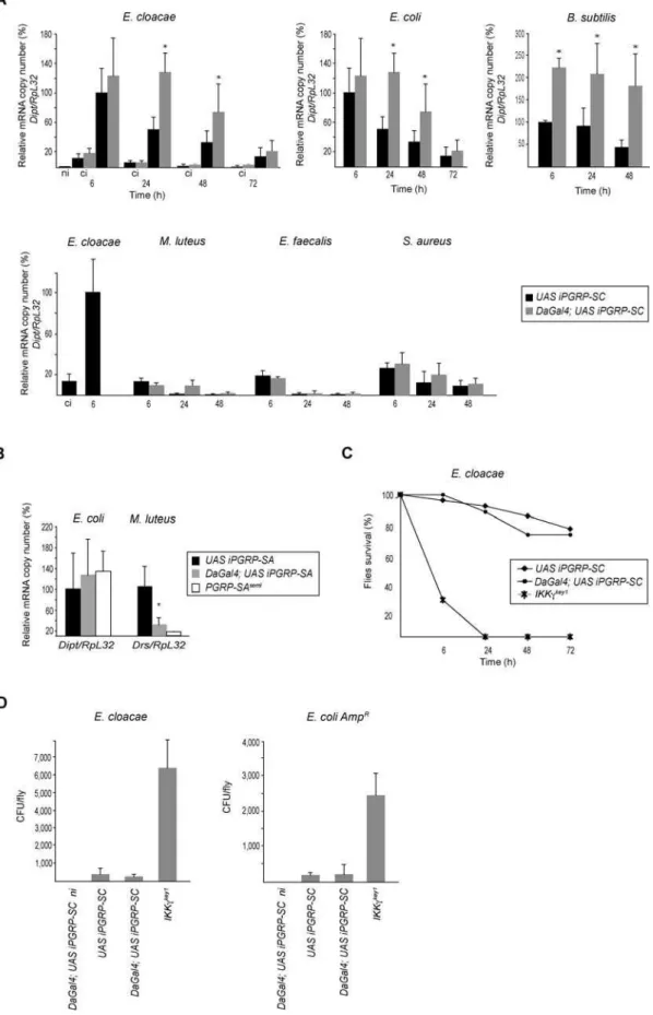
with gram-negative bacteria and measured diptericin tran-script levels as a conventional readout for IMD-pathway activation. Six hours after infection withEnterobacter cloacaeor
Escherichia coli(a time-point that corresponds to the peak of
diptericin mRNA kinetics in wild-type flies), no differences were noted between the levels of diptericin mRNA in UAS
iPGRP-SC and DaGal4;UAS iPGRP-SC flies (Figure 2A).
However, whereas thediptericinmRNA level dropped signifi-cantly at 24 and 48 h inUAS iPGRP-SCflies (as it usually does in the wild-type condition), it remained high inDaGal4;UAS iPGRP-SCflies. Similar results were obtained with another m-DAP-PGN–containing bacteria(Bacillus subtilis), although the differences betweenUAS iPGRP-SC and DaGal4;UAS iPGRP-SCcould be detected as soon as 6 h after infection (Figure 2A). Experiments performed with another ubiquitous Gal4
driver (ActinGal4) and an independent UAS iPGRP-SC
insertion generated identical results (data not shown). When gram-positive bacteria were used as inducers, two of them
(Micrococcus luteusandEnterococcus faecalis)did not activate the IMD pathway above clean injury levels. A third one, Staph-ylococcus aureus, triggered slightly higher diptericin- transcrip-tion levels in PGRP-SC–depleted flies than in controls, although the induction was mild, as expected from gram-positive bacteria (Figure 2A).
These results indicate that depletion of PGRP-SC induces an over-activation of the IMD pathway after bacterial infection. This phenotype was not observed in noninfected
DaGal4;UAS iPGRP-SC flies or after pricking with a clean needle (Figure 2A). Altogether, this indicates that the observed effects are dependent on the presence of bacteria and do not correspond to a constitutive activation of the IMD pathway in PGRP-SC–depleted flies. They also demonstrate that the other putative secretedDrosophilaamidases (PGRP-SB1 and PGRP-SB2) are not able to compensate for the absence of PGRP-SCs in vivo. In the absence of loss-of-function mutants for each of the threePGRP-SCgenes, we can obviously not rule out the possibility that only one or two of them are responsible for the observed phenotype. To obtain additional proof that the effects obtained in PGRP-SC–depleted flies were specific, we performed similar experi-ments in flies in which the non-amidase bacterial receptor PGRP-SA had been depleted by RNA interference. Whereas
DaGal4;UAS iPGRP-SA flies showed an expected reduced
ability to respond to infection by the gram-positiveM. luteus,
their response to E. coli remained wild-type (Figure 2B).
Therefore, DaGal4;UAS iPGRP-SA flies behave as classical
PGRP-SAseml loss-of-function mutants [7] and not like flies with reduced PGRP-SC levels.
PGPR-SC1/2–Depleted Flies Are Able to Clear Bacteria
Since PGRP-SC1 has been proposed to act as a scavenger molecule [27], we tested whether the IMD-pathway over-activation observed in PGRP-SC–depleted flies could reflect the inability of these flies to clear bacteria. If this were to be proved the case, accumulation of bacteria in the body cavity
of DaGal4;UAS iPGRP-SC flies could explain the
over-activation of the IMD pathway. To test this hypothesis, we compared bacterial loads and survival curves ofIKKckey1
, UAS iPGRP-SC, and DaGal4;UAS iPGRP-SC flies infected with E. cloacaeor with E. coli (Figure 2C and 2D). WhereasIKKckey1
mutant flies showed a very high bacterial load and a strong susceptibility to these bacteria, no such phenotypes were
observed inDaGal4;UAS iPGRP-SC flies. Similar results were obtained withB. subtilis(unpublished data).This suggests that the over-response observed in PGRP-SC–depleted flies did not result from an uncontrolled bacterial growth in the hemolymph, and that the role of PGRP-SC proteins in vivo is not to scavenge bacteria from the circulating hemolymph.
Toll-Pathway Activation Is Wild-Type inPGRP-SC1/2
Mutant Flies
It is notable that non-enzymatic PGRPs, such as PGRP-SA, PGRP-LC, or PGRP-LE are able to discriminate between Lys-type and m-DAP-Lys-type PGN [14–17] (Figure S1). Recent experiments have nevertheless demonstrated that PGRP-SC1b can act as a cleaving enzyme for both gram-positive and gram-negative bacterial PGN in vitro. We therefore asked whether reducing the endogenous levels of PGRP-SC1/2 could also have an effect on Toll-pathway activation by gram-positive bacteria. As illustrated in Figure 3, the effects were IMD-pathway–specific since Toll-dependent activation of
drosomycin by gram-positive or gram-negative bacteria (M. luteus, E. faecalis, S. aureus, E. cloacae, E. coli, B. subtilis) were similar inUAS iPGRP-SCandDaGal4;UAS iPGRP-SCflies. This difference between the role of PGRP-SC1/2 on Toll- and IMD-pathway activation could reflect functional redundancy between amidases for Lys-type PGN which might not exist for m-DAP-type PGN-cleaving enzyme. Alternatively, this could pertain to the difference in the mode of activation of the transmembrane receptors upstream of each pathway.
PGRP-SC1/2 Function in Larval Immune Response
We next addressed the role of PGRP-SC proteins in larvae. Previous qualitative analyses indicated that PGRP-SC1 and
PGRP-SC2genes are transcribed in almost identical patterns and mostly in the gut cells [25]. Using quantitative RT-PCR, we confirmed that the larval gut is strongly enriched in
PGRP-SC1 and PGRP-SC2 mRNA and represents the main site of
PGRP-SC amidase synthesis at this developmental stage (Figure 4A). In a previously established model of infection by ingestion [31], it was observed that larvae fed with the gram-negative bacteria Erwinia carotovora carotovora induce
diptericintranscription in the fat body. Surprisingly, most of the other gram-negative bacterial species tested in this assay failed to do so. We reasoned that the gut PGRP-SC amidases might act to reduce the PGN immunogenic potential of these bacteria, preventing them from activating a systemic immune response.
To test this hypothesis,UAS iPGRP-SCcontrol larvae and
DaGal4;UAS iPGRP-SClarvae, which exhibit a strong reduc-tion of PGRP-SC1 and PGRP-SC2mRNA levels in their gut (Figure 4A), were fed with various bacterial species. We then monitored diptericin expression in whole larvae or, more specifically, in the fat body. As previously reported [31], we found that ingestedE. carotovora carotovora,but notE. coli,was able to activate the IMD pathway inUAS iPGRP-SC control larvae (Figure 4B). Strikingly, reducing the PGRP-SC levels induced a strong increase in the expression level of the
diptericinmRNA at 6 and 24 h after feeding on E. carotovora carotovora,as compared to controls (Figure 4B). Under these conditions, E. coli now became a good inducer of diptericin
Figure 2.IMD-Pathway Activation Is Downregulated by PGRP-SC1/2
amidases act upstream of the IMD-pathway transmembrane receptor (Figure 4B). As shown above for the immune response in adults, reducing the PGRP-SC levels had no effect on Toll-pathway activation in larvae (Figure 4C).
PGRP-SC1/2 Are Required in the Larval Gut to Dampen the Immune Response
We tested whether similar results could be obtained by reducing PGRP-SC levels specifically in the larval gut using a tissue-specific driver(CadGal4)[32]. Consistent with previous reports indicating thatCadGal4is not a very strong driver[32], we noted that the reduction ofPGRP SC1/2 mRNA levels in the gut were not as pronounced in CadGal4;UAS iPGRP-SC
than inDaGal4;UAS iPGRP-SC larvae (Figure 4A). However, this reduction was sufficient to trigger an activation of the IMD pathway after feeding on E. coli (Figure 4B). Using a
DiptlacZreporter transgene, we could show that up to 40% of theCadGal4;UAS iPGRP-SClarvae activated the IMD pathway in the fat body after feeding onE. coli(Figure 4D and E). In wild-type control larvae, this percentage was only 5% (Figure 4E). In parallel experiments, we tested the effects of
over-expressing the PGRP-SC1b protein in the larval gut. Whereas 80% of thediptLacZ;UAS PGRP-SC1bcontrol larvae fed withE. carotorova carotorovaactivateddiptericintranscription in the fat body, this percentage dropped to 10% in larvae which specifically over-expressed PGRP-SC1b in the gut (Figure 4E). Altogether, these results are compatible with the hypothesis that an essential role of gut PGRP-SC1/2 amidases in larvae is to modulate activation of the IMD pathway. We propose that this modulation is achieved by lowering the amount of immunogenic PGN, most probably via an amidase-dependent degradation. However, we cannot rule out that it is also partly due to sequestration of the PGN.
Larvae with Reduced PGRP-SC1/2 Levels Are Highly Susceptible to Infection
To evaluate the consequences of the reduction of this immuno-modulatory function, we followed the fate of naturally infected wild-type and PGRP-SC–depleted larvae. We observed that mortality was three to four times higher in depleted larvae fed withE. coliorE. carotovora carotovorathan in controls (Figure 5A). This increase in larval lethality was
induction after gram-positive bacterial infections were compared to that obtained after infection byE. cloacae(100%).RpL32is used as an internal control. ci, clean injury; ni, noninfected.
(B) Quantification ofdiptericinmRNA levels inUAS iPGRP-SA,DaGal4;UAS iPGRP-SAandPGRP-SAseml
flies shows that reduction ofPGRP-SAmRNA levels does not influence IMD-pathway induction 6 h after infection byE. coli.Quantification ofdrosomycinmRNA levels 24 h afterM. luteusinfection indicates that PGRP-SA is efficiently knocked down by dsRNA interference.PGRP-SAsemlis a complete loss-of-function mutant for PGRP-SA. Each histogram corresponds to the mean value of five independent experiments (6standard deviation). Asterisk indicates that the difference betweenDaGal4;UAS iPGRP-SAand controlUAS iPGRP-SAvalues is statistically significant (p,0.05).
(C)DaGal4;UAS iPGRP-SCflies are as susceptible to infection byE. cloacaeas control flies.
(D)E. cloacaeandE. coli AmpRgrowth in various genetic backgrounds 24 h after infection. Flies with reduced levels of PGRP-SC1/2, unlikeIKKckey1
mutants, are able to clear bacteria from their hemolymph. Each histogram corresponds to the mean value of four independent experiments (6standard deviation).
DOI: 10.1371/journal.ppat.0020014.g002
Figure 3.Toll-Pathway Activation Is Wild-Type inDaGal4;UAS iPGRP-SCFlies
Kinetics ofdrosomycin mrna induction(Drs/RpL32)after infection by gram-positive (upper panel) and gram-negative (lower panel) bacteria. Each histogram corresponds to the mean value of six independent experiments (6standard deviation). Asterisk indicates that the difference between DaGal4;UAS iPGRP-SCand controlUAS iPGRP-SCvalues is statistically significant (p,0.05). One hundred percent corresponds to the level of activation 24 h after infection in control flies. In the lower panel,drosomycininduction after gram-negative bacterial infections is compared to that ofS. aureus infection which is set to 100%.RpL32is used as an internal control.
totally suppressed in aPGRP-LC mutant background, dem-onstrating that over-activation of the IMD pathway was indeed the cause of larval death. Interestingly, a small percentage of theDaGal4;UAS iPGRP-SC larvae that
pupari-ated and eclosed as pharate adults presented developmental defects such as wing notching (Figure 5B–5D). These phenotypes were never observed in control larvae fed with bacteria. To test whether this wing phenotype could be due to
Figure 4.Reduction of PGRP-SC1/2 Levels in the Larval Gut Increases IMD-Pathway Activation after Natural Infection
(A)PGRP-SC1andPGRP-SC2mRNAs(PGRP-SC/RpL32)are mainly expressed in the larval gut and are severely reduced inDaGal4;UAS iPGRP-SCand CadGal4;UAS iPGRP-SClarvae. Each histogram corresponds to the mean value of four independent experiments (6standard deviation).
(B)Diptericinmrna induction levels(Dipt/RpL32),measured 6 h and 24 h after natural infection. Each histogram corresponds to the mean value of variable numbers (shown in parentheses) of independent experiments (6 standard deviation). Asterisks indicate that the difference between DaGal4;UAS iPGRP-SCorCadGal4;UAS iPGRP-SCand controlUAS iPGRP-SCvalues is statistically significant (p,0.05).
(C)Drosomycinmrna induction levels(Drs/RpL32), measured 24 h after natural infection. Each histogram corresponds to the mean value of four independent experiments (6standard deviation).
(D) Three hours after natural infection withE. coliGFP, bacteria were found to be highly concentrated in the anterior half of the larval gut. In larvae with reduced gutPGRP-SClevels(CadGal4;UAS iPGRP-SC),feeding onE.coliis sufficient to trigger IMD-pathway activation in the fat body after 24 h (visualized here by the use of adiptericin-LacZtransgene).
(E) Percentage of larvae showingb-galactosidase activity in the fat body 24 h after natural infection. For each genotype, ten larvae were dissected and stained. Each histogram corresponds to the mean value of five independent experiments (6standard deviation). Asterisks indicate that the difference betweendiptLacZ;UAS iPGRP-SCanddiptLacZ;DaGal4;UAS iPGRP-SCordiptLacZ;CadGal4;UAS iPGRP-SCvalues and betweendiptLacZ,UAS iPGRP-SCand diptLacZ;CadGal4;UAS iPGRP-SCvalues is statistically significant (p,0.05).
increased cell death during larval development, imaginal discs from infected larvae were stained with acridine orange. Wing discs from PGRP-SC–depleted, bacteria-fed larvae showed higher levels of cell death than wing discs from control larvae fed with normal levels of PGRP-SC (Figure 5E and 5F).
Discussion
The need for a tight balance between initiation and resolution in the control of inflammation in vertebrates has been documented for a long time. Recent reports have reviewed the molecular mechanisms that are put in place to dampen inflammation and to prevent damaging effects associated with a prolonged immune response [33,34]. The data presented here suggest that immune response needs to be tightly regulated also in invertebrates.
Taken together, our data provide novel insights into the physiological roles of PGRPs inDrosophila.They show that in addition to the function as a pattern-recognition receptor of some PGRP family members, others can specifically control the level of activation of the IMD signaling pathway. Flies deficient for PGRP-SC1a, PGRP-SC1b, and PGRP-SC2 present a specific over-activation of the IMD pathway. A recent report described an effect of a PGRP-SC1 mutant
(picky)on Toll-pathway activation [35], a phenotype that we did not observe in PGRP-SC1/2–depleted flies (see Figure 3). This discrepancy is not yet fully understood but could be
explained by the fact that picky flies are mutant only for PGRP-SC1a and PGRP-SC1b, whereas PGRP-SC2 is also affected in our PGRP-SC–depleted flies.
Our results indicate that the gut is the main tissue in which the regulation by PGRP-SC proteins is taking place. However, the fact that IMD-pathway over-activation was also detected when bacteria were introduced directly into the circulating hemolymph suggests that these secreted proteins could also be present in the blood or in the circulating hemocytes. We further show that in the absence of a control of the immune response, infection can lead to developmental defects or death by over-activation of the immune pathway. Interest-ingly, recent reports indicate that other immune-induced pathways can have a harmful effect on fly survival.Salmonella typhimurium–infected flies produce a tumor necrosis factor (TNF)–like cytokine which has been shown to be damaging for the host [36]. In addition, flies in which the gut catalase level is experimentally reduced show high mortality rates after ingestion of microbe-contaminated foods. This has been interpreted as evidence that infection-mediated induction of reactive oxygen species (such as H2O2) must be tightly
balanced to avoid larval lethality [32]. In this respect, our data indicate that PGRP-SC1/2 may act as detoxifying proteins for bacterial PGN in flies. Although we did not demonstrate that the amidase function of these PGRPs is required for this effect, in vitro biochemical data strongly suggest that it is the case. Similar functions have recently
Figure 5.Reduction of PGRP-SC1/2 Levels Sensitizes Larvae to Bacterial Infection
(A) The percentage of dead larvae is measured 24 h after natural infection. Numbers in parentheses correspond to the total number of infected larvae. Each histogram corresponds to the mean value of six independent experiments (6standard deviation) forE. coliand five forE. carotovora carotovora. Asterisks indicate that the difference betweenUAS iPGRP-SCandDaGal4;UAS iPGRP-SCvalues is statistically significant (p,0.05).
(B) The percentage of adults showing wing notching is measured 7 d after natural infection. Numbers in parentheses correspond to the total number of infected larvae.
(C–F) Natural infection withE. carotovora carotorovaandE. colitriggers increased cell death and developmental defects inDaGal4;UAS iPGRP-SCflies. Wing imaginal discs dissected fromDaGal4;UAS iPGRP-SClarvae (F) show higher levels of cell death after natural infection than discs from infectedUAS iPGRP-SC control larvae (E). Consistently, some DaGal4;UAS iPGRP-SC adults derived from infected larvae exhibit wing notching (indicated by arrowheads) (D), which was never observed in infected controls (C).
been attributed to enzymes which reduce the immunogenic potential of lipopolysaccharide during vertebrate immune response [37].
The results presented here are consistent with previous data showing that over-expression of some components of the IMD pathway are larval lethal. However, the molecular mechanisms by which over-activation of the IMD pathway leads to lethality remain unknown. A number of observations may provide clues about this issue: (i) several components of the IMD pathway are homologous to mammalian proteins involved in signaling through the TNF receptor, a pathway known to trigger apoptosis [38]; and (ii) the MAP3 kinase TAK1, which is an essential component of the IMD pathway, has been shown to function both as an IjB kinase (regulating
diptericinexpression) and as a JNK (c-JunN-terminal kinase) kinase [39]. It is significant in this context that inappropriate activation of the JNK signaling cascade in the wing disc leads to apoptotic pathway-dependent morphological defects [40]. Further investigations will be needed to clarify the molecular links existing between the activation of the Drosophila IMD pathway and the developmental defects which we observed; in particular, a role of the apoptosis pathways in this process should be considered. Finally, it will be of interest to investigate whether amidases or other PGN-modifying enzymes are involved in modulating bacteria-induced im-mune response in mammals. In this respect, it is intriguing that one human PGRP family member (PGRP-Ib) is expressed in the esophagus [21], which evokes the gut expression of PGRP-SC.
Materials and Methods
Bacterial strains.The following microorganisms were used:E. coli, M. luteus, E. carotovora carotovora 15, B. subtilis, E. cloacae, E. faecalis, S.
aureus,andE. coli AmpR.
Drosophila strains.IKKckey1
is a loss-of-function mutation allele.
PGRP-LCDE is a complete deletion of the PGRP-LC locus. Flies
carrying either of these mutations are unable to activate the IMD pathway. PGRP-SAsemlis a point-mutation null allele which prevents Toll-pathway activation by some bacteria. All these alleles have been previously described [8]. Daughterless Gal4(DaGal4)and Caudal Gal4
(CadGal4)are transgenic strains in which all(DaGal4)or only the gut
(CadGal4)cells express the yeast Gal4 transcription factor. InHsGal4
flies, the production of the Gal4 protein is inducible by a heat pulse.
DaGal4;UAS iPGRP-SC are strains in which all the cells produced
PGRP-SCdsRNA targeting the endogenous PGRP-SC transcript to
degradation by RNA interference. Drosophila strains produced include: PGRP-SA, PGRP-SC1a, PGRP-SC1b, PGRP-SC2, PGRP-SD,
PGRP-LB, PGRP-LC, PGRP-LE, IKKc,andRpL32.
Septic injuries, bacterial growth, and fly survival experiments.Cells from overnight bacterial cultures were recovered by centrifugation at 3,000 g for 10 min at room temperature. The supernatant was discarded and the pellet was resuspended in fresh Luria-Bertani (LB) media. Cell suspensions were serially diluted in PBS, and the concentration of cells was determined by optical-density measure-ment. Flies were anaesthetized with CO2and were infected by pricking
the dorsal thorax with a thin tungsten needle that had previously been dipped into cultures of the appropriate bacterial strains.
In bacterial-growth experiments, five 4-d-old adults were infected
with E. colior with E. cloacae. Twenty-four hours later, flies were
homogenized in 400 ll of LB media, and various dilutions were spread on LB plates containing ampicillin forE. coli(50lg/ml) or on LB plates forE. cloacae.The number of colony-forming units per fly was determined through overnight growth on plates. For survival experiments, 25 flies of each tested genotype were pricked and transferred each day into new vials with fresh medium. Survival assays were repeated four times.
Generation ofUAS iPGRP-SC, UAS iPGRP-SA,andUAS PGRP-SC1b
constructs.TheUAS iPGRP-SCplasmid was constructed by inserting a 558-bp PCR fragment corresponding to the PGRP-SC1a coding
sequence flanked byBamHI andNheI sites between theNheI–BamHI sites (sense) and theXbaI–BglII sites (antisense), respectively, into the RNAi vector [41]; primers were as follows: forward 59-GGGGGATC CATGGTTTCCAAAGTGGCTCTC-39, reverse 59-GGGGCTAGCC TAGCCAGACCAGTGGGA CCA-39. The same strategy was used for plasmidUAS iPGRP-SAby inserting a 612-bp fragment corresponding to thePGRP-SAcoding sequence; primers were as follows: forward 59 -GGGGGATCCATGCAGCCGGTTCGATTCGGA-39, reverse 59 -GGGG CTAGCTCCGA TGGAAGTTTATCCACA-39. For plasmid
UAS PGRP-SC1b, the wild-type PGRP-SC1bcDNA was obtained by
PCR using the EST GH07464 (FlyBase, http://flybase.bio.indiana.edu) as a template. This fragment was then sub-cloned into the pUAST vector. Primers were as follows: forward 59-GGGGGAATT CATGGTTTCCAAAGTGGCTCT-39, reverse 59-GGGGTCTAGA CACTCTAACCAGACCAGTGG-39. After sequencing, the constructs were injected intow1118embryos.
Natural infection of larvae. Wandering third-instar larvae were placed in a tube containing a mixture of a concentrated overnight bacterial culture with 5 % sucrose, and were incubated for 30 min at 258C. For controls, larvae were incubated in LB broth supplemented by 5% sucrose. Larvae were then transferred to grape-juice plates and incubated at 258C. Larvae were monitored fordiptericintranscription by real-time Q-PCR 6 and 24 h after infection. For observation of developmental defects, larvae were transferred to standard cornmeal-agar medium and incubated at 258C until hatching.
Quantitative real-time PCR. Real-time Q-PCR was performed as described [8]. Primers were as follows: PGRP-SC1, forward 59 -aagcgatcgtcaactattacagc-39, reverse 59-gagagccactttggaaacCA-39;
PGRP-SC1b (for over-expression checking), forward 59
-A G C T T C C T G G G C -A -A C T -A C -A -A - 39, r e v e r s e 59- G A G A T CATGTTCGGCTCCAG-39; PGRP-SC2, forward 59-TGACCAT CATCTCCAAGTCG-39, reverse 59-CAGCGAGGTCTTGCTCGT-39;
PGRP-SA, forward 59-GCTTCGTTGGGACTCCACTA-39; reverse 59
-CGTGTGATGGATG ACCACAT-39.RpL32, diptericin, drosomycin,and
PGRP-SDprimers are described in [8,11].
Acridine orange staining. Imaginal discs were dissected in PBS, incubated in a 5lg/ml AO solution for 1 min, rinsed three times for 5 min in fresh PBS, and mounted in glycerol.
Supporting Information
Figure S1.Schematic Representation of the PGN Structure Found at DOI: 10.1371/journal.ppat.0020014.sg001 (578 KB PDF).
Figure S2.PGRP-SC Alignments and Phylogeny
Found at DOI: 10.1371/journal.ppat.0020014.sg002 (1.4 MB PDF).
Accession Numbers
The FlyBase (http://flybase.bio.indiana.edu) accession numbers for the
Drosophila strains produced include IKKc (CG16910), PGRP-LB
(CG14704), PGRP-LC (CG4432), PGRP-LE (CG8995), PGRP-SA
(CG11709),PGRP-SC1a(CG14746),PGRP-SC1b(CG8577),PGRP-SC2
(CG14745),PGRP-SD(CG7496), andRpL32(CG7939).
The Swiss-Prot Enzyme Nomenclature database (http://www.expasy. org/enzyme) accession number for NAMLAA is EC3.5.1.28.
Acknowledgments
We would like to thank Fanny Rueda and Sebahat Ozkan for their excellent technical work and Marie Meister for comments on the manuscript. TheCadGal4line was kindly provided by Dr Won Jae Lee. The help given by Laurent Troxler and Charles Hetru with sequence alignment is appreciated.
Author contributions.VB, CV, and JR conceived and designed the experiments. VB and CV performed the experiments. VB, CV, BD, and JR analyzed the data. VB, CV, BD, JAH, and JR wrote the paper. IGB provided biological material and analyzed the results.
Funding. This work was supported by CNRS, the Ministe`re de l’Education Nationale de la Recherche et de la Technologie, the Fondation pour le Recherche Me´dicale and the Agence National pour la Recherche (ACI jeunes Chercheurs and IUF to JR). Financial support from the National Institutes of Health (Bethesda, Maryland, United States) is acknowledged.
Competing interests.The authors have declared that no competing
References
1. Bulet P, Charlet M, Hetru C (2002) Antimicrobial peptides in insect immunity. In: Ezekowitz RAB, Hoffmann JA, editors. Infectious disease: Innate immunity. Totowa (New Jersey): Human Press. pp. 89–107. 2. Zasloff M (2002) Antimicrobial peptides of multicellular organisms. Nature
415: 389–395.
3. De Gregorio E, Spellman PT, Rubin GM, Lemaitre B (2001) Genome-wide analysis of the Drosophila immune response by using oligonucleotide microarrays. Proc Natl Acad Sci U S A 98: 12590–12595.
4. Irving P, Troxler L, Heuer TS, Belvin M, Kopczynski C, et al. (2001) A genome-wide analysis of immune responses inDrosophila. Proc Natl Acad Sci U S A 98: 15119–15124.
5. Hoffmann JA (2003) The immune response ofDrosophila. Nature 426: 33–38. 6. Brennan CA, Anderson KV (2004) Drosophila: The genetics of innate
immune recognition and response. Annu Rev Immunol 22: 457–483. 7. Michel T, Reichhart JM, Hoffmann JA, Royet J (2001)DrosophilaToll is
activated by gram-positive bacteria through a circulating peptidoglycan recognition protein. Nature 414: 756–759.
8. Bischoff V, Vignal C, Boneca IG, Michel T, Hoffmann JA, et al. (2004) Function of theDrosophilapattern-recognition receptor PGRP-SD in the detection of gram-positive bacteria. Nat Immunol 5: 1175–1180. 9. Gobert V, Gottar M, Matskevich AA, Rutschmann S, Royet J, et al. (2003)
Dual activation of theDrosophilaToll pathway by two pattern recognition receptors. Science 302: 2126–2130.
10. Pili-Floury S, Leulier F, Takahashi K, Saigo K, Samain E, et al. (2004) In vivo RNA interference analysis reveals an unexpected role for GNBP1 in the defense against gram-positive bacterial infection inDrosophilaadults. J Biol Chem 279: 12848–12853.
11. Gottar M, Gobert V, Michel T, Belvin M, Duyk G, et al. (2002) TheDrosophila
immune response against gram-negative bacteria is mediated by a peptidoglycan recognition protein. Nature 416: 640–644.
12. Ramet M, Manfruelli P, Pearson A, Mathey-Prevot B, Ezekowitz RA (2002) Functional genomic analysis of phagocytosis and identification of a
Drosophilareceptor forE. coli. Nature 416: 644–648.
13. Choe KM, Werner T, Stoven S, Hultmark D, Anderson KV (2002) Requirement for a peptidoglycan recognition protein (PGRP) in Relish activation and antibacterial immune responses inDrosophila. Science 296: 359–362.
14. Takehana A, Katsuyama T, Yano T, Oshima Y, Takada H, et al. (2002) Overexpression of a pattern-recognition receptor, peptidoglycan-recog-nition protein-LE, activates IMD/Relish-mediated antibacterial defense and the prophenoloxidase cascade inDrosophilalarvae. Proc Natl Acad Sci U S A 99: 13705–13710.
15. Takehana A, Yano T, Mita S, Kotani A, Oshima Y, et al. (2004) Peptidoglycan recognition protein (PGRP)-LE and PGRP-LC act synergisti-cally inDrosophilaimmunity. EMBO J 23: 4690–4700.
16. Kaneko T, Goldman WE, Mellroth P, Steiner H, Fukase K, et al. (2004) Monomeric and polymeric gram-negative peptidoglycan but not purified LPS stimulate theDrosophilaIMD pathway. Immunity 20: 637–649. 17. Leulier F, Parquet C, Pili-Floury S, Ryu JH, Caroff M, et al. (2003) The
Drosophilaimmune system detects bacteria through specific peptidoglycan recognition. Nat Immunol 4: 478–484.
18. Schleifer K, Kandelr P (1972) Peptidoglycan types of bacterial cell walls and their taxonomic implications. Bacteriol Rev 36: 407–477.
19. Stenbak CR, Ryu JH, Leulier F, Pili-Floury S, Parquet C, et al. (2004) Peptidoglycan molecular requirements allowing detection by theDrosophila
immune deficiency pathway. J Immunol 173: 7339–7348.
20. Yoshida H, Kinoshita K, Ashida M (1996) Purification of a peptidoglycan recognition protein from hemolymph of the silkworm,Bombyx mori. J Biol Chem 271: 13854–13860.
21. Liu C, Gelius E, Liu G, Steiner H, Dziarski R (2000) Mammalian peptidoglycan recognition protein binds peptidoglycan with high affinity, is expressed in neutrophils, and inhibits bacterial growth. J Biol Chem 275: 24490–24499.
22. Liu C, Xu Z, Gupta D, Dziarski R (2001) Peptidoglycan recognition proteins: A novel family of four human innate immunity pattern recognition molecules. J Biol Chem 276: 34686–34694.
23. Kang D, Liu G, Lundstrom A, Gelius E, Steiner H (1998) A peptidoglycan recognition protein in innate immunity conserved from insects to humans. Proc Natl Acad Sci U S A 95: 10078–10082.
24. Tydell CC, Yount N, Tran D, Yuan J, Selsted ME (2002) Isolation, characterization, and antimicrobial properties of bovine oligosaccharide-binding protein. A microbicidal granule protein of eosinophils and neutrophils. J Biol Chem 277: 19658–19664.
25. Werner T, Liu G, Kang D, Ekengren S, Steiner H, et al. (2000) A family of peptidoglycan recognition proteins in the fruit flyDrosophila melanogaster. Proc Natl Acad Sci U S A 97: 13772–13777.
26. Steiner H (2004) Peptidoglycan recognition proteins: On and off switches for innate immunity. Immunol Rev 198: 83–96.
27. Mellroth P, Karlsson J, Steiner H (2003) A scavenger function for a
Drosophilapeptidoglycan recognition protein. J Biol Chem 278: 7059–7064. 28. Kim MS, Byun M, Oh BH (2003) Crystal structure of peptidoglycan recognition protein LB fromDrosophila melanogaster. Nat Immunol 4: 787– 793.
29. Gelius E, Persson C, Karlsson J, Steiner H (2003) A mammalian peptidoglycan recognition protein withN-acetylmuramoyl-L-alanine ami-dase activity. Biochem Biophys Res Commun 306: 988–994.
30. Wang ZM, Li X, Cocklin RR, Wang M, Fukase K, et al. (2003) Human peptidoglycan recognition protein-L is an N-acetylmuramoyl-L-alanine amidase. J Biol Chem 278: 49044–49052.
31. Basset A, Khush RS, Braun A, Gardan L, Boccard F, et al. (2000) The phytopathogenic bacteriaErwinia carotovorainfectsDrosophilaand activates an immune response. Proc Natl Acad Sci U S A 97: 3376–3381. 32. Ha EM, Oh CT, Ryu JH, Bae YS, Kang SW, et al. (2005) An antioxidant
system required for host protection against gut infection inDrosophila. Dev Cell 8: 125–132.
33. Han J, Ulevitch RJ (2005) Limiting inflammatory responses during activation of innate immunity. Nat Immunol 6: 1198–1205.
34. Serhan CN, Savill J (2005) Resolution of inflammation: The beginning programs the end. Nat Immunol 6: 1191–1197.
35. Garver LS, Wu J, Wu LP (2006) The peptidoglycan recognition protein PGRP-SC1a is essential for Toll signaling and phagocytosis ofStaphylococcus aureusinDrosophila. Proc Natl Acad Sci U S A 103: 660–665.
36. Brandt SM, Dionne MS, Khush RS, Pham LN, Vigdal TJ, et al. (2004) Secreted bacterial effectors and host-produced Eiger/TNF drive death in a
Salmonella-infected fruit fly. PLoS Biol 2: e418. DOI: 10.1371/journal.pbio. 0020418
37. Munford RS (2005) Detoxifying endotoxin: Time, place and person. J Endotoxin Res 11: 69–84.
38. Micheau O, Tschopp J (2003) Induction of TNF receptor I-mediated apoptosis via two sequential signaling complexes. Cell 114: 181–190. 39. Silverman N, Zhou R, Erlich RL, Hunter M, Bernstein E, et al. (2003)
Immune activation of NF-kappaB and JNK requiresDrosophilaTAK1. J Biol Chem 278: 48928–48934.
40. Adachi-Yamada T, Fujimura-Kamada K, Nishida Y, Matsumoto K (1999) Distortion of proximodistal information causes JNK-dependent apoptosis inDrosophilawing. Nature 400: 166–169.
41. Naitza S, Rosse C, Kappler C, Georgel P, Belvin M, et al. (2002) The