Work performed in theLife Sciences Center, Catholic Pontifical University in Campinas, Campinas-SP.
Correspondence address: Dr. José Roberto Ortale. Centro de Ciências da Vida, Pontifícia Universidade Católica de Campinas - Campus II.Av. John Boyd Dunlop, s/nº - Jardim Ipaussurama. Campinas, SP CEP 13059-900. Tel: (0xx19) 3729-8466. Fax: (0xx19) 3729-8517. E-mail: [email protected]
José Roberto ORTALE, José MECIANO FILHO, Ana Maria Ferreira PACCOLA, Júlia Guedes Pereira Garcia LEAL, Carolina Alves SCARANARI
Article received in March, 2005 Article accepted in May, 2005
RBCCV 44205-746
Abstract
Objective: The objective of the present report was to describe the lateral, diagonal and anterosuperior arterial branches in the epicardial adipose tissue of the left ventricle and to analyze their frequency and diameters according to the type of coronary circulation. The precious knowledge of these branches has surgical application in their revascularization or during the injection of the cardioplegic substances into these branches.
Method: Fifty hearts obtained at autopsy from adult cadavers were dissected and fixed in formalin and the left ventricle was divided into three thirds: superior, middle and inferior. The lateral branch originated from the circumflex branch, the diagonal branch from the division of the left coronary artery and the anterosuperior branch from the anterior interventricular branch in the superior third of the left ventricle. The length in the epicardium and the diameter of each branch were measured and the blood flow was related to the type of coronary circulation.
Results: The diameter of the lateral branch, present in 88% of the cases, ranged from 0.6 to 4.5 mm (mean: 2.1 ±±±±± 0.7
mm). The diameter of the diagonal branch, present in 50% of cases, ranged from 1.0 to 3.8 mm (mean: 2.2 ±±±±± 0.7 mm). The diameter of the anterosuperior branch, present in 84% of cases, ranged from 1.0 to 4.1 mm (mean: 2.5 ±±±±± 0.8 mm). We detected 30/50 (60%) cases of dominance of the right coronary artery, 14/50 (28%) cases of the balanced type, and 6/12 (12%) cases of dominance of the left coronary artery. Mean blood flow in the anterosuperior branch presented a decreasing value in the following types: dominance of the right coronary artery, balanced and dominance of the left coronary artery. Conversely, the lateral branch showed respectively increasing values, while the diagonal branch presented a greater flow in the balanced type.
Conclusion: The results demonstrated the complementarity of the lateral, diagonal and anterosuperior arterial branches, as well as the correlation among these branches with the following types of coronary circulation: right dominance, balanced and left dominance.
Descriptors: Heart, anatomy & histology. Coronary circulation.
Anatomia dos ramos lateral, diagonal e ântero-superior no ventrículo esquerdo do coração humano
Anatomy of the lateral, diagonal and
INTRODUCTION
Initially, let us present the concepts adopted in respect to the coronary artery branches of the anterior face of the left ventricle, the focus of the present study.
The lateral branch originates from the circumflex branch of the left coronary artery, before the root of the left marginal branch. The diagonal branch is one of the terminal branches of the left coronary artery, as well as the circumflex and anterior interventricular branches, when it splits into three or four branches.
The anterosuperior branch of the left ventricle is the one that originates from the anterior interventricular branch in the upper third of the left ventricle
Detailed knowledge about the coronary artery branches has already been greatly studied, however further study is necessary due to incessant advances in the diagnosis and treatment methods of heart diseases [1-3]. This work is justified as, according to Oliveira et al. [4], the diagonal, lateral and anterosuperior branches are particularly important, due to the frequent use of their epicardial sections in coronary artery bypass surgery, as well as the internal thoracic artery and the saphenous vein.
Among the authors who have researched the subject of the diagonal, lateral and anterosuperior branches of the left ventricle, we must mention: Bianchi [5], Crainicianu [6], Smith [7], James [8], De Paula [9], Mac Alpin et al. [10], Gensini et al. [11], Kalbfleisch & Hort [12], Carvalho [13], Leguerrier et al. [14], Di Dio & Rodrigues [15], Henriquez Pino et al. [16] and Baptista et al. [17].
The aim of the present study is to evaluate the frequency and the absolute and relative lengths, but specifically the
diameter of the diagonal, lateral and anterosuperior branches to determine the co-existence among them. Additionally, the objective of this article is to investigate the possibility of correlation among the distribution of the aforementioned branches in the anterior face of the left ventricle and the three traditional types of circulation: dominance of the right coronary artery, balanced circulation and dominance of the left coronary artery. These three types are based on the analysis of the ramification of the coronary arteries exclusively on the posterior face of the heart [18].
METHOD
The coronary arteries and their branches in the epicardium of 50 adult cadaver hearts were carefully dissected. They were fixed in 10% formalin solution and conserved at 5% formalin solution, in the Anatomy Laboratory of the Life Sciences Center of the Pontifical Catholic University of Campinas.
For each investigated branch (diagonal, lateral and anterosuperior) the length on the surface of the heart was measured, that is, the distance between the root of the branch and its point of penetration into the myocardium. According to Baptista et al. [17] the relative length is calculated as the result of the division between the length of the branch on the heart surface and the length of the left ventricle, considered to be the distance between the point where the left coronary artery splits and the apex of the heart. This result is multiplied by 100 to form a percentage. Each branch was classified as short, medium or long, depending on the relative length being up to 33.3%, from 33.4% to 66.6%; and more than 66.6%, respectively.
Resumo
Objetivo: O objetivo deste trabalho é a descrição dos ramos lateral, diagonal e ântero-superior, no tecido adiposo epicárdico do ventrículo esquerdo, e a análise da freqüência e do diâmetro destes, conforme o tipo de circulação coronariana. O conhecimento preciso desses ramos tem aplicabilidade na abordagem cirúrgica para a sua revascularização ou durante a injeção de substâncias cardioplégicas nos mesmos.
Método: Dissecados 50 corações obtidos de necropsias de adultos, fixados em solução de formol e o ventrículo esquerdo dividido em três terços: superior, médio e inferior. O ramo lateral originou-se do ramo circunflexo; o ramo diagonal, do ponto de divisão da artéria coronária esquerda e o ramo ântero-superior, do ramo interventricular anterior no terço superior do ventrículo esquerdo. Para cada ramo foram medidos o comprimento no epicárdio e o diâmetro, além disso foi relacionado o fluxo sangüíneo com o tipo de circulação coronariana.
Resultados: O diâmetro do ramo lateral, presente em 88% dos casos, variou de 0,6 a 4,5 mm (média 2,1 ±±±±± 0,7mm). O
diâmetro do ramo diagonal, presente em 50% dos casos, variou de 1,0 a 3,8 mm (média 2,2 ±±±±± 0,7 mm). O diâmetro do ramo ântero-superior, presente em 84% dos casos, variou de 1,0 a 4,1 mm (média 2,5 ±±±±± 0,8 mm). Foram encontrados: 30/50 (60%) casos de dominância da artéria coronária direita, 14/50 (28%) casos de tipo balanceado e 6/12 (12%) casos de dominância da artéria coronária esquerda. A média do fluxo sangüíneo do ramo ântero-superior apresentou valor decrescente nos tipos: dominância da artéria coronária direita, balanceado e dominância da artéria coronária esquerda. Inversamente, o ramo lateral mostrou valor crescente, enquanto o ramo diagonal apresentou maior fluxo no tipo balanceado.
Conclusão: Os resultados demonstraram a complementaridade entre os ramos lateral, diagonal e ântero-superior, bem como a correlação entre a distribuição dos mesmos e os tipos de circulação coronariana.
For the schematic representation of the distribution territory of the studied branches, the heart was viewed from the left pulmonary face with the coronary groove placed horizontally and two transversal lines drawn parallel to the coronary groove to divide the left ventricle into three parts: the upper, middle and lower parts (Figure 1).
anterosuperior arterial branches and the type of coronary artery domination was studied. With the aim of determining the blood flow through the arterial branches, Poiseuille’s law was utilized, that is, the blood flow per unit of time is equal to α x diameter 4/
vessel length. When more than one branch was assessed the sum of the diameters 4 was considered, for example, two branches
with diameters of 2 and 3 mm would equal 24 + 34. We did not take
into consideration the length of each branch, as the difference of length is practically negligible, according to Souza [19].
RESULTS
The left coronary artery branch was present in all cases; its diameter varied from 3.3 to 7.4 mm (mean 5.0 ± 0.9 mm)
and its length varied from 7.2 to 20.0 mm (mean 13.l ± 2.8
mm). In 50% of the cases there was bifurcation of the left coronary artery into anterior and circumflex interventricular branches. In 46% of the cases there was trifurcation, with the presence of the anterior, circumflex interventricular and diagonal branches. In 4% of the cases there were four divisions of the left coronary artery, with the presence of the bifurcated branches and of two diagonal branches.
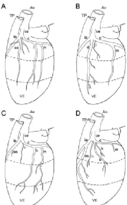
The diagonal branch was present in 25/50 (50%) cases, with one branch in 23/25 cases and two branches in 2/25 cases. The anterosuperior branch was present in 42/50 (84%) cases with one branch in 36/42 cases and two branches in 6/ 42 cases. The frequency of the lateral branch was 44/50 (88%) cases with one branch in 18/44 cases, two branches in 12/44 cases, three branches in 10/44 cases and four branches in 4/44 cases (Figure 1A-D)
Fig. 1 - Diagram showing the coexistence of the anterosuperior, diagonal and lateral branches from the left side view of the heart (4 cases; in A, B and D right coronary artery dominance; in C -balanced coronary circulation). A. coexistence of the three branches: long anterosuperior (as); short diagonal (d) and long lateral (l). B. Coexistence of a long diagonal with a short lateral branch. C. In this case there are two lateral branches, one short (l1) and one long (l2), by the side of a short anterosuperior. D. The long anterosuperior branch is by the side of three lateral branches: two medium (l1 and l2) and one short (l3)
Ao= aorta; TP= pulmonary trunk; VE= left ventricle; ce= left coronary artery; ia= anterior interventricular branch; c= circumflex branch; m= left marginal branch; cd= right coronary artery
Fig. 2 - Left lateral view of the heart in two cases of right coronary artery dominance. The auricula was removed using a pin A. Anterosuperior branch (as) is short, the diagonal branch (d) is medium and the two lateral branches are short (l1 and l2). B. Two anterosuperior branches, one medium (as1) and one long (as2), are by the side of one lateral branch (l)
Ao= aorta; TP= pulmonary trunk; VE= left ventricle; ce= left coronary artery; ia= anterior interventricular branch; c= circumflex branch; m= left marginal branch; cd= right coronary artery
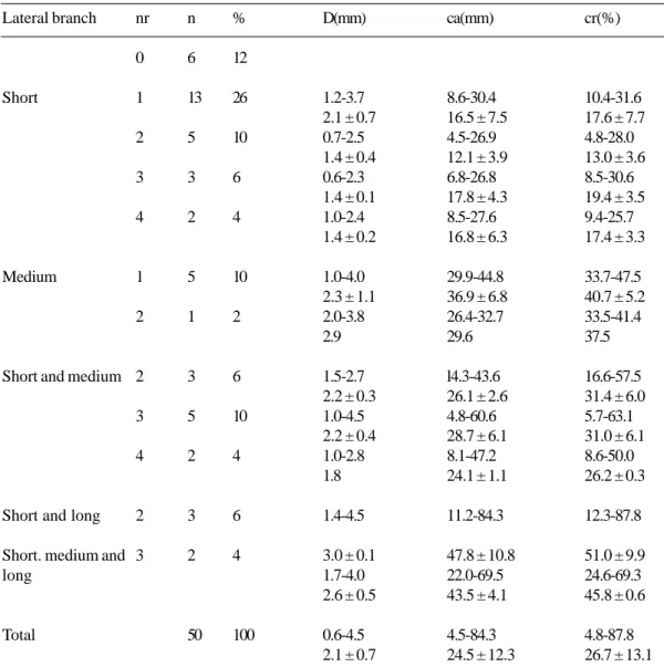
Table 1. Distribution of frequency, number of branches, diameters and absolute and relative lengths of the diagonal branch according to the type of dominance
nr= number of branches; al= absolute length; rl = relative length
Diagonal branch
Short
Medium
Long
Short and medium
Total
nr
0
1
2
1
1
2
n
25
13
1
6
4
1
50 %
50
26
2
12
8
2
100
D(mm)
1.0-3.8 2.1 ± 0.8
1.3-1.5 1.4
1.7-3.4 2.5 ± 0.6
1.7-3.4 2.6 ± 0.7
1.5-2.6 2.1
1.0-3.8 2.2 ± 0.7
al(mm)
6.6-30.2 21.3 ± 6.9
18.3-24.7 21.5
32.5-70.3 41.9 ± 13.5
55.3-103.7 77.8 ± 19.5
12.9-31.4 22.2
6.6-103.7 35.3 ± 23.8
rl(%)
6.9-31.1 23.3 ± 7.5
19.2-25.9 22.5
35.6-68.5 43.5 ± 12.8
68.9-99.6 82.2 ± 15.2
14.2-34.6 24.4
6.9-99.6 37.6 ± 23.6
Table 2. Distribution of frequency, number of branches, diameters and absolute and relative lengths of the anterosuperior branch according to the type of dominance
Anterosuperior branch
Short
Medium
Long
Short and medium
Medium and long
Total
nr
0
1
2
1
2
1
2
2
n
8
20
2
13
1
3
2
1
50 %
16
40
4
26
2
6
4
2
100
D(mm)
1.1-3.8 2.3 ± 0.8
1.0-2.9 1.8 ± 0.9
1.2-3.9 2.8 ± 0.8
1.9-2.5 2.2
2.9-4.1 3.6 ± 0.6
1.7-3.2 2.5 ± 0.4
1.7-2.1 1.9
1.0-4.1 2.5 ± 0.8
al(mm)
6.1-32.6 20.4 ± 7.0
8.0-27.6 17.5 ± 8.8
28.0-56.2 39.9 ± 7.7 48.2-62.1
55.2
64.0-82.5 70.5 ± 10.4
17.7-57.1 36.5 ± 1.3
55.0-64.4 59.7
6.1-82.5 32.4 ± 16.9
rl(%)
6.9-32.8 22.1 ± 7.6
7.7-29.2 18.0 ± 10.0
33.7-63.4 44.3 ± 8.7 45.0-57.9
51.4
71.3-82.3 77.3 ± 5.6
20.2-65.2 40.9 ± 2.5
63.5-74.4 68.9
6.9-82.3 35.4 ± 18.5
Table 3. Distribution of frequency, number of branches, diameters and absolute and relative lengths of the lateral branch according to the type of dominance
Lateral branch
Short
Medium
Short and medium
Short and long
Short. medium and long
Total
nr
0
1
2
3
4
1
2
2
3
4
2
3 n
6
13
5
3
2
5
1
3
5
2
3
2
50 %
12
26
10
6
4
10
2
6
10
4
6
4
100
D(mm)
1.2-3.7 2.1 ± 0.7 0.7-2.5 1.4 ± 0.4 0.6-2.3 1.4 ± 0.1 1.0-2.4 1.4 ± 0.2
1.0-4.0 2.3 ± 1.1 2.0-3.8 2.9
1.5-2.7 2.2 ± 0.3 1.0-4.5 2.2 ± 0.4 1.0-2.8 1.8
1.4-4.5
3.0 ± 0.1 1.7-4.0 2.6 ± 0.5
0.6-4.5 2.1 ± 0.7
ca(mm)
8.6-30.4 16.5 ± 7.5 4.5-26.9 12.1 ± 3.9 6.8-26.8 17.8 ± 4.3 8.5-27.6 16.8 ± 6.3
29.9-44.8 36.9 ± 6.8 26.4-32.7 29.6
l4.3-43.6 26.1 ± 2.6 4.8-60.6 28.7 ± 6.1 8.1-47.2 24.1 ± 1.1
11.2-84.3
47.8 ± 10.8 22.0-69.5 43.5 ± 4.1
4.5-84.3 24.5 ± 12.3
cr(%)
10.4-31.6 17.6 ± 7.7 4.8-28.0 13.0 ± 3.6 8.5-30.6 19.4 ± 3.5 9.4-25.7 17.4 ± 3.3
33.7-47.5 40.7 ± 5.2 33.5-41.4 37.5
16.6-57.5 31.4 ± 6.0 5.7-63.1 31.0 ± 6.1 8.6-50.0 26.2 ± 0.3
12.3-87.8
51.0 ± 9.9 24.6-69.3 45.8 ± 0.6
4.8-87.8 26.7 ± 13.1
nr= number of branches; al= absolute length; rl = relative length
The distribution of the frequency, the diameter, the length in the epicardium and the length in respect to the length of the left ventricle, according to the number of branches and the type (short, medium or long) for the diagonal, anterosuperior and lateral branches are shown in Tables 1 to 3 respectively. We detected that for each branch (diagonal, anterosuperior or lateral) the greater the mean diameter of the vessel, the greater was the mean length at the surface of the ventricle and the mean relative length.
In regards to the type of coronary circulation, in 30/50 (60%) cases the right coronary artery was dominant, in 14/ 50 (28%) cases the dominance was balanced and in 6/50
(12%) cases the left coronary artery was dominant. Table 4 shows the frequency of the types of coexistence and diameters of the anterosuperior, diagonal and lateral branches, distributed according to the type of coronary circulation.
Table 4. Frequency of the type of coexistence and diameter of the anterosuperior, diagonal and lateral branch distribution according to the type of coronary circulation
diagonal
D(mm)
-1.0-3.8
2.3 ± 0.9
1.7-2.8
2.3 ± 0.5
1.3-3.4
2.4 ± 1.0
1.7
1.0-3.8
2.3 ± 0.8
lateral
D(mm)
0.6-4.0
2.1±0.7
0.7-4.5
2.2± 0.8
1.1-1.5
1.6 ± 0.3
-0.6-4.5
2.1 ± 0.7
n
(%)
6
(12)
4
(8)
1
(2)
1
(2)
2
(4)
14
(28)
antero
superior
D(mm)
1.0-3.8
2.7 ± 0.9
2.0-3.7
2.7 ± 0.6
-2.0
-1.0-3.8
2.6 ± 0.8
diagonal
D(mm)
-1.7-2.4
1.9 ± 0.3
1.5
1.5-2.6
2.1 ± 0.6
2.0-3.4
2.7 ± 0.7
1.5-3.4
2.1 ± 0.6
lateral
D(mm)
1.0-4.5
2.0 ± 0.6
0.9-3.6
2.1± 0.9
1.0
-0.9-4.5
2.0 ± 0.8
n
(%)
4
(8)
2
(4)
6
(12) antero
superior
D(mm)
1.7-4.1
2.7 ± 0.7
1.2-3.9
2.5 ± 0.9
-1.4-2.9
2.2 ± 0.8
-1.2-4.1
2.6 ± 0.8
Right dominance Balanced
Conjunto
de ramos
as + l
as + d + l
d + l
as + d
d
Total
n
( %)
25
(50)
14
(28)
5
(10)
3
(6)
3
(6)
50
(100) n
( %)
15
(30)
10
(20)
2
(4)
2
(4)
1
(2)
30
(60)
Left dominance
as= anterosuperior branch; d= diagonal branch; l= lateral branch
antero
superior
D(mm)
1.1-1.9
1.5 ± 0.4
-1.1-1.9
1.5 ± 0.4
diagonal
D(mm)
-2.4-2.6
2.5 ± 0.1
-2.4-2.6
2.5 ± 0.1
lateral
D(mm)
1.7-3.9
2.5 ± 0.4
-1.1-2.2
1.6
-1.1-3.9
2.2 ± 0.5
AL
-
Anatomy of the lateral, diagonal and
dominance, 9/15 anterosuperior branch dominance and in 6/15 cases lateral branch dominance. In 6/25 cases of balanced circulation, the anterosuperior branch was dominant in 4/6 cases and the lateral branch was dominant in 2/6 cases. In all the 4/25 cases of left dominance, the dominant branch was the lateral branch. In respect to the mean blood flow, the anterosuperior branch presented a decreasing flow, in right dominance and balanced circulation and left dominance, respectively, while the lateral branch showed an increasing value respectively and the diagonal branch presented greater flow in the balanced circulation. These outcomes demonstrate the coexistenceamong these three coronary branches. Consequently, the greater the blood flow of the lateral branch, the longer was the length of the
circumflex branch of the left coronary artery in the diaphragmatic face of the heart.
COMMENTS
The length of the left coronary artery trunk is important for the cannulae used in myocardial perfusion, during aortic valve surgery [1,2]. In our results, the length was from 7.2 to 20.0 mm (mean of 13.1 ± 2.8 mm), values practically identical
to those reported by Henriquez Pino et al. [16] and a mean value a little greater than the one reported by Kronzon et al. [20]. These authors established a correlation between significantly shorter left coronary artery lengths and left dominance and balanced circulation, when compared with
Table 5. Distribution of the dominant branch and the mean flow according to the type of coronary circulation
Set of branches
as + l
as + d + l
d + l
as + d
d
Total
n ( %)
25 (50)
14 (28)
5 (10)
3 (6)
3 (6)
50 (100)
antero superior
9
5
-1
-15
diagonal
-3
2
1
1
7
lateral
6
2
-8
antero superior
4
3
-7
diagonal
-1
1
2
4
lateral
2
1
-3
antero superior
-0
diagonal
-2
-2
lateral
4
-4 Right dominance
Dominant branch
Balanced Dominant branch
Left dominance Dominant branch
Mean blood flow of the branch (mm4)
Right dominance Balanced Left dominance
antero superior
67.83
diagonal
27.16
lateral
69.86
antero superior
64.94
diagonal
19.15
lateral
71.98
antero superior
2.75
diagonal
13.16
lateral
108.56
patients with right coronary artery dominance. We did not confirm this correlation.
Our outcomes showed the left coronary artery was bifurcated in 50% of the cases, giving only anterior and circumflex interventricular branches, in 46% of the cases it
had three divisions with an addition of the diagonal branch and in 4% of the cases four divisions with two diagonal branches. These percentages are intermediate to those found by Banchi [5], Crainicianu [6], De Paula [9], Leguerrier et al. [14], Henriquez Pino et al. [16] and Baptista et al. [17].
Table 7. Variations of the lengths of the diagonal, anterosuperior and lateral branches in respect to the left ventricle
Relative lenght (%)
Less than 10 10 a 20 20 a 30 30 a 40 40 a 50 50 a 60 60 a 70 70 a 80 80 a 90 90 a 100 Total
Diagonal Branch (%)
3.9 19.5 44.1 11.7 9.1 2.6 3.9 -1.3
Diagonal Branch (%)
4.0 12.0 32.0 28.0 4.0
-12.0
-8.0 100.0
Antero-superior Branch
(%)
4.8 11.9 28.6 23.8 11.9 7.1 4.8 4.8 2.3 -100.0
Lateral Branch (%)
2.3 38.6 20.5 22.7 11.4 4.5
-100.0 Baptista et al.
(1991) Current study
Table 6. Variations of the lengths of the diagonal, anterosuperior and lateral branches in the left ventricle epicardium
Absolute length (mm)
Less than 10 10.1 a 20.0 20.1 a 30.0 30.1 a 40.0 40.1 a 50.0 50.1 a 60.0 60.1 a 70.0 70.1 a 80.0 80.01 a 90.0 90.1 a 100.0 Greater than 100 Total
Diagonal Branch (%)
1.3 7.8 27.3 39.0 13.0 5.2 2.6 1.3 1.3 -1.3
Diagonal Branch (%)
4.0 16.0 36.0 20.0 4.0 4.0 4.0 4.0 4.0 -4.0 100.0
Antero-superior Branch (%)
4.8 19.0 28.6 23.8 9.5 7.1 4.8 -2.4
-100.0
Lateral Branch (%)
6.8 36.4 31.8 11.4 9.1 4.5 -100.0 Baptista et al.
(1991)
The diagonal branch was baptized of diagonal artery by Crainicianu [6]. We identified one diagonal branch in 46% of the cases and two branches in 4% of the cases, frequencies very close to those reported by Baptista et al. [17] who found 46.1% and 7.5%, respectively.
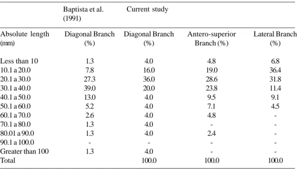
According to these authors, the variation in length of the diagonal branch is significant in heart surgery, due to the use of its external portion in the implantation of saphenous vein grafts in coronary artery bypass grafting. In our results, the length of the diagonal branch was consistent with the lengths of these authors. The relative length ranged from 6.9% to 99.6% (mean 38.8%) however, on average the diagonal branch reaches the middle third of the left ventricle, as reported by Baptista et al. [17] and De Paula [9]. Tables 6 and 7 show the distribution of the absolute length and the relative length of the external part of the diagonal branch at 10-mm intervals respectively, giving similar to the results to Baptista et al. [17]. In these tables, we added data concerning at variation of the length of the anterosuperior and lateral branches at 10-mm intervals.
The anterosuperior branch of the left ventricle was so denominated by Baptista et al. [21] due to its origin in the anterior interventricular branch in the upper third of the left ventricle, to differentiate it from the branches that originate from the lower and middle thirds. Banchi [5] denominated all of them descending collateral branches.
The circumflex branch, before giving rise to the marginal branch, derives the lateral branch, which was denominated the lateral artery for the first time by Leguerrier et al. [14]. Banchi [5] denominated it the left ventricle anterior branch, the name used by Smith [7]. The length of the lateral branch on the surface of the ventricle ranged from 4.5 to 84.3 mm (mean: 24.5 ± 12.3 mm), which is 30 mm longer than that
reported by Leguerrier et al. [14]. According to these authors, normally there was a delicate 2 to 3-mm thick myocardial bridge that easily allowed arteriotomy during surgery. We did not find any myocardial bridge in the lateral or anterosuperior branches. In only two cases we identified bridges on the diagonal branch, in one case it was 70.3 mm in length covered by a bridge of 29.6 mm and in the other it was 89.0 mm with a bridge of 9.5 mm.
According to Leguerrier et al. [14] the lateral branch had a diameter of more than l.5 mm in 82.1% of the cases which made the implantation of a saphenous graft for coronary artery bypass grafting possible. In our cases, the diameter of the lateral branch ranged from 0.6 to 4.5 mm (mean 2.1 ±
0.7 mm) and was greater than l.5 mm in 38/44 cases. In respect to the anterosuperior and diagonal branches, in our series the diameters were greater than 1.5 mm in 36/42 and 21/25 cases, respectively, which according to Leguerrier et al. make coronary artery bypass grafting possible [14].
In respect to the coronary circulation, a dominance of
the right coronary artery was seen in 60% of the cases, balanced circulation in 28% and left coronary artery dominance in 12% of the cases, percentages similar to those reported by Hadziselimovic et al.[12], (63%, 24% and 13%, respectively).
According to the Kalbfleisch & Hort [12], there is a small correlation between the diameter of the coronary artery and the myocardial area it supplies, which is in agreement with Poiseuille’s law, that says that the blood flow is proportional to the fourth power of the diameter of the vessel (diameter
4). We calculate the flow in each type of coronary circulation
and identified coexistence among the studied branches. This coexistence was suggested by Banchi [5] and De Paula [9]. In respect to the relationship among the coexistence of the branches and the type of coronary circulation, we verified that, as the flow in the lateral branch increased from right coronary artery dominance, balanced circulation and left coronary artery dominance, the anterosuperior branch decreased with the diagonal branch presenting with higher arterial flow in the balanced circulation.
CONCLUSIONS
We came to the following conclusions:
a) The frequencies of the lateral, anterosuperior and diagonal branches seen in this study were 88%, 84% and 50%, respectively;
b) The greater the diameter of the lateral, anterosuperior or diagonal branches, the greater was their length in the epicardium;
c) There was coexistence among the diagonal and anterosuperior or lateral branches in the arterial irrigation of the left ventricle;
d) A simple observation of the lateral, diagonal and anterosuperior branches in the anterior face of the left ventricle, allows a prediction of the type of coronary circulation: right coronary artery dominance, balance circulation or left coronary artery dominance, classically obtained by an analysis of the arteries exclusively in the posterior face of the heart.
ACKNOWLEDGEMENTS
12. Kalbfleisch H, Hort W. Quantitative study on the size of coronary artery supplying areas postmortem. Am Heart J. 1977;94(2):183-8.
13. Carvalho RG. Nomenclatura das artérias coronárias. Arq Bras Cardiol. 1978;31(6):415-20.
14. Leguerrier A, Bourgin T, Marcade E, Duval JM, Rioux C, Logeais Y et al. Les branches ventriculaires de l'artère circonflexe du coeur. Bull Assoc Anat. 1980;64(186):415-23.
15. Di Dio LJA, Rodrigues H. Cardiac segments in the human heart. Anat Clin. 1983;5:115-24.
16. Henriquez Pino J, Riffo EO, Vargas FM, Vargas JE. Dispositión de las ramas arteriales ventriculares en corazones de individuos chilenos. An Anat Norm. 1987;5(5):67-72.
17. Baptista CA, Di Dio LJ, Prates JC. Types of division of the left coronary artery and the ramus diagonalis of the human heart. Jpn Heart J. 1991;32(3):323-35.
18. Ortale JR, Keiralla LC, Sacilotto L. The posterior ventricular branches of the coronary arteries in the human heart. Arq Bras Cardiol. 2004;82(5):468-72.
19. Sousa OM. Anatomia topográfica: parte geral. 3a ed. São Paulo:Rossolillo;1970. p.142-7.
20. Kronzon I, Deutsch P, Glassman E. Length of the left main coronary artery: its relation to the pattern of coronary arterial distribution. Am J Cardiol. 1974;34(7):787-9.
21. Baptista CA, Di Dio LJ, Davis JT, Teofilovski-Parapid G. The cardiac apex and its superficial blood supply. Surg Radiol Anat. 1988;10(2):15-60.
22. Hadziselimovic H, Dilberovic F, Ovcina F. Blood vessels of the human heart: coronarography and dissection. Acta Anat. 1980;106(4):443-9.
BIBLIOGRAPHIC REFERENCES
1. Green GE, Bernstein S, Reppert EH. The length of the left main coronary artery. Surgery. 1967;62(6):1021-4.
2. Fox C, Davies MJ, Webb-Peploe MM. Length of left main coronary artery. Br Heart J. 1973;35(8):796-8.
3. Falci Jr. R, Prates NEVB. Anatomia das artérias coronárias. Rev Med. 1994;72(1/4):21-4.
4. Oliveira SA, Lemos PCP, Dallan LAO. Cirurgia das artérias coronárias. In: Goffi FS, editor. Técnica cirúrgica: bases anatômicas, fisiopatológicas e técnicas da cirurgia. 4a ed. São Paulo:Atheneu;1996. p.422-33.
5. Banchi A. Morfologia delle arteriae coronariae cordis. Arch Ital Anat Embriol. 1904;3:87-164.
6. Crainicianu A. Anatomische studien über die coronaraterien und experimentelle untersuchungen über die durchgängigkeit. Virch Arch Path Anat. 1922;238:1-75.
7. Smith GT. The anatomy of the coronary circulation. Am J Cardiol. 1962;9(Suppl.):327-42.
8. James TN. Anatomy of the coronary arteries in health and disease. Circulation. 1965;32(3):1020-33.
9. De Paula W. Estudo estatístico sobre irrigação coronariana no coração humano em brancos e negros. Fol Clin Biol. 1972;1(1):18-40.
10. MacAlpin RN, Abbasi AS, Grollman Jr. JH, Eber L. Human coronary artery size during life: a cinearteriographic study. Radiology. 1973;108(3):567-76.