Simone da Silva FREITAS(a)
Lucas Lopes Araújo SOUSA(b)
José Machado MOITA NETO(c)
Regina Ferraz MENDES(d)
Raimundo Rosendo PRADO JUNIOR(d)
(a)Universidade Federal do Piauí – UFPI, School of Dentistry, Health Sciences Center,Teresina, PI, Brazil.
(b)Universidade Federal do Piauí – UFPI, School of Dentistry, Post-Graduate Program in Dentistry, Teresina, PI, Brazil.
(c)Universidade Federal do Piauí – UFPI, Natural Science Center, Chemistry Department, Teresina, PI, Brazil.
(d)Universidade Federal do Piauí – UFPI, Health Sciences Center, Department of Restorative Dentistry, Teresina, PI, Brazil.
Dentin hypersensitivity treatment
of non-carious cervical lesions – a
single-blind, split-mouth study
Abstract: This study aims to compare the in vivo effect of a desensitizing therapy associated with a restorative technique for the treatment of cervical dentin hypersensitivity (CDH) in non-carious lesions. The sample consisted of 68 teeth with moderate or severe dentin hypersensitivity in 17 individuals (one tooth per quadrant). The sensitivity levels of the teeth were scored, and the teeth were randomly distributed into four groups: T1 – desensitizing gel applied once per week until remission of pain; T2 – desensitizing gel applied once per week followed immediately by restoration with resin composite (Filtek Z250, 3M Espe); T3 – desensitizing gel once per week until remission of pain and then restoration with resin composite; and T4 - restoration with resin composite. Dentin hypersensitivity was assessed at 0, 7, 30, 90 and 180 days. The Kruskal-Wallis, Wilcoxon and Mann-Whitney (p < 0.05) tests were used to compare the treatments. The mean baseline CDH scores were T1 - 2.41, T2 - 2.41, T3 - 2.47, and T4 - 2.70 (p > 0.05). At seven and 180 days, the mean CDH scores were as follows: T1 - 1.47/0.65, T2 - 1.35/0.71, T3 - 0.71/0.53, and T4 - 1.12/0.59, all of which were
signiicantly lower (p < 0.001) than the baseline scores. The scores at
30, 90 and 180 days were not signiicantly different when compared to
the score of the previous period. At 180 days, CDH scores were similar among groups (p > 0.05). Teeth with moderate or severe hypersensitivity
that required a illing responded similarly regardless of whether the desensitizing procedure was carried out prior to the illing.
Keywords: Tooth Wear; Dentin Sensitivity; Dental Materials.
Introduction
Cervical dentin hypersensitivity (CDH) may be a result of gingival recession or abfraction, erosion and abrasion that lead to the characteristic loss of dental tissue and dentin exposure.1 CDH is characterized by an
acute and short-term pain, which can be triggered by the touch of an instrument, brush bristles, and cold or sweet food and drinks.2
The population prevalence of CDH ranges from 10% to 20% at age 40, and it affects mainly canines and premolars.1,3 To date, no method has been
developed to resolve CDH satisfactorily, and recurrences are common.4
Patients with CDH must be thoroughly evaluated for the diagnosis and control of etiological factors, because the treatment must achieve rapid and permanent pain relief.4,5
Declaration of Interests: The authors certify that they have no commercial or associative interest that represents a conflict of interest in connection with the manuscript.
Corresponding Author: Raimundo Rosendo Prado Junior E-mail: [email protected]
DOI: 10.1590/1807-3107BOR-2015.vol29.0045
Submitted: Jul 05, 2014
Despite numerous studies and available commercial products, CDH treatment still lacks a standard protocol to address patient discomfort.6 Ideally, the design
of CDH studies should include the severe cases, otherwise, the conclusions may be limited.7 However,
few studies have included such cases.
Thus, the aim of this study was to compare different types of therapeutic approaches (the use of a desensitizing agent associated or not with a restorative technique) to treat non-carious cervical lesions (NCCL) with moderate or severe CDH. The hypothesis was that the tested therapeutic approaches would affect CDH differently.
Methodology
This study was carried out in Teresina, Brazil, at the Dental Clinic of the Universidade Federal do Piauí –UFPI after approval by the university’s ethics committee (CAAE No. 0324.0.045.000-10) and the Brazilian Clinical Testing Registration – ReBEC (UTNREQ385UTN: U111-1125-7425). The participants gave signed consent for the study.
Sample Selection and Power
Three hundred and thirty-one patients sought treatment during the study period and were considered eligible. CDH prevalence was 17.8% (59 individuals) and 17 met the inclusion criteria. Individuals were not included if they had fewer than 20 teeth, had undergone periodontal scaling, had received desensitizing treatment or tooth whitening in the previous six months, had active bruxism, had systemic diseases, were drug users or pregnant women.
Teeth were not included if they had incipient NCCL, lacked the antagonistic teeth, had been previously restored, had questionable pulp vitality or were not eligible for rubber dam isolation.
The sample consisted of 8 females and 9 males between 29 and 57 years old (mean age 44.4±8.5). Each individual had four teeth (N = 68 teeth) with NCCL and moderate or severe CDH: one tooth did
not require a illing, and three teeth did require a
filling. The power analysis of the sample, which was carried out after the calculation of means and standard deviations of the CDH score using the software G*Power 3.1.9.2,8 was higher than 97% in
all cases. The power of the sample between T1 and T3 for seven days was 69%.
Clinical Examinations and Interventions Anamnesis, visual and tactile clinical examination of teeth were carried out. Afterwards, the CDH severity of each tooth was recorded as follows: 0 = no discomfort, 1 = mild, 2 = moderate, and 3 = severe.5,9
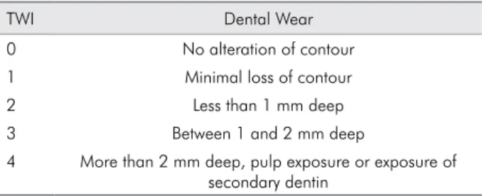
Only teeth with CDH scores of 2 or 3 were included. Each tooth received a clinical probing (tactile stimulus) and air blast (thermal-evaporative stimulus). Air blast was applied with an air syringe for 1s at a distance of 1 cm.7,10 The Tooth Wear Index (TWI)11 was used
to score the lesions’ depth from 1 to 4 (Table 1). The depth was measured by placing a periodontal probe perpendicular to the tooth’s long axis at the center of
the lesion. Those with score 3 or 4 required a illing.
The operator was calibrated for the clinical interventions by attending presentations on how the treatments should be carried out and how to use the restorative materials. Any questions regarding the
treatment protocol were clariied during the presentation.
In each patient, each tooth randomly received one type of treatment. A list of selected teeth was prepared for each patient, and they were arranged in a sequence according to a randomized approach. Then, a list of the treatments was randomly paired with the teeth.
The patient was not informed of the treatment for each tooth, and the clinical procedures were performed so that the patient could not distinguish among them, except for treatment 1.
The four treatments were:
• Treatment 1 (T1): Desensibilize KF 2% desensitizing gel (Dentscare Ltda., Joinville, Brazil), with active ingredients 5% potassium nitrate and 2% sodium
Table 1. The tooth wear index (TWI).
TWI Dental Wear
0 No alteration of contour 1 Minimal loss of contour
2 Less than 1 mm deep
3 Between 1 and 2 mm deep
luoride, was applied once per week according
to the manufacturer’s recommendations until complete remission of pain. Teeth were not restored in this group;
• Treatment 2 (T2): desensitizing gel was applied once, followed immediately by a restoration with Filtek Z250 resin composite and Adper Single Bond 2 adhesive (both from 3M Espe, Saint Paul, USA) following the manufacturers’ recommendations;
• Treatment 3 (T3): desensitizing gel was applied once per week until complete remission of pain and a subsequent restoration with Filtek Z250 resin composite and Adper Single Bond 2 adhesive;
• Treatment 4 (T4): only restoration was performed with Filtek Z250 resin composite and Adper Single Bond 2 adhesive.
Resin increments no thicker than 2 mm were applied and photoactivated using an LED-type device (Ultraled-Dabi Atlante, Ribeirão Preto, Brazil) with frequency 50/60 Hz and minimum power 500 mW/cm2.
The main outcome of the study was the patient’s discomfort or pain relief – a patient-reported outcome (PRO-type).12
Clinical Assessments
Clinical assessments were conducted by a calibrated examiner, other than the operator, at baseline and at intervals of 7, 30, 90 and 180 days after the treatments were performed. To assess the CDH intensity, an air blast was used.7,13
The calibration was carried out at the Universidade Federal do Piauí – UFPI using a dental mannequin, an air-water syringe and an explorer. The duration of the calibration process (training, adjustments and calibration exercises) was approximately 16 h, during which 10 patients with CDH were examined and their CDH scores were measured. The intra-examiner weighted kappa value was calculated by using the baseline values for CDH and by reexamining the patients seven days later. The kappa value was determined to be 0.76.
For the clinical assessments, the examiner received a form with a list of the teeth whose CDH should be scored. He was blinded to the group to which each tooth belonged and the treatments that were being
assessed. The order in which the teeth were evaluated was maintained during each observation period.
To process the data, SPSS 15 (SPSS Inc., Chicago, USA) for Windows was used. The results were analyzed using the Wilcoxon, Kruskal Wallis and Mann-Whitney tests (p < 0.05).
Results
The sample consisted of 8.8% maxillary incisors, 10.2% mandibular incisors, 7.35% maxillary canines, 35.3% maxillary premolars, 30.9% mandibular premolars, 34.4% maxillary molars and 3% mandibular molars. At baseline, the mean CDH scores of each
group did not signiicantly differ from one another
(p > 0.05) (Table 2). The recall rates were 100% for treatments 1, 2 and 4. For treatment 3, two patients
(12%) missed the inal assessment.
The mean CDH scores of groups and observation periods were compared using the Kruskal-Wallis
test. A statistically signiicant difference between
the treatments (p < 0.001) was found. At 7 days, there
was a statistically signiicant difference between all
treatments and the baseline: T1 (p = 0.001), T2 (p = 0.001), T3 (p < 0.001) and T4 (p < 0.001) (Table 2). Furthermore,
there was a statistically signiicant difference for T1
(p = 0.006), T2 (p = 0.04), and T4 (p = 0.023) when the mean CDH score at 180 days was compared with that at 7 days.
At seven days, there was a statistically signiicant
percent reduction of the CDH scores for all groups (p < 0.05) (Table 2). No subsequent reduction was observed (p = 0.221 at 30 days; p = 0.825 at 90 days; and p = 0.683 at 180 days).
At 7 days, there was no signiicant difference in
the mean CDH scores of T2, T3 and T4; furthermore,
there was no signiicant difference among T1, T2 and
T4. However, the mean CDH score of T3 was lower than that of T1 (p = 0.009). At 180 days, no difference was observed among treatments (p > 0.05) (Table 2).
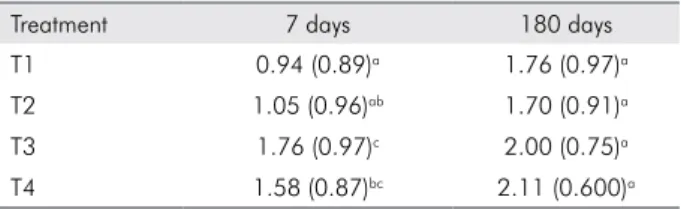
The mean CDH score at 7 days was subtracted from that of the baseline to calculate the CDH score variation. This variable represents the early effect of each therapy, theoretically eliminating the subjectivity
of the pain interpretation. There was a signiicant
T1 (p = 0.01) and T2 (p = 0.03). However, the mean CDH score variation after T4 was not statistically
signiicant from that after T2 (Table 3).
The mean CDH score at 180 days of observation was subtracted from the baseline score to calculate the CDH score variation. This variable represents the
late effect of each therapy. No signiicant CDH score
variation was observed at 180 days (p > 0.05) (Table 3).
Discussion
The aim of this study was to contribute to the methods of treatment of NCCL with CDH. A pain scale was applied at the beginning and at the end of the
experiment, so that pain intensity could be quantiied
and analyzed. The verbal scale used here has been adopted in previous studies to successfully measure CDH.3,5,7,9 However, because pain is an individual
experience, this study was limited by the psychological profile of participants and by the materials and
techniques tested. Thus, patients who found it dificult
to interpret the discomfort felt after treatments were performed were not included in the sample.
The use of desensitizing agents has been previously tested.13,14 Fluorides are one of the substances
recommended for CDH treatment because they occlude the dentinal tubules by forming insoluble precipitates. Dentin permeability decreases, but
the effect is short lived because luoride interacts
with calcium and calcium fluoride subsequently dissociates.14,15 However, potassium nitrate reduces
nervous excitability.16 This effect is the mechanism of
action currently accepted for the material tested in T1. T1 had the highest mean sensitivity score at 7 days. Furthermore, T1 was able to gradually
reduce sensitivity over time, leading to signiicant
differences after 7 and 180 days (Table 2), despite
having the lowest mean sensitivity score variation (Table 3). Because its effect is cumulative, the gel must be reapplied at weekly intervals. The limitation of this technique is that it requires that patients abide by the professional recommendations in the interval between applications. Traumatic brushing technique or dietary habits are risk factors that should be diagnosed during anamnesis and
modiied during the therapy. T1’s comparatively
poor performance may have occurred because, once CDH remitted, therapy ceased, and there was
likely a recurrence due to the dificult control of
the etiological factors of CDH. This factor was one limitation of this study that could have interfered
with the inal outcome. However, the operator kept
close telephone contact with patients during the study to clarify any questions and to remind them of this drawback.
T2 had a relatively high mean CDH score and a similar variation to T1 at seven days (Tables 2 and 3). This technique was tested to simulate the potential for achieving a desensitizing effect and bonding technique in the same clinical session. Although there is no current clinical recommendation for such
a procedure, the authors did not ind any scientiic
evidence that it is contraindicated.
One single application of the gel may not have
been suficient to induce the formation of insoluble
precipitates in the dentinal tubules. Furthermore, phosphoric acid etching increases dentin permeability. The latter may have been predominant because the two procedures occurred in the same clinical session. Dentin that is devoid of a smear layer is highly sensitive because products and stimuli are easily disseminated to the pulp.2,14,16 Furthermore,
an incompatibility between the gel and the adhesive
Table 2. Mean CDH scores at observation periods, according to treatment.
Treatment Mean (S.D.)
Baseline 7 days 180 days T1 2.41 (0.5)a 1.47 (0.8)b 0.65 (0.9)d T2 2.41 (0.5)a 1.35 (0.9)bc 0.71 (0.6)d T3 2.47 (0.5)a 0.71 (0.9)c 0.53 (0.9)d T4 2.71 (0.5)a 1.12 (0.6)bc 0.59 (0.5)d The same letter indicates a lack of significant difference (p < 0.05).
Table 3. Mean CDH scores variation (SD) at 7 and 180 days of observation when compared with baseline, according to treatment.
Treatment 7 days 180 days
T1 0.94 (0.89)a 1.76 (0.97)a T2 1.05 (0.96)ab 1.70 (0.91)a
system would have resulted in a poor resinous
iniltration and inadequate CDH control.
T3 and T4 had the lowest mean CDH score (Table 2) and the highest mean score variation (Table 3) at seven days of observation, respectively, which suggests that the synergistic effect between the desensitizing
gel and subsequent illing with resin composite (T3)
has the same effect as a filling with only a resin
composite (T4). However, at least ive consecutive
weekly applications of the desensitizing gel were necessary for a CDH reduction to a mild or absent
level. In the case of T4, the effect was signiicantly better at the end of the irst observation period (7 days).
Furthermore, the mean number of sessions required for T3 to achieve such a result was the same (p > 0.05) as for T1 (data not shown).
The choice of the desensitizing agent is important
when the NCCL will subsequently be illed with
a composite because the desensitizing agent that
contain sodium and calcium luoride may reduce
the bond strength of adhesives.9,17 However, none
of the illings were lost, probably due to the use of
a cavosurface bevel.18 The reduction in sensitivity
may have been the result of the effect of potassium nitrate on nervous excitability.15 Furthermore, any
agent that blocks the tubules reduces luid low,
thereby reducing hypersensitivity.19 Thus, T3’s
results may relect the effect of the gel on dentin
permeability and nervous excitability, and the
restorative technique could block the dentin luid
flow with the composite acting as a mechanical barrier against thermal stimuli.20 In the case of T4,
the latter effect can be hypothesized.
Dentists frequently use bonding agents and composites to treat CDH due to their familiarity with composites. However, one study has shown that composites provided a longer lasting desensitizing effect when compared with home treatments.5 A
different study found that dentists who were asked about CDH treatment reported a low success rate for the method.21 Movement of intratubular luid can be
prevented by using restorative resins and adhesive systems, and provide comfort to CDH patients.22
The results suggest that combining the two
techniques (desensitizing and composite illing) had
the same effect as performing an immediate composite
restoration, which is recommended when there is a substantial loss of tooth structure and teeth do not respond to other treatments.23 Restoring the tooth
reduced the CDH quickly. Therefore, the hypothesis of the study was rejected.
Although there is a general consensus that a
lexible restorative material in the cervical area is important, inite element analysis has raised awareness
about the role of elasticity. One argument is that the elastic modulus of restorative materials would
have to be signiicantly lower than the current level (1 GPa) to have a clinically signiicant impact on its
performance. Furthermore, adhesion quality plays a role in the behavior of restorations under tensile stress. Therefore, as long as the adhesive technique is carefully performed, the type of composite used is not as important as the choice of a material that has demonstrated clinical evidence of success.18 Thus, a
microhybrid composite resin was selected for this study, all the restorations were placed under rubber dam isolation and the manufacturers’ instructions were strictly followed.
An important limitation of this study is that etiology of NCCL were not taken into consideration. However,
the patients were clariied about the etiological factors
in order to control such factors whenever possible. Other studies that take into account the different clinical situations involved could be designed to compare different techniques and operatory conditions or the use of desensitizing substances and restorative materials in an attempt to resolve CDH.
The clinical relevance of this study lies in the fact that evidence is in favor of carrying out an immediate
illing of teeth with considerable tooth wear and
moderate to severe CDH. Further knowledge about dentin permeability is needed to understand the dynamics of CDH.
Conclusion
The results of this study demonstrate the following:
a. The response of the teeth with CDH was more pronounced after 7 days of observation but
without signiicant improvement thereafter;
b. It is more eficient to restore a tooth with moderate
to severe CDH as soon as possible rather than
1. Orchardson R, Gangarosa LP, Holland GR, Pashley DH, Trowbr idge HO, Ash ley FP, et a l. Dent i ne hypersensitivity-into the 21st century. Arch Oral Biol. 1994;39(Suppl):113S-119S.
2. Mjör IA. Dentin permeability: the basis for understanding pulp reactions and adhesive technology. Braz Dent J. 2009;20(1):3-16.
3. Rees JS, Addy M. A cross-sectional study of dentine hypersensitivity. J Clin Periodontol. 2002 Nov;29(11):997-1003. 4. Dababneh RH, Khouri AT, Addy M. Dentine hypersensitivity:
an enigma? A review of terminology, epidemiology, mechanisms, aetiology and management. Br Dent J. 1999 Dec;187(11):606-11
5. Miglani S, Aggarwal V, Ahuja B. Dentin hypersensitivity: recent t rends i n ma nagement. J Con ser v Dent. 2010 Oct;13(4):218-24.
6. Porto IC, Andrade AK, Montes MA. Diagnosis and treatment of dentinal hypersensitivity. J Oral Sci. 2009 Sep;51(3):323-32. 7. Holland GR, Narhi MN, Addy M, Gangarosa L, Orchardson
R. Guidelines for the design and conduct of clinical trials on dentine hypersensitivity. J Clin Periodontol. 1997 Nov;24(11):808-13.
8. Faul F, Erdfelder E, Lang AG, Buchner A. G*Power 3: a flexible statistical power analysis program for the social, behavioral, and biomedical sciences. Behav Res Methods. 2007 May;39(2):175-91.
9. Yu X, Liang B, Jin X, Fu B, Hannig M. Comparative in vivo study on the desensitizing efficacy of dentin desensitizers and one-bottle self-etching adhesives. Oper Dent. 2010 May-Jun;35(3):279-86.
10. Rees JS, Jin LJ, Lam S, Kudanowska I, Vowles R. The prevalence of dentine hypersensitivity in a hospital clinic population in Hong Kong. J Dent. 2003 Sep;31(7):453-61. 11. Smith BG, Knight JK. An index for measuring the wear of
teeth. Br Dent J. 1984 Jun;156(12):435-38.
12. Acquadro C, Berzon R, Dubois D, Leidy NK, Marquis P, Revicki D, et al. Incorporating the patient’s perspective into drug development and communication: an ad hoc task force report of the patient-reported outcomes (PRO) harmonization
group meeting at the food and drug administration, February 16, 2001. Value Health. 2003 Sep-Oct;6(5):522–31.
13. Kielbassa AM, Attin T, Hellwig E, Schade-Brittinger C. In vivo study on the effectiveness of a lacquer containing CAF2/ NaF in treating dentine hypersensitivity. Clin Oral Investig. 1997 Jun;1(2):95-9.
14. Aranha AC, Pimenta LA, Marchi GM. Clinical evaluation of desensitizing treatments for cervical dentin hypersensitivity. Braz Oral Res. 2009 Jul-Sep;23(3):333-9.
15. Oda M, Matos AB, Liberti EA. Morphology of dentin t reated with desensit izi ng substa nces: sca n n i ng electron microscopy study. Rev Odontol Univ São Paulo. 1999 Oct-Dec;13(4):337-42. Portuguese.
16. Pashley DH. Dentin permeability, dentin sensitivity, and treatment through tubule occlusion. J Endod. 1986 Oct;12(10):465-74.
17. Külünk S, Saraç D, Külünk T, Karakaş O. The effects of different desensitizing agents on the shear bond strength of adhesive resin cement to dentin. J Esthet Restor Dent. 2011 Dec;23(6):380-7.
18. Pecie R, Krejci I, García-Godoy F, Bortolotto T. Noncarious cervical lesions (NCCL) – a clinical concept based on literature review. Part 2: restoration. Am J Dent. 2011 Jun;24(3):183-92. 19. Zaimoglu A, Aydin AK. An evaluation of smear layer with
various desensitizing agents after tooth preparation. J Prosthet Dent. 1992 Sep;68(3):450-7.
20. Santiago SL, Franco EB, Mendonça JS, Lauris JR, Navarro MF. One-year clinical evaluation of tooth-colored materials in non-carious cervical lesions. J Appl Oral Sci. 2003 Sep;11(3):175-80.
21. Cunha-Cruz J, Wataha JC, Zhou L, Manning W, Trantow M, Bettendorf MM, et al. Treating dentin hypersensitivity: therapeutic choices made by dentists of the northwest precedent network. J Am Dent Assoc. 2010 Sep;141(9):1097-105 22. Pashley DH, Pashley EL, Carvalho RM, Tay FR. The effects
of dentin permeability on restorative dentistry. Dent Clin North Am. 2002 Apr;46(2):211-45, v-vi.
23. King PA. Adhesive techniques. Br Dent J. 1999 Apr 10;186(7):321-6.