• Nenhum resultado encontrado

Braz. Dent. J. vol.14 número1

N/A
N/A
Protected

Academic year: 2018

Share "Braz. Dent. J. vol.14 número1"

Copied!
4
0
0

Texto

(1)

Braz Dent J 14(1) 2003

12 N. Magro-Érnica et al.

Histologic Study of Use of Microfibrillar Collagen

Hemostat in Rat Dental Sockets

Natasha MAGRO-ÉRNICA Osvaldo MAGRO-FILHO Idelmo RANGEL-GARCIA

Department of Oral Surgery and Integrated Clinic, School of Dentistry of Araçatuba, UNESP, Araçatuba, SP, Brazil

The aim of this paper was to evaluate if the placement of microfibrillar collagen hemostat (MCH) into a dental socket interfered with healing. General anesthesia was administered to 30 adult male Albinus Wistar rats and the maxillary right central incisor was extracted. In the control group after each tooth was extracted, the socket was sutured. In the MCH group after each tooth was extracted, MCH was placed into the socket before suturing. Postoperatively, 5 animals were sacrificed from each group at 7, 21 and 28 days. The right maxilla was removed from each animal and histologic slides were stained with Masson’s trichromic and hematoxylin and eosin. Quantitative and qualitative analyses were done. The percentage of bone area in the dental socket was quantified using the Image Lab 98 image analysis system. The bone area formation for the control and MCH groups was: 8.1% and 3.3% at 7 days, 34.4% and 33% at 21 days and 41% and 41.3% at 28 days, respectively. We concluded that MCH interferes with the beginning of dental socket healing but does not interfere with the final healing of the dental socket.

Key Words: tooth extraction, hemostat, bleeding, dental socket.

Correspondence: Dr. O. Magro-Filho, Caixa Postal 341, 16015-050 Araçatuba, SP, Brasil. Fax: +55-18-622-2136. e-mail: dinho@foa.unesp.br ISSN 0103-6440

INTRODUCTION

Microfibrillar collagen hemostat (MCH) is a topical agent composed of resorbable microfibrillar collagen prepared to preserve the native bovine col-lagen molecules. The MCH attracts platelets and al-lows for the formation of a blood clot when it comes into contact with blood (1).

The manufacturer (MedChem Products Inc., Woburn, MS) suggests the use of MCH Avitene to achieve hemostasis with or without conventional meth-ods or in situations in which conventional methmeth-ods are not practical. This hemostatic agent is contraindicated as the sole hemostatic control in patients who have systemic problems causing hemorrhage.

To date, there is no experimental research in the literature relating the wound healing of a dental socket to MCH. The aim of this study was to determine whether or not the placement of MCH into a dental socket interferes with socket healing.

MATERIAL AND METHODS

(2)

Braz Dent J 14(1) 2003 Microfibrillar collagen hemostat in dental sockets 13

gentially to the distal surface of the last molar. The specimens were fixed in 10% formalin for 24 h, decalcified in formic acid and sodium citrate solution, clarified and embedded in paraffin so the sockets could be cut in a longitudinal plane (3). The blocks were semi-serially sectioned at 6 µm thickness and the sections were stained with Masson’s trichromic and hematoxylin and eosin.

Qualitative and quantitative analyses of bone formation were done. The percentage of bone area in each dental socket was quantified using the IMAGE LAB 98 image analysis system. Data were analyzed with the Tukey test and analysis of variance.

RESULTS

Qualitative Analysis

Control Group: After 7 days, the alveolus was partially filled with newly formed connective tissue, blood clot and showed thin osseous trabeculae princi-pally in the apical third (Figure 1). On day 21, the alveolus exhibited thick osseous trabeculae and some areas of mature connective tissue (Figure 2). On day 28, the alveolus was filled with mature osseous trabeculae (Figure 3).

MCH Group: At day 7, osteoid tissue and thin osseous trabeculae were present at the apical third. There was part of the MCH material in some samples (Figure 4) and polymorphonuclear neutrophil infiltra-tion. Thick osseous trabeculae were present at the apical and middle thirds at 21 days (Figure 5). The mature osseous trabeculae present on the 28th day filled almost all alveoli (Figure 6).

Quantitative Analysis

The quantification of the trabecular bone area in the dental sockets was done by carefully following all of the dental sockets linearly. The mean percentile values of trabecular bone area in the dental socket at 7, 21 and 28 days were: control group: 8.1%, 34.4% and 41.0%, respectively; MCH group: 3.3%, 33.0% and 41.3%, respectively.

The Tukey test was applied and the interaction values were then subjected to analysis of variance. There was a significant statistical difference between the experimental and the control groups only at

postop-erative day 7 and no significant statistical difference between the groups at postoperative days 21 and 28.

DISCUSSION

It is well-documented that MCH has local hemostatic action. Kubo et al.(4) placed it in contact with bleeding surfaces after great vessel hemostasis during urologic surgery. They concluded the mate-rial quickly controlled hemorrhage in all of the patients studied. Low et al. (5) used MCH in laparoscopy because of its effectiveness as a hemo-static agent. Nakamura et al.(6) used MCH on the brain cortex and reported that the hemostatic action was 95% effective without any side effects in all of their experimental surgeries. Low et al.(5) and Na-kamura et al. (6) concluded that MCH is biocompa-tible in their areas of research. The dental socket, however, presents its own peculiarities when com-pared with other areas of the body (7).

An in vitro coagulation mechanism of com-mercialized products was evaluated by Wagner et al. (8). They classified from better to worse the follow-ing hemostatic agents: microfibrillar collagen (Avitene), collagen sponges (Helistat, Instatand

Actifoam), gelatin sponge (Gelfoam) and

oxi-dized regenerated cellulose (Surgicel). As a result,

the use of MCH for postoperative bleeding in dental sockets appears to be an interesting option. The aim of this paper was not to evaluate hemostasis; how-ever, we did notice that the MCH group did not show the same amount of bleeding as the control group.

Barbolt et al. (9) carried out a comparative study with gelatin sponge, oxidized regenerated cel-lulose and MCH implanted in the subdural space after craniotomy in rabbits. They reported that the tissue reaction to MCH was characterized by moder-ate to marked granulomatous inflammation with an acute inflammatory component indicating a greater degree of tissue irritation. Sequelae of this reaction were still observed at 92 days post-implantation.

(3)

Braz Dent J 14(1) 2003

14 N. Magro-Érnica et al.

Figure 1. Control Group. 7 days postoperative. Newly formed connective tissue, blood clot and thin osseous trabeculae in the apical third. H&E. Original magnification, 63X.

Figure 2. Control Group. 21 days postoperative. Presence of thick osseous trabeculae and some areas of mature connective tissue. H&E. Original magnification, 63X.

Figure 4. MCH Group. 7 days postoperative. Connective tissue, osteoid tissue and thin osseous trabeculae in the apical third. MCH material is still present. H&E. Original magnification, 63X.

Figure 5. MCH Group. 21 days postoperative. The apical third presents a thick osseous trabeculae. H&E. Original magnification, 63X.

Figure 6. MCH Group. 28 days postoperative. Mature osseous trabeculae fills the alveolus. H&E. Original magnification, 63X. Figure 3. Control Group. 28 days postoperative. Mature osseous

(4)

Braz Dent J 14(1) 2003 Microfibrillar collagen hemostat in dental sockets 15

Okamoto (7) who reported after numerous studies that the use of materials in the dental socket should be restricted to those cases in which the need for hemosta-sis outweighs the potential morbidity.

In our study, MCH was not totally absorbed at 7 days postoperatively and there was formation of thin trabeculae bone in the apical third of the alveolus; however, the control group showed a greater quantity of new bone at 7 days, most likely due to the presence of MCH in the other group. The final healing quality, however, was similar in both groups. The initial inter-ference of MCH did not influence the quantity of trabecular bone in the dental socket..

We conclude that the hemostatic agent presented here appears to be a viable alternative in controlling intraoperative and postoperative hemorrhage from the dental alveolus because it is both biocompatible and effective.

ACKNOWLEDGMENTS

This study was supported by CNPq. The authors wish to thank Dr. Tetuo Okamoto for his assistance with the histological analysis and Dr. Eloi Dezan Jr. for his assistance with the statistical analysis (School of Dentistry of Araçatuba, UNESP, Brazil).

RESUMO

O objetivo deste trabalho é avaliar se a colocação do MCH no interior do alvéolo dental interfere ou não no reparo alveolar. Foi feita anestesia geral nos trinta ratos machos adultos Albinus

Wistar e o incisivo central superior direito foi extraído. No grupo controle, após a exodontia o alvéolo foi suturado. No grupo MCH, após a exodontia introduziu-se MCH dentro do alvéolo que foi suturado. No pós-operatório, cinco animais de cada grupo foram sacrificados aos 7, 21 e 28 dias. Retirou-se a maxila

direita de cada animal e cortes histológicos foram confeccionados. Foi feita análise qualitativa e quantitativa. A porcentagem de área óssea no interior dos alvéolos dentais foi quantificada usando o sistema de análise de imagens ImageLab 98. A área óssea neoformada no grupo controle e no grupo MCH foi: 8.1% e 3.3% aos 7 dias, 34.4% e 33% aos 21 dias e 41% e 41.3% aos 28 dias, respectivamente. Dentro das condições experimentais deste trabalho foi possível concluir que o MCH interfere durante o início do reparo alveolar e não interfere no reparo final do alvéolo dental.

REFERENCES

1. Nakajima M, Kamei T, Tomimatu K, Manabe T. An intraperito-neal tumorous mass caused by granulomas of microfibrillar col-lagen hemostat (Avitene). Arch Pathol Lab Med 1995;119:1161-1163.

2. Okamoto T, Russo MC. Wound healing following tooth extrac-tion. Histochemical study in rats. Rev Fac Odont Araçatuba 1973;2:153-169.

3. Morse A. Formic acid sodium citrate descalcification and butyl alcohol deydration of teeth and bone for sectioning in paraffin. J Dent Res 1945;24:143.

4. Kubo S, Ishibashi A, Koshiba K. Clinical studies on topical hemostat. Avitene in urological surgery. Hinyokika Kiyo 1984;30:131-134.

5. Low RK, Moran ME, Goodnight JE. Microfibrillar collagen he-mostat during laparoscopically direct liver biopsy. J Laparoendosc Surg 1993;3:415-420.

6. Nakamura N, Hashimoto T, Sakai H, Yasue S, Nagayama I, Sasaki T, Basugi N, Kim K, Kitamura K, Yoshimasu M. Clinical application of microfibrillar collagen hemostat (Avitene) in neu-rosurgical field. No Shinkei Geka 1998;16:933-938.

7. Perri de Carvalho AC, Okamoto T. Cirurgia Bucal, Fundamentos Experimentais Aplicados à Clínica. 1st ed. São Paulo: Editora Panamericana, 1987.

8. Wagner WR, Pachence JM, Ristich J, Johnson PC. Comparative in vitro analysis of topical hemostatic agents. J Surg Res 1996;66:100-108.

9. Barbolt TA, Odin M, Kangas L. Pre-clinical subdural tissue reaction and absorption study of absorbable hemostatic devices. Neurol Res 2001;23:537-542.

10. Carvalho ACP, Okamoto T. Implantes intra-alveolares. Considerações sobre estudos experimentais. Rev Ass Paul Cirurg Dent 1978;32:11-15.

Imagem

Figure 2. Control Group. 21 days postoperative. Presence of thick osseous trabeculae and some areas of mature connective tissue

Referências

Documentos relacionados

The structure of the lung tissue in group OA+IX was damaged; the alveolar septal capillaries were markedly expanded and congested, the alveolar cavity was partially

The animals of group III showed a lower level of radioactivity in the spleen (P<0.0006) than the control group and a higher level in the liver, lung and blood clot than the

Segue o acórdão citado: Importante ressaltar, ainda, que o Sistema Único de Saúde funda-se no princípio da co-gestão, pela participação simultânea dos entes estatais dos

O valor de “fz” (ou de “hm“ , que é o valor que deve ser colocado na equação 2.7 mencionada no capitulo 2, em lugar de “ae” para se estimar o valor da rugosidade

In treated rats, however, the volume fraction of blood clot and connective tissue was greater, causing a smaller quantity of trabecular bone (Figure 1).. Histometric data

defect filled by newly formed bone, characterized by the presence of globules or thin interconnecting bone trabeculae and the presence of osteocytes and parallel basophilic lines

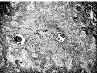
The mean relative volume density of the inflammatory cells (Table 2) was higher for the control group in comparison with the test group both at 7 and at 14 days.. This

The parameters evaluated were remaining biomaterial, loose connective tissue and newly formed bone in a standard area.. Statistical analysis was performed by Mann-Withney and