Abstract
Submitted: February 20, 2017
Modiication: June 19, 2017 Accepted: June 22, 2017
Alveolar bone repair with
strontium-containing nanostructured carbonated
hydroxyapatite
Objective: This study aimed to evaluate bone repair in rat dental sockets after implanting nanostructured carbonated hydroxyapatite/sodium alginate (CHA) and nanostructured carbonated hydroxyapatite/sodium alginate containing 5% strontium microspheres (SrCHA) as bone substitute materials. Methods: Twenty male Wistar rats were randomly divided into two experimental groups: CHA and SrCHA (n=5/period/group). After one and 6 weeks of extraction of the right maxillary central incisor and biomaterial implantation, 5 µm bone blocks were obtained for histomorphometric evaluation. The parameters evaluated were remaining biomaterial, loose connective tissue and newly formed bone in a standard area. Statistical analysis was performed by Mann-Withney and and Wilcoxon tests at 95%
level of signiicance. Results: The histomorphometric results showed that the
microspheres showed similar fragmentation and bio-absorbation (p>0.05). We observed the formation of new bones in both groups during the same
experimental periods; however, the new bone formation differed signiicantly
between the weeks 1 and 6 (p=0.0039) in both groups. Conclusion: The CHA and SrCHA biomaterials were biocompatible, osteoconductive and bioabsorbable, indicating their great potential for clinical use as bone substitutes.
K e y w o r d s : Hydroxyapatite. Bone repair. Rats. Strontium. Histomorphometric evaluation.
André Boziki Xavier do CARMO1 Suelen Cristina SARTORETTO1 Adriana Terezinha Neves Novellino
ALVES1 José Mauro GRANJEIRO2 Fúlvio Borges MIGUEL3
Jose CALASANS-MAIA1
Monica Diuana CALASANS-MAIA1
http://dx.doi.org/10.1590/1678-7757-2017-0084
1Universidade Federal Fluminense, Faculdade de Odontologia, Laboratório Associado de Pesquisa Clínica em Odontologia, Niterói, RJ, Brasil.
2Instituto Nacional de Metrologia, Qualidade e Tecnologia, Programa de Bioengenharia, Duque de Caxias, RJ, Brasil
3Universidade Federal do Recôncavo da Bahia, Centro de Ciências da Saúde, Santo Antônio de Jesus, BA, Brasil.
Corresponding address: Mônica Diuana Calasans-Maia Laboratório Associado de Pesquisa Clínica em Odontologia, Faculdade de Odontologia
Introduction
Implant-supported restoration has been increasingly performed by dentists for both aesthetic and functional
reasons. However, when infections, pathological
processes, extractions, or congenital and traumatic
injuries on the maxilla and the mandible lead to bone loss, dental implant installation might not be the best
opition20. Therefore, to minimize the loss of alveolar
bone, or even restore it, different types of alloplastic
grafts have been used, and new biomaterials have been the focus of research aiming to develop bone
substitutes.
Among these grafts, HA Ca10(PO4)6(OH)2 has been widely used as a bone substitute for approximately 8 0 y e a r s1 8. T h i s c e ra m i c i s b i o c o m p a t i b l e ,
osteoconductive15, similar to the bone and tooth tissue
inorganic portions1, bioactive, and allows substitutions
in its molecular formula and periodic monitoring via imaging because of its radiopacity. Additionally, it
is mechanically tough and bioactive, and it is not
antigenic, carcinogenic, or toxic.
However, the clinically used HA is not biodegradable and remains at the implantation site for long periods23,
which limits bone regeneration. The lack of degradation
is probably due to the high temperatures during
ceramics production16 and treatment after synthesis
(sintering), which increases the crystallinity and
hinders biosorption. With this in mind, nanostructured
materials composed of particles smaller than 100 nm
with low crystallinity show to be potential alternatives to grafts when produced with non-sintered materials
at low temperatures17, considering they can imitate
biological apatite12.
Researchers have chemically modified HA by substituting phosphate groups (PO4) or hydroxyl groups (OH) with carbonate (CO3) to develop a nanostructured carbonated hydroxyapatite at low
temperatures9. Under these conditions, the produced
biomaterial is similar to stoichiometric HA, but with
lower crystallinity and higher solubility, which favors
rapid bioabsorption and bone regeneration17.
Furthermore, the stability and lexibility of the HA structure enables different ionic substitutions3.
It is possible to induce the exchange of many
cations and anions by modifying the structure of
stoichiometric HA to resemble biological apatite1.
In these techniques, calcium frequently substitutes
strontium (Sr2+)10; despite still being present in lower
amounts compared to strontium, calcium alters the
crystal structure and some HA properties, including phase stability, solubility, and reactivity4, thus
decreasing ceramic mechanical strength. Additionally,
strontium reduces bone resorption and increases bone
formation. The high solubility of HA combined with Sr2+ increases the number of interconnected pores,
promoting cell migration, interfacial bonding13, and
osseointegration11,18.
Thus, this study evaluated histomorphometric bone repair in rat tooth sockets after implanting CHA and
SrCHA synthesized at low temperatures.
Material and methods
Animal experiments and breeding were performed
according to the institutional review board (CEUA/ UFF), N°179/2012, the NIH Guide for Care and Use
of Laboratory Animals and the Brazilian legislation on
animal research.
Biomaterials
The nanostructured carbonated hydroxyapatite powder and nanostructured carbonated hydroxyapatite
containing strontium were prepared using a
precipitation wet method with average temperature
of 5°C and 6% wt CO3. The synthesized solids were iltered and washed with deionized water (MilliQ®,
Millipore Corporation, Billerica, MA, USA) until reaching
neutral pH (pH=7). Then, the material was lyophilized
in a FreeZone 1 lyophilizer (Labconco®, Kansas City,
Missouri, USA) and separated on sieves according to particle sizes from 74 μm to 37 μm.
To produce microspheres, the obtained powders
were individually mixed with a solution of sodium alginate (Sigma Aldric®/Fluka Biochemika®, Buchs,
Switzerland) diluted in ultrapure water (MilliQ®,
Millipore Corporation, Billerica, MA, USA) at a ratio of
15:1. The mixture was then extruded into 0.15 molar calcium chloride (0.15 M CaCl2), in which instant microsphere formation was observed. The mixture was
kept at rest for 24 h until complete gelation. After this
step, the microspheres were washed with ultrapure water until fully eliminating saline. Immediately
after the washing, the microspheres were dried by
an Eppendorf tube and sterilized with gamma radiation
in a cobalt-60 irradiator (Gamma Cell) for 760 m with total dose of 15 kGy and dose rate of 19.72 Gy/m.
Electron microscopy scanning of the microspheres
showed similar morphology and surface texture.
SrCHA presented fewer surface pores than CHA, as previously shown25.
The XRD patterns revealed that the microspheres had low crystallinity, as indicated by the broad and poorly deined peaks. However, the XRD pattern of SrCHA (Figure 1) showed narrower peaks than that
of CHA because of the presence of Sr.
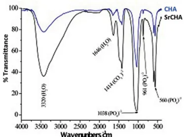
The spectra in Figure 2 show the vibrational bands
correspond to CHA. The 3435 and 1639 cm−1 regions,
which are large and intense, represent water bands,
indicating that the material is not ceramic. In Figure
2, regions 867, 868, 1415, 1425 and 1482 cm−1 show
carbonate ions, indicating that replacement occurred
as expected. The other bands that can be observed in the igure show phosphate ions. Because of the high hydration of the samples, it was not possible to identify
hydroxyl ions representative bands.
Animals and surgical procedure
A total of 20 male Wistar rats with average weight
of 300 g were randomly divided into two groups (CHA and SrCHA) with ive animals for each experimental period. The sample size was based previous studies that followed the same animal protocol16. According
to the CONCEA and 3R’s program, we should reduce
the number of animals in experimentation as much as
possible without loosing the accuracy of the statistical
analysis. Five animals for each group is the minimum
to perform the normality test21. The animals were
evaluated after one and six weeks.
After anesthesia with intramuscular injection of 75
Figure 1- XRD pattern of CHA and SrCHA. The peaks of SrCHA are narrower than those of CHA, indicating low cristallinity for SrCHA when compared to the CHA group
mg/kg ketamine hydrochloride (Ketalar®, Veltbrands,
São Paulo, Brazil) and sedation with 1.5 ml/kg xylazine (Rompun®, Veltbrands, São Paulo, Brazil),
the antisepsis of the perioral region and oral mucosa
was achieved with 2.0% and 0.12% chlorhexidine,
respectively. Then, sindesmotomy and extraction of the upper right central incisor were performed with a dental explorer no. 5 (Dulex®, São Paulo, SP, Brazil)
and a pediatric forceps no. 151 (Dulex®, São Paulo, SP,
Brazil), respectively (Figure 3A and 3B), and 0.2 mg of the biomaterials (Figure 3C) was implanted in the tooth socket, followed by suturing (Figure 3D). After the surgical procedures were completed, postoperative analgesia with 1 mg/kg meloxicam (Duprat®, Rio de
Janeiro, Brazil) was administered subcutaneously
every 24 h for three d since the day of the surgery.
After the experimental period, the animals were
euthanized by applying a lethal dose of thiopental 150 mg/kg [(Thiopentax® (Cristália), Itapira, São
Paulo, Brazil)] to collect bone blocks containing the
biomaterials and surrounding tissues. The specimens
were fixed in 4% formaldehyde, decalcified in decalciication solution (Allkimia®, Campinas, São
Paulo, Brazil) for 48 h and embedded in parafin. The blocks were cut at 5-μm thickness, stained with hematoxylin and eosin (HE) and examined by light microscopy (Eclipse E400, Nikon®, Tokyo, Japan).
Histomorphometric analysis
Histomorphometric analysis was performed to
quantify the remaining biomaterial, the connective
tissue loose and the bone newly formed in the standard area. The morphometric measurements were
performed using the Image-Pro Plus® software, version
4.5.0.29 (Media Cybernetics, Silver Spring, EUA) with 5 microscopic ields under 20x augmentation with a 1-blinded examiner (SCS). In each histological slice
stained with HE, 5 non-superimposing microscopic ields obtained by scanning at 20x magniication were captured in the medium third region of the socket
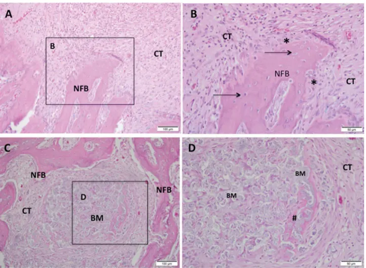
Figure 3- Representative photomicrographs of alveolar socket after 7 days. A and B: CHA group and C and D: SrCHA group. In A and B: presence of connective tissue surrounding the biomaterial (BM) microsphere and peripheric newly formed bone (NFB). C and D: presence of newly formed bone surrounding the connective tissue area, containing particulate biomaterial with peripheric osteoid (#).
after biomaterial implantation. With the Image-Pro
Plus® 6.0 (Media Cybernetics, Silver Spring, Maryland,
USA), a grid of 250 points superimposed on the area
under analysis allowed the determination of the
volume density of the newly formed bone, of the
connective tissue and of the residual biomaterial. The 250 points superimposed on each photomicrograph were considered as 100%, so each point was classiied and the percentage of each parameter was obtained.
Statistical analysis
The results are presented as percentages, and values are presented as the mean value (±) standard
deviation. The mean values and standard deviations
obtained in each group were tested for normality according to D’Agostino-Pearson’s omnibus test. The data did not present a normal distribution, this way
both the non-parametric statistical analysis of
Mann-Withney for inter-group and and the Wilcoxon for
intra-group were performed using the Prism Graph Pad 6.3
software (Inc. La Jolla, California, USA), with p≤0.05 being considered statistically signiicant.
Results
Histological results
1 week
At the irst experiment, both biomaterial groups presented the dental socket illed with fragmented biomaterial spheres surrounded by granulation tissue with remnants of blood clots, neoformed capillaries, and ibroblasts. In both groups, we observed moderate mononuclear inlammatory iniltrate as lymphocytes and macrophages between the components of the conjunctive tissue and the surroundings of partially
bioabsorbed microspheres. We also observed giant
cells permeating the biomaterial particles. New bone
formation occurred centripetally in patches during this period and was more evident in the apical region of
the socket (Figure 3).
6 weeks
In both groups, we observed a discrete chronic inlammatory response, with a few giant cells close to the microspheres, which were evidently fragmented.
The biomaterials were fragmented and bioabsorbed differently. The quantity of remaining material in the
SrCHA group was slightly lower than that from the
CHA group. Bone formation process replaced the inlammatory cell content in the irst experiment at the same time that there was a decrease in mature
connective tissue composed by collagen fibers.
The newly formed bone characterized by the thick trabecular bone was similar in both groups and occurred near the remaining bone and in direct contact
with microspheres (Figure 4).
The histological evaluation showed that in the
interstitial spaces formed between the microspheres
with no bone formation had a highly vascularized loose of connective tissue after one week that showed
increased organization after six weeks with newly
formed bone. This angiogenesis is essential for bone
regeneration because these new blood vessels provide oxygen, nutrients, and cells, all considered essential
for bone formation.
Histomorphometric evaluation
H i s t o m o r p h o m e t r i c a n a l y s i s s h o w e d n o signiicant differences after 1 (CHA=14.6±2.50 and SrCHA=18.9±1.69) and 6 weeks (CHA=16.5±2.41
and SrCHA=10.4±2.33) between groups regarding
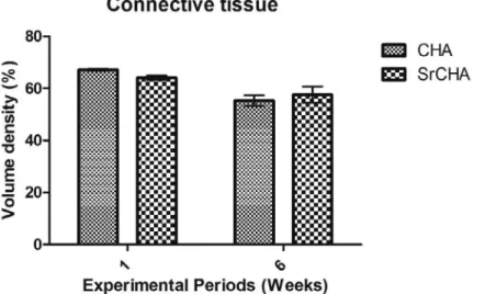
biomaterials bioabsorption (Figure 5). The amount
of connective tissue formed in the tooth socket permeating the microspheres was similar for both
Figure 5- Volume density of the remaining biomaterial in the dental alveolus after 1 and 6 weeks of implantation. The values were similar for both groups in both experimental periods. We observed no signiicant difference between groups. Results are shown as mean percentages ± conidence intervals (vertical bars)
groups and periods (Figure 6). Regarding the newly formed bone, morphometric analysis showed that the
percentage of mineralized tissue was similar between
groups at 1 (CHA=18.2±2.04 and SrCHA=17±17,
and 6 weeks (CHA=28.2±3.82 and SrCHA=32±4.15). However, we observed a higher percentage of newly
formed bone in both groups after 6 weeks when
compared to1 week (p=0.0039) (Figure 7).
Discussion
Calcium ions in biological apatite have been
partially substituted with other ions, such as Sr2+,
Mg2+, and Zn2+. This change affects the crystallinity,
solubility, surface energy, and dissolution rate of the
material, thus improving its bioactivity. Based on these indings, this study evaluated a promising bone substitute based on a synthesized at low temperature nanostructured carbonated hydroxyapatite containing
strontium10.
According to a previous study5, the sintering
of nanostructured hydroxyapatite causes crystal densiication and nanostructured features loss, thereby increasing crystallinityand reducing solubility15.
Additionally, the sintering process is responsible for
the removal of sodium alginate. In this study, we did not sintered the materials, retaining their nanometric
characteristics, low crystallinity and sodium alginate
content.
The non-strontium-containing hydroxyapatite was used as control group, as we aimed to evaluate the inluence of strontium on hydroxyapatite in bone
repair. We did not perform the dental socket illing with blood clot, as these results have already been
published with the same experimental periods22 and
are already established in the literature19. In addition,
according to the recommendation by the CONCEA and the 3R’s Program21, the number of animals used in
experimental studies should be reduced.
The biomaterials evaluated in this study were
biocompatible, despite the presence of a limited chronic inlammatory response with giant cells that diminished with time. The giant cells, considered
as foreign body type giant cells, are of
monocytic-phagocytic lineage and are important in the tissue repair mechanism, as they carry out cellular and
tissue debris phagocytose, as well as contribute to
the bioabsorption of biomaterial fragments, besides
secreting cytokines that favor essential cellular events in tissue repair. The presence of multinucleated
giant cells modulated by the chemical surface of
the biomaterials2 has demonstrated the importance
of macrophage subsets in the reaction to foreign bodies and, consequently, in the biocompatibility of
biomaterials6,7. Therefore, the presence of these cells
can occur as an attempt to reabsorb the material,
which not necessarily implies lack of biocompatibility. From a biological point of view, the material
fragmentation caused by lower crystallinity and smaller
particles susceptible to phagocytosis could justify the
presence of these cells around the particles. This type of tissue response is considered inherent to the healing
mechanism after the implantation of biomaterials14
and has been observed in other studies on bone
regeneration with biomaterials14.
A previous study observed that at the nanoscale,
the biomaterial resembled biological apatite5 and
presented bioabsorption similar to that observed in other studies15,19. Such bioabsorption is an essential
for a suitable bone substitute because biosorption
is important for bone physiology after biomaterial
implantation15. In areas where the microspheres
were bioabsorbed, we observed the formation of
mineralized tissue, characterized by more cellular and non-lamellar bone associated with microspheres
and at contact with the remaining alveolar bone, delimiting the dental alveolus, which conirms that the nanostructured carbonated HA here evaluated was
bioactive, osteoconductive16,19 and highly crystalline5,23.
The use of biomaterial microspheres has been considered preferable because the interstitial space
between the implanted spheres provides macropores
for tissue invasion and also because microsphere
implantation can be performed with minimally invasive
surgical techniques. In addition, the spheres do not have surface edges or dimensions that could lead to inlammation. To produce the microspheres, we mixed nanostructured carbonated HA powders with sodium
alginate, an inert and biodegradable polymer. The histological results showed that there were interstitial
spaces between the microspheres. In areas where
there was no new bone formation, we observed a
highly vascularized and loose connective tissue after one week that showed increased organization after
six weeks. This angiogenesis is essential for bone
regeneration because these new blood vessels provide
oxygen, nutrients, and cells considered essential for bone formation24.
In the histological evaluation, we observed a
greater fragmentation of SrCHA compared to the CHA group. We did not sinter the biomaterials used
in this study, so they are considered low crystalline
materials. However, the incorporation of strontium in
the hydroxyapatite by partially replacing it with calcium
changed crystallinity, morphology, lattice parameters, crystal size, stability, bioactivity, biocompatibility,
and osteoconductivity of CHA. This set of physical
and chemical changes alter the fragmentation and
bioabsorption of biomaterials25.
Sr2+ has been widely used for partial substitutions
of HA because of its dual ability to stimulate bone
formation and reduce bone resorption. However, in our study, we observed no increase in newly formed
mineralized tissue in the SrCHA group, indicating
that Sr2+ did not inluence the osteogenic potential
of nanostructured carbonated HA, possibly because of the low Sr2+ content (1.7%) identiied by atomic
absorption spectrometry after synthesis and prior to
the implantation, regardless of an initial theoretical
Sr2+ concentration of 5%. These results are similar to
those obtained by other study8 aimed at producing
1% ZnHA, obtaining a maximum incorporation of 0.4%. Similarly, Resende, et al.23 (2013) showed a
reduction of approximately 50% in the experimental zinc concentration of ZnHA compared to the initial
theoretical concentration.
Conclusion
Our results suggest that both CHA and SrCHA,
produced at low temperature and not sintered, were biocompatible, bioactive, ostecondutive
osteoconductive, and bioabsorbable, indicating its
great potential for clinical use as bone substitutes.
Further studies with a higher content of Sr2+ associated
with nanostructured carbonated hydroxyapatite
are necessary to evaluate the effect of Sr2+ on the
biological response.
Acknowledgements
The authors are grateful to FAPERJ – Rio de Janeiro Research Foundation for inancing this study and making it possible.
References
1- Aina V, Bergandi L, Lusvardi G, Malavasi G, Imrie FE, Gibson, et al. Sr-containing hydroxyapatite: morphologies of HA crystals
and bioactivity on osteoblast cells. Mater Sci Eng C Mater Biol Appl. 2013;33(3):1132-42.
2- Anderson JM, Rodriguez A, Chang DT. Foreign body reaction to biomaterials. Semin Immunol. 2008;20(2):86-100.
3- Bigi A, Boanini E, Capuccinia C, Gazzano M. Strontium-substituted hydroxyapatite nanocrystals. Inorganica Chim Acta.
2007;360(3):100-16.
4- Brook I, Freeman C, Grubb S, Cummins N, Curran D, Reidy C, et al. Biological evaluation of nano-hydroxyapatite-zirconia (HA-ZrO2) composites and strontium- hydroxyapatite (Sr-HA) for load-bearing
applications. J Biomater Appl. 2012;27(3):291-8.
5- Calasans-Maia MD, Melo BR, Alves AT, Resende RF, Louro RS, Sartoretto SC, et al. Cytocompatibility and biocompatibility of nanostructured carbonated hydroxyapatite spheres for bone repair. J
6- Carneiro E, Garcia RB, Oliveira RC, Moraes FG, Menezes R, Letra A, et al. Microscopic and radiographic analysis of the effect of particle size of demineralized bovine cancellous bone matrix on the repair of
bone defects in femurs of rabbits. J Appl Oral Sci. 2005;13(2):157-62. 7- Cestari TM, Oliveira RC, Sanada JT, Garlet GP, Taga R, Granjeiro JM. Biocompatibility evaluation of a new bioresorbable pin for membrane ixation. Braz Dent J. 2010;21(6):482-90.
8- Costa AM, Soares GD, Calixto R, Rossi AM. Preparation and properties of zinc containing bioceramics. Key Eng Mater. 2004;254-256:119-22. 9- Cox SC, Jamshidi P, Grover, LM, Mallick KK. Low temperature aqueous
precipitation of needle-like nanophase hydroxyapatite. J Mater Sci Mater Med. 2014;25(1):37-46.
10- Cox SC, Jamshidi P, Grover LM, Mallick KK. Preparation and characterisation of nanophase Sr, Mg, and Zn substituted
hydroxyapatite by aqueous precipitation. Mater Sci Eng C Mater Biol Appl. 2014;35:106–14.
11- Dagang G, Kewei X, Yong H. The inluence of Sr doses on the in vitro biocompatibility and in vivo degradability of single-phase Sr-incorporated HAPcement. J Biomed Mater Res A. 2008;86(4):947-58. 12- Dorozhkin SV. Nanosized and nanocrystalline calcium orthophosphates. Acta Biomater. 2010; 6(3):715-34.
13- Guo D, Xu K, Zhao X, Han Y. Development of a strontium-containing hydroxyapatite bone cement. Biomaterials. 2005;26(19):4073-83. 14- Hankenson KD, Dishowitz M, Gray C, Schenker M. Angiogenesis in bone regeneration. Injury. 2011;42(6):556-61.
15- Hasegawa M, Doi Y, Uchida A. Cell-mediated bioresorption of sintered carbonate apatite in rabbits. J Bone Joint Surg Br. 2003;85(1):142-7.
16- Hesaraki S, Nazarian H, Pourbaghi-Masouleh M, Borhan S. Comparative study of mesenchymal stem cells osteogenic
differentiation on low-temperature biomineralized nanocrystalline carbonated hydroxyapatite and sintered hydroxyapatite. J Biomed Mater Res B Appl Biomater. 2014;102(1):108-18.
17- Liao S, Watari F, Xu G, Ngiam M, Ramakrishna S, Chan CK. Morphological effects of variant carbonates in biomimetic
hydroxyapatite. Mater Lett. 2007;61(17):3624-8.
18- Machado CP, Sartoretto SC, Alves AT, Lima IB, Rossi AM, Granjeiro JM, et al. Histomorphometric evaluation of strontium-containing nanostructured hydroxyapatite as bone substitute in sheep. Braz Oral Res. 2016;30(1):e45.
19- Menezes LR Junior, Gaujac C, Trento CL. Inluência das alterações locais sobre o processo de reparo alveolar. Rev Saude Pesq. 2009;2(3):411-16.
20- Moraschine V, Barboza ES. Quality assessment of systematic reviews on alveolar socket preservation. Int J Oral Maxillofac Surg.
2016;45(9):1126-34.
21- National Centre for the Replacement, Reinement and Reduction of Animals in Research - NC3Rs. Animal Research - Reporting in vivo experiments: the ARRIVE guidelines. J Physiol. 2010;588(Pt 14):2519-21.
22- Okamoto T, Russo MC. Wound healing following tooth extraction: histochemical study in rats. Rev Fac Odontol Aracatuba. 1973;2(2):153-69.
23- Resende RF, Fernandes GV, Santos SR, Rossi AM, Lima I, Granjeiro JM, et al. Long-term biocompatibility evaluation of 0.5 % zinc containing